
Proguithera caspersi, Chen & Li & Cai, 2021
|
publication ID |
https://doi.org/ 10.11646/zootaxa.4963.2.6 |
|
publication LSID |
lsid:zoobank.org:pub:07661229-9D28-40EC-AA20-9746720DB42D |
|
DOI |
https://doi.org/10.5281/zenodo.4730746 |
|
persistent identifier |
https://treatment.plazi.org/id/03B787D3-FFF8-4F0A-2088-AC82FC1B1DD9 |
|
treatment provided by |
Felipe |
|
scientific name |
Proguithera caspersi |
| status |
sp. nov. |
Proguithera caspersi sp. nov.
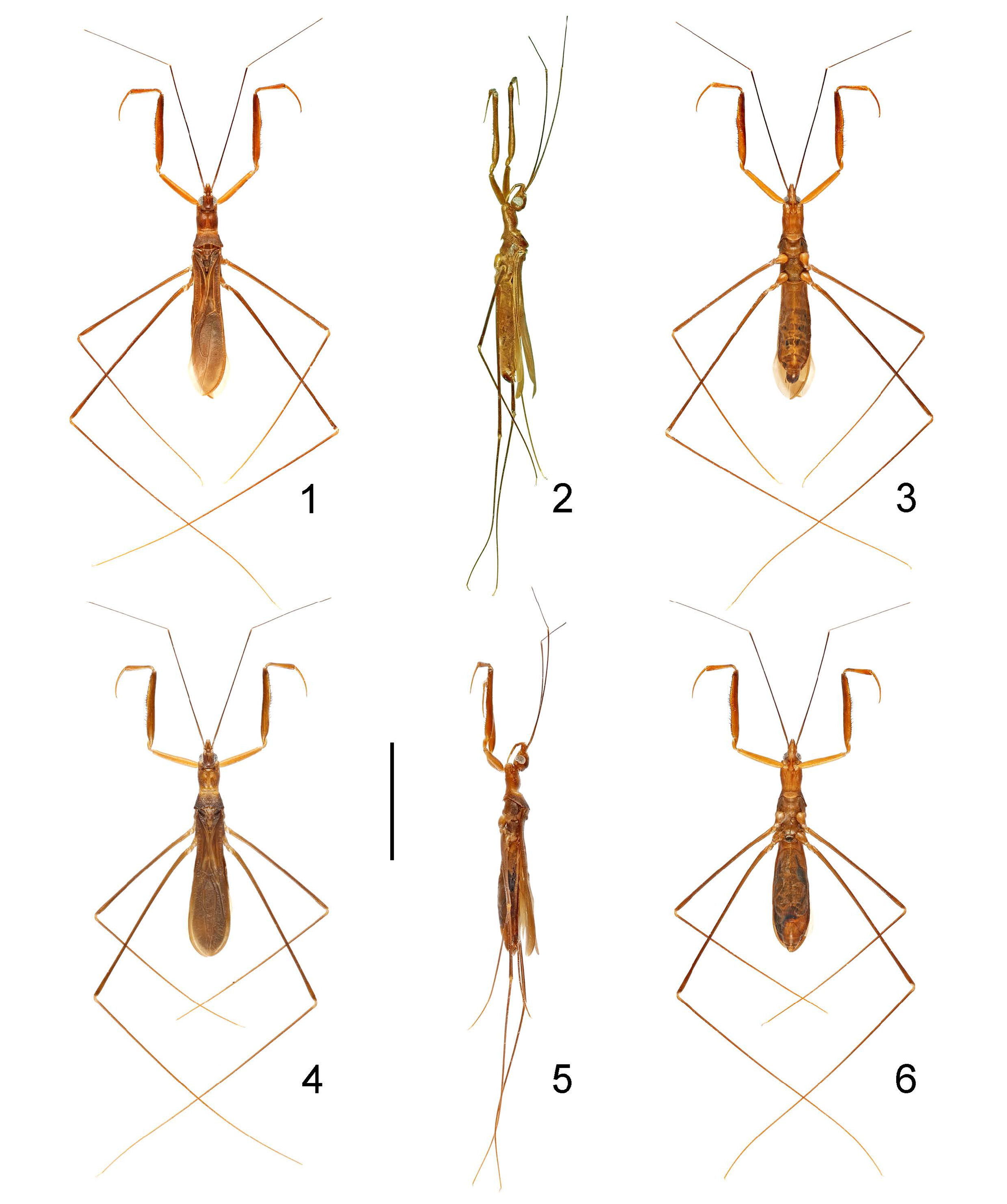
( Figs. 1–29 View FIGURES 1–6 View FIGURES 7–18 View FIGURES 19–21 View FIGURES 22–29 )
Diagnosis. Recognized within the genus by the following combination of characters: pronotum along midline 1.1 times as long as its width across humeral angles, with deep median longitudinal sulcus; anteroventral series of fore femur composed of about 25 spine-like setae; fore wing with cross vein between M + Cu and submarginal vein basad of discal cell weakly developed, oblique, inserted directly from basal angle of discal cell; Rs vein poorly developed; discal cell elongate, longer than M extending from its apex; paramere short and slender, rod-shaped.
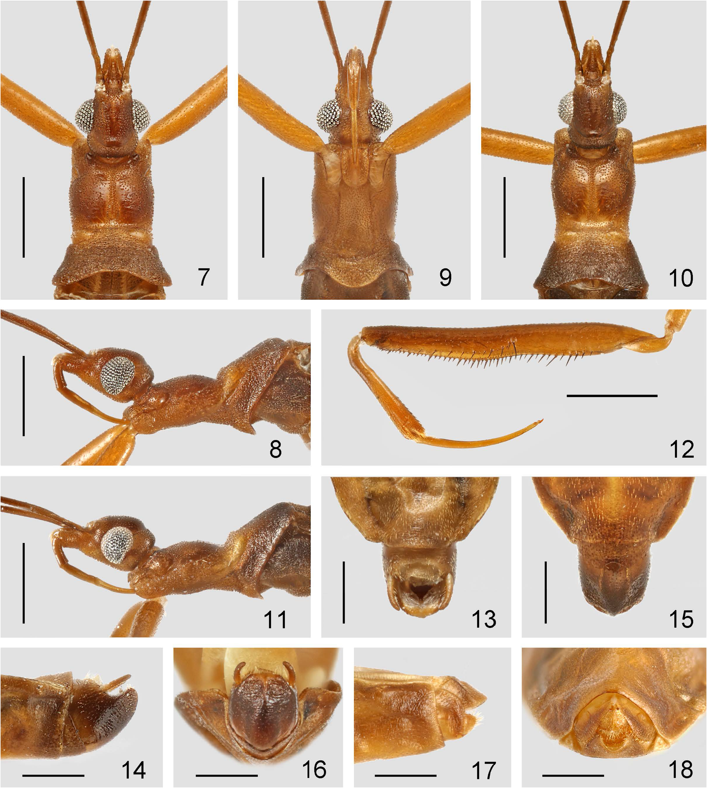
Description. Macropterous male ( Figs. 1–3 View FIGURES 1–6 ). Coloration. Body generally brown. Head slightly darker on dorsum ( Fig. 7 View FIGURES 7–18 ); labrum whitish-yellow ( Figs. 7, 8 View FIGURES 7–18 ); antennal scape and pedicel dark-brown, the former with basal part slightly lighter, flagellomeres absent; labial segment III light-brown, segment IV yellowish-brown ( Fig. 8 View FIGURES 7–18 ). Pronotum slightly darkened on both sides of anterior lobe ( Fig. 7 View FIGURES 7–18 ); tubercle of each anterolateral angle blackish-brown ( Fig. 7 View FIGURES 7–18 ); midline of anterior lobe, transverse sulcus and anterolateral bulges of posterior lobe light-brown ( Fig. 7 View FIGURES 7–18 ); posterior half of posterior lobe dark-brown ( Fig. 7 View FIGURES 7–18 ). Scutellum, metanotum and metapleuron slightly darkened; mesopleuron dark-brown. Fore leg light-brown, coxa yellowish-brown, femur slightly darkened on dorsum with apex dark-brown ( Fig. 12 View FIGURES 7–18 ); mid and hind coxae and trochanters as well as bases of tibiae yellowish-brown, femora and tibiae dark-brown, tibiae gradually becoming paler towards apexes, tarsi light-yellowish-brown. Fore wing with veins slightly paler; area basad of discal cell tinged with obscure pale suffusion ( Fig. 19 View FIGURES 19–21 ). Abdomen suffused with dark-brown laterally; apical half of pygophore dark-brown to blackish-brown, apex yellowish ( Figs. 14, 16 View FIGURES 7–18 ).
Vestiture. Body surface densely covered with tiny, decumbent setae arising from tiny granulations on head, thorax, legs and abdomen; antennae with tiny, oblique, dark setae and short, decumbent, whitish pubescence; labium with sparse, tiny setae. Pronotum with anterior lobe partially glabrous; posterior lobe of pronotum, scutellum and metanotum finely wrinkled. Dorsal surface of apical two thirds of fore tibia and ventral surface of extreme base of fore tarsomere I with short, oblique, golden setae; tarsi and apical parts of tibiae of mid and hind legs with short, slender, decumbent setae. Fore wing weakly rugose.
Structure. Head ( Figs. 7–9 View FIGURES 7–18 ) teardrop-shaped, porrect, 1.5 times as long as width across eyes; anteocular part 2.4 times longer than postocular part; interocular space 1.8 times as broad as single eye in dorsal view; interocular furrow strongly curved backwards, median portion reaching far behind level of posterior margin of eyes. Eyes moderately protruding laterally, elliptical in lateral view, far removed from dorsal and ventral outlines of head in lateral view ( Fig. 8 View FIGURES 7–18 ). Antennal insertion closer to anterior margin of eye than to apex of head; scape 2.1 times as long as length of head and pronotum combined, 1.5 times as long as pedicel, pedicel slenderer than scape. Labium ( Fig. 8 View FIGURES 7–18 ) long and generally slender; labial segment II stout, segment III surpassing anterior margin of eyes, segment IV longest, gradually tapering.
Pronotum ( Figs. 7, 8 View FIGURES 7–18 ) along midline 1.2 times as long as head, 1.1 times as long as its width across humeral angles, vaguely divided into anterior and posterior lobes by transverse sulcus at middle; anterior margin concave, anterolateral angles forming small but distinct tubercles; anterior lobe slightly swollen laterally, with clear, deep, median longitudinal sulcus ( Fig. 7 View FIGURES 7–18 ); posterior lobe with one pair of transverse, anterolateral bulges, lateral margins diverging posteriorly, posterior margin broadly concave. Posterior margin of prosternum rounded ( Fig. 9 View FIGURES 7–18 ). Scutellum subtriangular, with apical nodule and weakly developed lateral ridges. Metanotum with an apical nodule.
Fore leg ( Fig. 12 View FIGURES 7–18 ) stout; coxa cylindrical, 1.5 times as long as pronotum, very faintly narrowed towards apex; trochanter simple; femur long, laterally flattened, 1.5 times as long as coxa, 8 times as long as its maximum width, ventral surface armed with two series of short to long, spine-like setae arising from small tubercles, and an accessory series composed of minute setae and small denticles; anteroventral series ( Fig. 12 View FIGURES 7–18 ) composed of about 25 spine-like setae, beginning beyond basal third of segment, with basal most setae longest; posteroventral series ( Fig. 12 View FIGURES 7–18 ) composed of about 46 spine-like setae, with short and long setae in basal two thirds and small peg-like ones in apical third; tibia short and stout, less than half as long as femur, widened towards apex, armed ventrally with a row of about 15 deflexed spine-like setae on apical three fifths; tarsus arched, nearly as long as tibia, tarsomere I about six times longer than tarsomere II. Mid and hind legs long and slender; mid and hind tibiae 1.45 and 1.5 times as long as respective femora.
Fore wing ( Fig. 19 View FIGURES 19–21 ) elongate, conspicuously surpassing apex of abdomen; cross vein connecting M + Cu and submarginal vein basad of discal cell weakly developed, oblique, inserted directly from basal angle of discal cell ( Fig. 20 View FIGURES 19–21 ); discal cell elongate, longer than remaining M extending from its apex ( Fig. 19 View FIGURES 19–21 ); Rs situated extremely apically, rudimentary and poorly defined ( Fig. 19 View FIGURES 19–21 ). Hind wing ( Fig. 21 View FIGURES 19–21 ) with hamus approaching Sc + R with a moderate angle but not conjoined; m-cu cross vein long and slightly oblique; section of M connecting m-cu to R + M short; Cu connected with R + M, forming a large cell, with long, backwardly directed branch extending from its apex; 1A and 2A present; anal lobe broad, about half as long as hind wing.
Abdomen elongate oval, slightly widened posteriorly, 3.2 times as long as its maximum width. Abdominal segment VIII narrowly exposed in lateral and ventral views ( Figs. 14, 15 View FIGURES 7–18 ), with posteromedial margin medially concave ( Fig. 15 View FIGURES 7–18 ).
Male genitalia: Pygophore ( Figs. 22–24 View FIGURES 22–29 ) elongate oval, apical fifth of ventral surface with a weakly developed, median longitudinal ridge ( Fig. 16 View FIGURES 7–18 ); anterior dorsal sclerotization wide; posterior margin broadly rounded, without posterosuperior process. Paramere ( Figs. 13, 14, 16 View FIGURES 7–18 , 25, 26 View FIGURES 22–29 ) short and slender, evenly bent, apically rounded. Phallus ( Figs. 27–29 View FIGURES 22–29 ) elongate, somewhat laterally flattened; articulatory apparatus stout and wide; basal plates widely separated, nearly parallel, basal foramen small, ponticulus basilaris thick, dorsal connectives short; basal plate extension small; phallotheca with struts long and slender, extending to apex of phallus, apexes acute; endosoma with large bursa basally and paired, narrow, sclerotized stripes along entire length ventrally, apex with a pair of vesiclelike processes inside.
Macropterous female ( Figs. 3–6 View FIGURES 1–6 ) similar to male in general appearance; eyes somewhat smaller ( Figs. 10, 11 View FIGURES 7–18 ); fore wing just reaching apex of abdomen, with veins almost same color as remainder. Female genitalia ( Figs. 17, 18 View FIGURES 7–18 ): Tergite VIII semicircular, apical two thirds yellowish; tergite IX declining, basal half yellowish, lateral sides blackish brown; valvifer I broad, apical half dark brown, apical margin rounded; valvula I triangular, rounded at apex, covered with dense setosity; styloids small, separated from each other, apically rounded.
Measurements [in mm, ♂ (n = 1, holotype) / ♀ (n = 2, paratypes)]. Length of body: to apices of fore wings 9.30 / 9.20, to apex of abdomen 8.70 / 9.20; length of head 1.25 / 1.25; length of anteocular part 0.60 / 0.60; length of postocular part 0.25 / 0.25; width across eyes 0.85 / 0.80–0.90; interocular space 0.40 / 0.40–0.50; length of antennal segments I–IV = 5.60 / 5.30–5.40, 3.80 / 3.50–3.55,? (missing) /? (missing),? (missing) /? (missing); length of labial segments II–IV = 0.35 / 0.35, 0.50 / 0.50, 0.55 / 0.55; length of anterior pronotal lobe 0.75 / 0.75–0.80; length of posterior pronotal lobe 0.70 / 0.70–0.75; width of anterior pronotal lobe 0.95 / 1.00–1.10; width of posterior pronotal lobe 1.30 / 1.30–1.40; median length of scutellum 0.40 / 0.40; basal width of scutellum 0.50 / 0.50; length of fore coxa, femur, tibia, tarsus = 2.15 / 2.10, 3.20 / 3.20–3.25, 1.30 / 1.20–1.30, 1.30 / 1.20–1.30; maximum width of fore femur 0.40 / 0.40; length of mid femur, tibia, tarsus = 5.30 / 5.20–5.40, 7.70 / 7.80, 0.30 / 0.30; length of hind femur, tibia, tarsus = 7.70 / 7.20–7.40, 11.90 / 11.30–11.70, 0.30 / 0.40; length of fore wing = 6.40 / 6.30–6.40; length of abdomen 4.50 / 4.80–4.90; maximum width of abdomen 1.40 / 1.60–1.80.
Type material. Holotype (♂): CHINA, Hainan, Baisha, Nankai , N19°4′37″, E109°25′6″, in cave, 25 Dec. 2019, Q. Zhao leg. ( CAU) GoogleMaps . Paratypes (2♀): same collection data as holotype ( CAU) GoogleMaps .
Additional material examined. One nymph (5 th instar): same collection data as type material ( CAU, in ethanol) .
Etymology. The specific epithet is dedicated to Dr. Max Caspers (Naturalis Biodiversity Center, Leiden) for his kind support in our study of Reduviidae .
Distribution. CHINA: Hainan.
Biology. The specimens of this species, including a 5 th- instar nymph, were collected on the walls of a cave. A male of Myiophanes tipulina was collected with them simultaneously.


| CAU |
China Agricultural University |
No known copyright restrictions apply. See Agosti, D., Egloff, W., 2009. Taxonomic information exchange and copyright: the Plazi approach. BMC Research Notes 2009, 2:53 for further explanation.
|
Kingdom |
|
|
Phylum |
|
|
Class |
|
|
Order |
|
|
Family |
|
|
SubFamily |
Emesinae |
|
Genus |