
Coeliccia paludensis, Dow, Rory A., 2016
|
publication ID |
https://doi.org/ 10.11646/zootaxa.4184.1.5 |
|
publication LSID |
lsid:zoobank.org:pub:2312F650-EB9C-457E-B3AD-71BC3984A30A |
|
DOI |
https://doi.org/10.5281/zenodo.6087473 |
|
persistent identifier |
https://treatment.plazi.org/id/03B7F636-FFBA-FFFB-4CB6-8840FCF2D7FC |
|
treatment provided by |
Plazi |
|
scientific name |
Coeliccia paludensis |
| status |
sp. nov. |
Coeliccia paludensis View in CoL spec. nov.
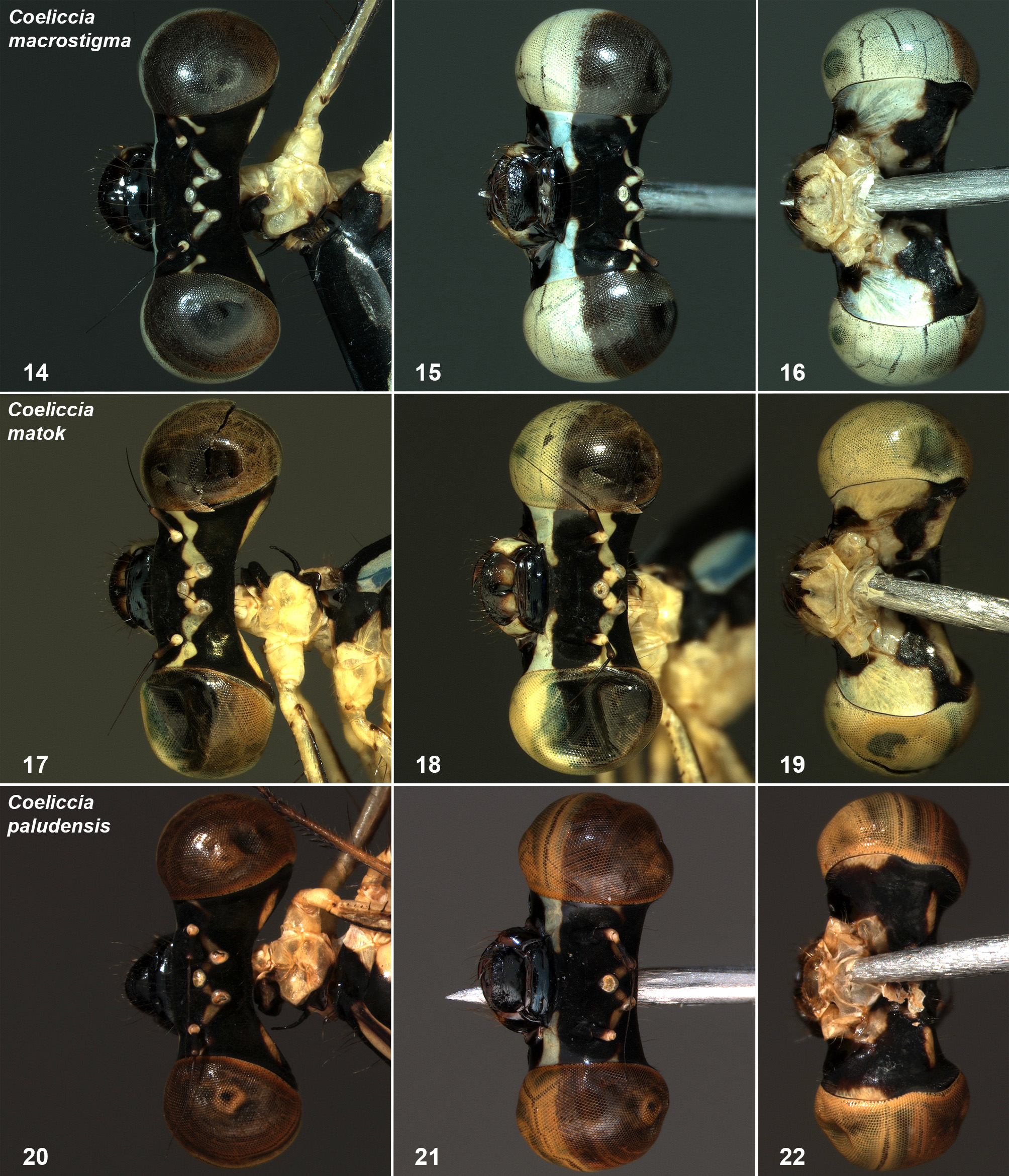
( Figs. 11, 12, 13 View FIGURES 5 – 13 , 20, 21, 22 View FIGURES 14 – 22 , 27, 28 View FIGURES 23 – 28 , 33, 34 View FIGURES 29 – 34 , 39, 40 View FIGURES 35 – 40 , 45, 46 View FIGURES 41 – 46 , 51, 52 View FIGURES 47 – 52 , 57, 58. 63, 64 View FIGURES 53 – 58 View FIGURES 59 – 64 , 67 View FIGURES 65 – 67 , 70, 71 View FIGURES 68 – 71 , 72 View FIGURE 72 )
Coeliccia View in CoL new sp. 1;— Dow & Silvius 2014: 12, Fig. 11 View FIGURES 5 – 13 .
Type material. Holotype: Ƌ (KAL12_PCD72; RMNH.INS.506696), peat swamp forest in ex Mega Rice Project Block E (1.936S, 114.056E), Kalimantan Tengah, Indonesia, 18 vi 2012, in RMNH, leg. M. Silvius GoogleMaps .
Paratypes: All Kalimantan Tengah, Indonesia (39 Ƌ, 8 ♀); in collection Dow unless noted otherwise: 1 Ƌ (KAL12_PCD73; RMNH.INS.506687), location and date as holotype, in RMNH, leg. R.A. Dow ; 1 Ƌ (KAL12_PCD64), same location, date and collector; 1 Ƌ (KAL12_PCD75, RMNH.INS.506727), peat swamp forest, CIMTROP Natural Laboratory Block C, Sebangau (1.664S, 113.902E), 21 vi 2012, in RMNH, leg. R.A. Dow GoogleMaps ; 3 Ƌ (KAL12_PCD57–59), 1 ♀ (KAL12_PCD60), same location and date; 1 Ƌ (KAL12_PCD49), peat swamp forest, Tuanan (2.152S, 114.441E), 24 vi 2012, leg. R.A Dow; 22 Ƌ (KAL12_PCD2, 4–20, 27–29, 61) GoogleMaps ; 1 ♀ (KAL12_PCD30, RMNH.INS.506896, in RMNH); 1 ♀ (KAL12_PCD3, in tandem with KAL12_PCD2); 1 ♀ (KAL12_PCD62, in tandem with KAL12_PCD61), same location, 25 vi 2012, leg. R.A. Dow GoogleMaps ; 6 Ƌ (KAL12_PCD21–26), same location and date, leg. M. Silvius GoogleMaps ; 4 Ƌ (KAL12_PCD42, 65–67), 1 ♀ (KAL12_PCD43, in tandem with KAL12_PCD42), 3 ♀ (KAL12_PCD68–70), same location, 26 vi 2012, leg. R.A. Dow GoogleMaps .
Etymology. paludensis , from palus, a Latin noun meaning marsh or swamp, named in reference to the peat swamp forest habitats of this species.
Description of holotype male. Head: Labium pale cream except hooks of labial palps, which are black. Labrum, mandible bases, clypeus and genae largely shining black, blue band running from eye margin to clypeus and narrowly for short distance above clypeus ( Fig. 12 View FIGURES 5 – 13 ). Antenna with white ring at top of scape and bottom of pedicel, remainder dark brown and black. Frons and vertex mostly black, with short stripe based on outer edge of lateral ocellus, directed towards rear of antenna base, tiny pale mark at eye margin, even smaller pale spot to rear of this, at ca half distance between antennae and eye margin ( Fig. 11 View FIGURES 5 – 13 ). Ocelli white. Yellow, elongate oval, transverse postocular spots. Underside of head mostly black with small whitish marks at eye margin, at same level as pair of bluish markings below point of attachment of prothorax, behind mandible bases ( Fig. 13 View FIGURES 5 – 13 ).
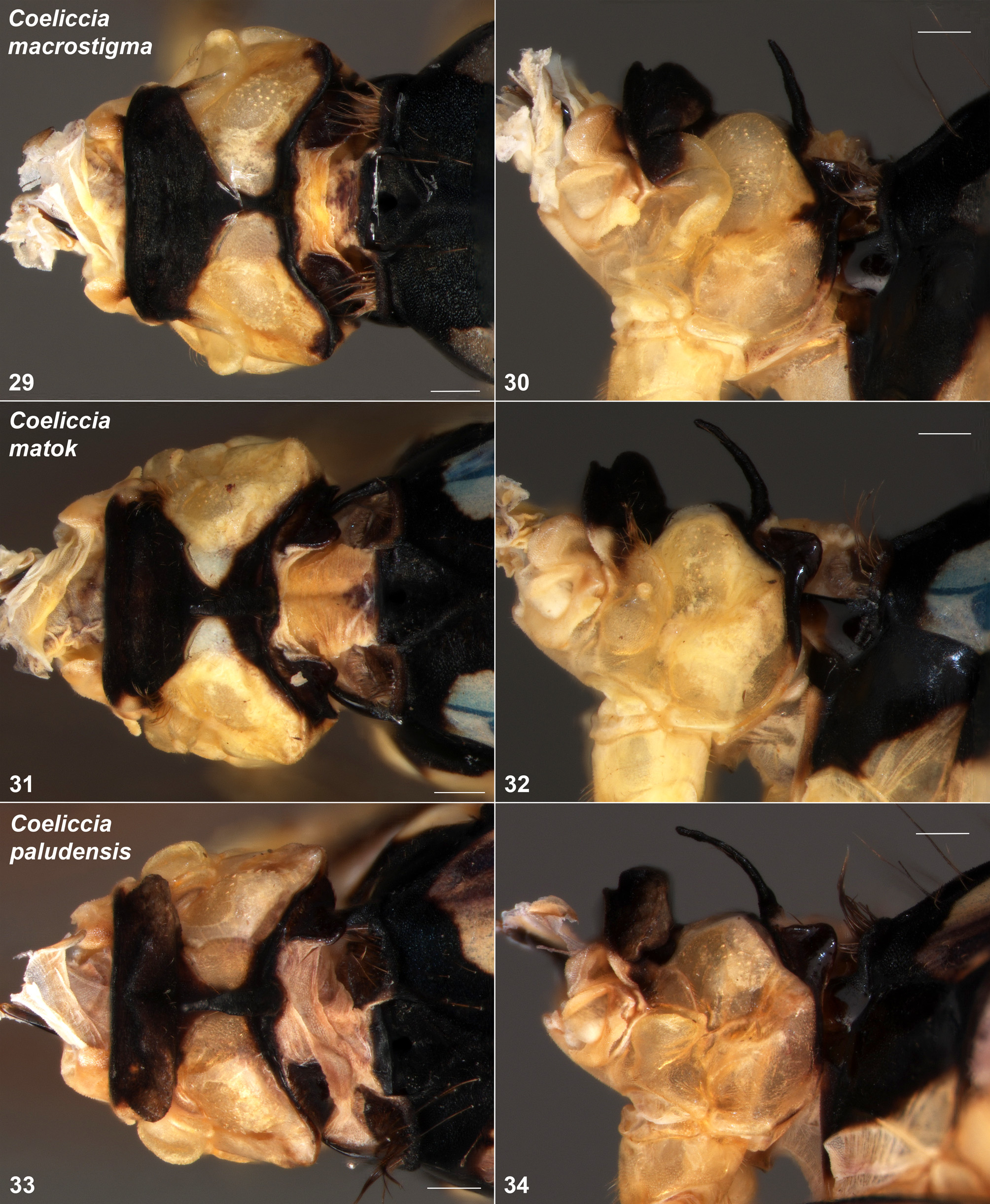
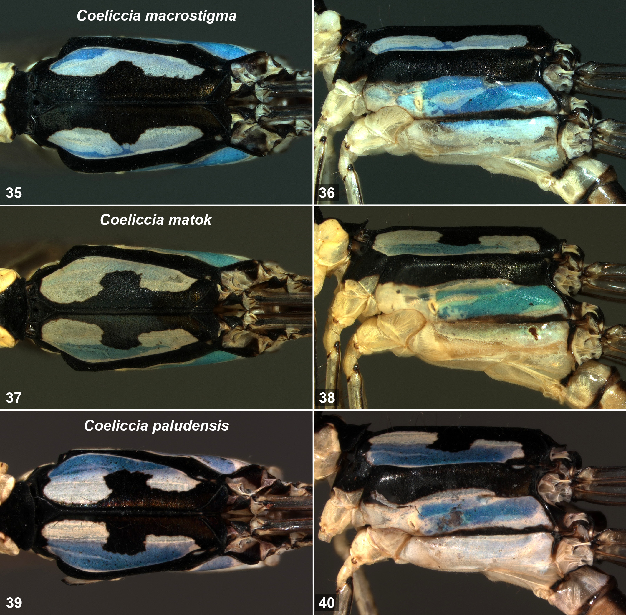
Thorax: Prothorax with propleuron entirely pale except a narrow black band at rear. Posterior and anterior pronotal lobes black, except narrowly laterally on anterior lobe, where pale. Anterior pronotal lobe erect, anterior carina lower than main part ( Fig. 28 View FIGURES 23 – 28 ). Upper part of notopleural projection barely present. Middle lobe pale laterally, black above, pale colour extending narrowly towards, but not reaching, centre ( Fig. 27 View FIGURES 23 – 28 ). Posterior pronotal lobe simple, collar-like. Synthorax with mesepisternum black with large blue antehumeral markings ( Fig. 45 View FIGURES 41 – 46 ), extending from mesostigmal plates beyond apex of antealar triangle, occupying entire width of mesepisternum near prothorax, outer margin following mesepleural suture for most of length, inner margin ca squarely excised centrally, marking narrower after excision. Mesepimeron black except narrowly blue above the interpleural suture for some distance, and small blue streak below mesepleural suture near antealar carina ( Fig. 46 View FIGURES 41 – 46 ). Metepisternum largely blue with black stripe running from antealar carina most of way towards spiracle. Metepimeron very pale blue. Venter of synthorax pale. Mesinfraepisternum black except lower corner adjacent to metepisternum, metinfraepisternum entirely pale. Legs (only right middle and left posterior legs remaining after trochanters) with coxae and trochanters entirely pale, femur greyish pale with black stripe along extensor surface, tibia pale with black stripe along flexor surface. Wings with arc situated slightly distal to Ax 2. 16 Px in Fw, 15 in Hw. Three postquadrilateral cells in all wings. R4 arising just proximal to subnodus, IR3 arising distal to subnodus, joined to R4 by short stalk. Pt dark brown with very narrow, incomplete, white margin, broad, ca rhomboidal, covering more than one underlying cell.
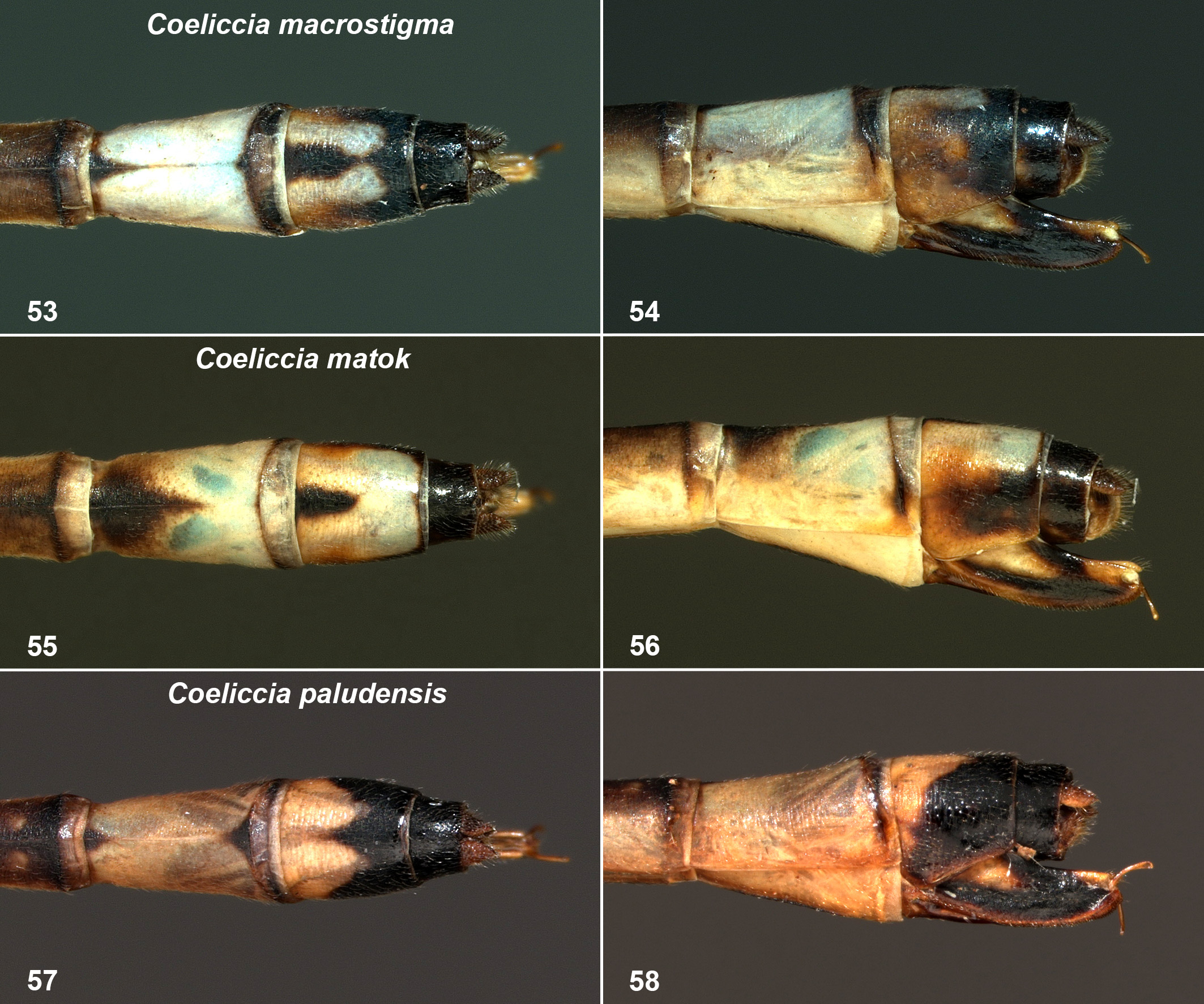
Abdomen: S1 pale laterally except narrow apical annulus including posterior carina and area behind, narrowly dark dorsally. S2 pale to sides, irregularly dark brown dorsally. S3–7 brown above, darker apically, pale brownish laterally with narrow whitish basal annulus interrupted dorsally. S8 pale blue lower laterally, black above, dorsally pair of apical blue markings, narrowly divided, subtriangular ( Fig. 51 View FIGURES 47 – 52 ). S9 almost entirely pale blue, except small black basal dorsal triangle. S10 pale blue except narrowly apically. Cerci large, blue dorsally and laterally, black interiorly; basal tooth not present, central tooth shortly before half length, visible in dorsal and lateral views. In dorsal view ( Fig. 63 View FIGURES 59 – 64 ) cercus subrectangular, inner margin turning rather sharply towards pointed apex, outer margin more gently; central ridge running almost from apex to ca level of central tooth. In lateral view ( Fig. 64 View FIGURES 59 – 64 ) expanding from base on lower margin to just before central tooth, then running straight and parallel to upper margin until just before apex, where hooked slightly down; upper margin running straight and upwards for most of length, at ca level of tips of paraprocts turning down towards tip; whole cercus with blocky, upward-directed appearance. Paraprocts black interiorly, mostly blue otherwise, relatively short, tips well before those of cerci; in ventral view of almost even width after base until hooked abruptly inwards at apices, which are sharp, black, pointing towards each other and slightly to rear ( Fig. 67 View FIGURES 65 – 67 ). In lateral view paraprocts appear rounded at apices. Genital ligula typical for the membranipes -group.
Measurements (mm): Abdomen without terminal appendages 33, cerci ca 0.8, Hw 21.
Description of female paratype (KAL12_PCD62). As male except as noted. Head: Pale markings around ocelli more extensive: a pale mark between each lateral ocellus and the median one, mark based at lateral ocellus running almost to antenna, marks near eye margin joined to form streak running parallel to that between lateral ocellus and antennae ( Fig. 20 View FIGURES 14 – 22 ).
Thorax: Prothorax with propleuron pale yellowish, anterior and posterior pronotal lobes black, middle lobe pale except for narrow black band joining anterior and posterior lobes. Upper cervical spur well defined ( Fig. 34 View FIGURES 29 – 34 ), triangular, apex just above notopleural suture, lower cervical spur short, not overlapping propleuron. Lower part of notopleural projection large, almost flat at top, somewhat hollowed inside in dorsal view ( Figs. 33 View FIGURES 29 – 34 ), upper part smaller, not apparent in dorsal view, apparently fused to lower lateral part of anterior pronotal lobe. Anterior pronotal lobe very erect and angular in appearance, anterior carina lower than top of main part, which is relatively narrow, more so centrally, hind margin straight in lateral view, apex with prominent dorsal lateral ridge running to just above and in front of junction with upper part of notopleural projection. Posterior pronotal lobe with short lapels ( Fig. 34 View FIGURES 29 – 34 , that on left broken, so the figure shows the right-hand side, flipped to left for consistency with other figures), dorsally flattened, apex just below level of highest part of middle lobe, in dorsal view extending over front part of mesostigmal plates, square-sided interiorly, rounded at rear ( Fig. 33 View FIGURES 29 – 34 ); horn narrow, longer than middle lobe, curved forward so apex just to rear of, but well above, anterior lobe ( Fig. 34 View FIGURES 29 – 34 ) in lateral view. Synthorax with subrectangular mesostigmal plates, depressed at front, with fringe of long setae at rear. Antehumeral markings ( Fig. 45 View FIGURES 41 – 46 ) similar to male but more widely separated from each other anteriorly, with excision narrower. Black stripe along metepleural suture reduced compared to male. Legs with coxae and trochanters entirely pale, femur pale with black stripe along extensor surface, anterior femur also with a black stripe along outer edge of flexor surface in lower ca two-thirds, tibia pale with black stripe along flexor surface. Wings with arc situated at (right Hw) or slightly distal to Ax 2. All wings with 16 Px. Pt greyish brown with very narrow white margin except on costal side, broad, ca rhomboidal, covering ca one underlying cell in Fw, more than one underlying cell in Hw.
Abdomen: S8 almost entirely pale except brown behind posterior carina. S9 black, pale basal marking, subtriangular in lateral view, extending dorsally where partially divided centrally, apices at more than half segment length. S10 black. Cerci shorter than S10, pale. Ovipositor mostly black, basal pale patch, brown along lower margin, small pale mark dorsally near apex, extending well beyond cerci.
Measurements (mm): Abdomen without anal appendages or ovipositor 33, Hw 21.
Variation in paratypes. In both sexes there is sometimes an indistinct grey area (difficult to see at low magnification) on the anteclypeus. The prothoracic horn of the female varies between almost straight upward pointing to that seen in the female described. The exact shape of the excision in the antehumeral stripes is slightly variable in both sexes, and in females occasionally the excision almost divides the marking into two, in one individual just dividing the marking. The small pale mark on the mesepimeron is often absent. R4 is variably at or proximal to Sn (even in different wings of the same individual), in one male from Tuanan distal to Sn in the left Fw. Very occasionally there are two or two-and-a-half postquadrilateral cells in one wing. The markings on S8 of males are highly variable in their exact shape, frequently smaller than in the holotype (e.g. Fig. 70 View FIGURES 68 – 71 in life); in many individuals they are tiny and occasionally entirely absent. In a few individuals they are joined dorsally and/or joined to the lateral pale area apically. The depth of division of the S9 marking in the females is variable but the division is present in all paratypes. There is no basal tooth on the cerci of any male; in some individuals a slight bulge is visible in dorsal view at the base of the cercus.
Measurements (mm): Males 14–17 Px in Fw, 14–17 in Hw; abdomen without anal appendages 31–34.5; Hw 19–22. Females 14–16 Px in Fw, 14–15 in Hw; abdomen without anal appendages and ovipositor 32–34.5; Hw 20– 22.
Diagnosis. The male of C. paludensis is readily distinguished from all other members of the C. membranipes - group by its inflated cerci lacking basal teeth. The female can be distinguished from all named species except C. macrostigma by the extremely large lower part of the notopleural projection, which lacks any spur or nipple-like projection, and from C. macrostigma by the very erect and narrow anterior pronotal lobe and by having the lower part of the notopleural projection almost flat at top, compared to that in C. macrostigma which forms a vertical or almost vertical rounded flap, concave to front.
Remarks. Among the species of the C. membranipes -group, C. paludensis is most notable for the lack of basal teeth on the cerci. Excluding the borneeensis -group, C. lieftincki from Java, the Philippine species and the highly unusual C. suoitia Dow, 2016 , from Vietnam, all of which are very unlikely to really belong in Coeliccia (Dow 2016) , it is the only member of Coeliccia that I have examined or seen adequate illustrations of that lacks a basal tooth. The very erect form of the anterior pronotal lobe in both sexes is also unusual.
Coeliccia paludensis is known only from three peat swamp forest sites in the south of Borneo in the Indonesian province of Kalimantan Tengah ( Fig. 72 View FIGURE 72 ). Of these sites only that at Sebangau has any protected status, and all sites are extremely vulnerable to forest fires during dry periods.


| RMNH |
National Museum of Natural History, Naturalis |
No known copyright restrictions apply. See Agosti, D., Egloff, W., 2009. Taxonomic information exchange and copyright: the Plazi approach. BMC Research Notes 2009, 2:53 for further explanation.
|
Kingdom |
|
|
Phylum |
|
|
Class |
|
|
Order |
|
|
Family |
|
|
Genus |
Coeliccia paludensis
| Dow, Rory A. 2016 |
Coeliccia
| Dow 2014: 12 |