
Photonectes cyanogrammicus, Prokofiev & Klepadlo, 2019
|
publication ID |
https://doi.org/ 10.11646/zootaxa.4590.2.4 |
|
publication LSID |
lsid:zoobank.org:pub:5BB7CA86-65F1-4F1E-854C-02F9DAA7A1E5 |
|
DOI |
https://doi.org/10.5281/zenodo.5924187 |
|
persistent identifier |
https://treatment.plazi.org/id/03B81E52-FFCF-FFDB-FF5D-63A0FAD5FBFA |
|
treatment provided by |
Plazi |
|
scientific name |
Photonectes cyanogrammicus |
| status |
sp. nov. |
Photonectes cyanogrammicus new species
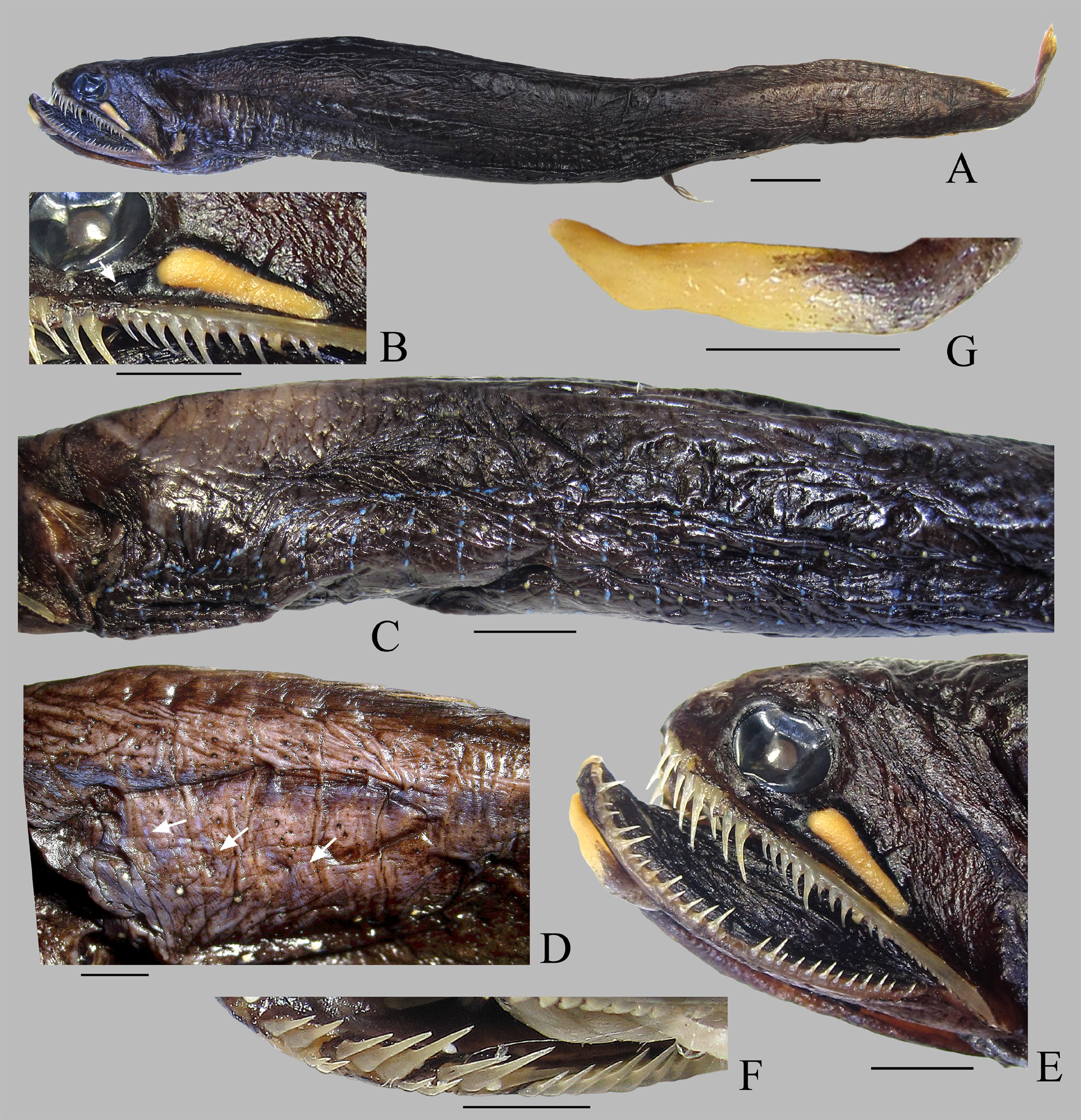
Fig. 1 View FIGURE 1
Holotype. MNHN 2002-3685, 140 mm SL, 9° 10' 4.8'' S, 159° 52' 58.8'' E, 749–799 m, R/ V Alis, camp. Salomon 1, sta. cp1751, 25 September 2001. GoogleMaps
Diagnosis. A species of Photonectes based on the following combination of unique derived characters: mental barbel distally expanded, lacking bulbs or appendages but with distal portion unpigmented and apparently luminous, and two posteriormost photophores VAV distinctly raised (vs. all photophores VAV on the same level in other Photonectes spp.).
Description. Body elongate, tapering toward caudal fin ( Fig. 1A View FIGURE 1 ). Snout 1.6 times shorter than eye. Morphometric values, in % of SL: head length 13.6; snout length 1.8; eye diameter 2.8; least interorbital width 3.9; barbel length 5.3; upper jaw length 13.6; postorbital organ length 3.9; ditto, greatest depth 0.9, entire black surrounding area 4.6; predorsal, preanal and prepelvic distances 76.4, 76.4 and 60.7, respectively; dorsal- and analfin base length each 18.9; pelvic-fin length 14.0; greatest body depth 11.4; least depth of caudal peduncle 2.1. Opercle slightly concave dorsally, its posteroventral corner somewhat produced; gill filaments extending slightly beyond gill cover. Gill filaments in upper half of first ceratobranchial moderately shortened (1.5 times shorter than filaments of lower half). Color of skin uniformly black.
Dorsal-fin rays 21; anal-fin rays 23; pelvic-fin rays 7; pectoral fins absent. Black fleshy skin extends on basal 1/3 of dorsal and anal fins. Pelvic fins reaching slightly before vent. Caudal fin forked, with lower lobe longer. Vertebrae 53. Photophores: IP 9; PV 22; IV 31; VAV 16 [last 5 over anal-fin base, last 2 raised]; AC 9; IC 56; OV 22; VAL 12 [last 2 over anal-fin base]; OA 34; BR 10. Anteriormost six IP photophores smaller than remaining ones, closely spaced, with distances between them not exceeding diameter of organ; remaining three IP photophores well-separated from each other [distance between 6 th and 7 th photophores and 8 th and 9 th photophores equal to two diameters of organ; distance between 7 th and 8 th photophores equal to 1.5 diameter of organ]. Distance between anterior tip of isthmus and first pair of IP photophores about 3 times greater than combined length of anteriormost six IP photophores (66.7% of isthmus length). Secondary photophores very small, scattered over head and body, most densely clustered on cheeks, along dorsum, between OV and PV organs and between PV organs of neighbouring sides, absent on fin-rays. Postorbital organ large, elongate, tapered posteriad, 1.4 times eye diameter. Antero-ventral corner of jet-black tissue encircling the photogenic gland of postorbital organ extends anteriad to a vertical through the center of eye ( Fig. 1B View FIGURE 1 ).
Blue luminous tissue on lower sides of body arranged as follows: a longitudinal line on each side, 1.5 times closer to PV than to OV organs, consisting of interrupted streaks between isthmus and anal-fin origin, conspicuous up to 18 th photophore PV, less obvious behind; transverse interrupted streaks connecting longitudinal lines of left and right sides of body between photophores, originating from space between 6 th and 7 th organs of IP and discernible until 10 th photophore of VAV, alternating with photo phores ( Fig. 1C View FIGURE 1 ). Diffuse indistinct superficial band of blue luminous tissue originating somewhat behind a level of pelvic-fin origin, extending toward caudal-fin base, having wavy extensions downward forming reticulate appearance in anterior portion (up to a level of mid-base of anal fin: Fig. 1D View FIGURE 1 ). Small spots of white luminous tissue scattered along anal-fin base. Clusters of small pale bluish to whitish luminous spots on opercle and behind posterior end of maxillary; no luminous tissue inside mouth.
Jaw dentition heterogeneous; premaxillary teeth biserial, teeth in outer row much shorter ( Figs. 1E, 1F View FIGURE 1 ). Premaxillary with 9 + 3 caniniform teeth on left side and 8 + 4 teeth on right side, their tips barbed. Inner premaxillary teeth distinctly longer than those on maxillary and dentary. Maxillary with 11/12 erect caniniform teeth, following by a comb-like row of inclined needle-like teeth (13 in number); erect maxillary teeth increasing in size from 1 st to 5 th, becoming smaller backward. Dentary teeth 30/32, consisting of long and short canines ( Fig. 1E View FIGURE 1 ). Vomer with 3 + 3 teeth, gradually increasing in size from first to third, third tooth very long, extending behind 2 nd palatal tooth. Palatine with 2 teeth on each side, second longest.
Barbel rather short (39% of head length), lacking a conspicuous bulb or any appendages, but unpigmented and apparently luminous in distal portion (60% of full barbel length); no sharp boundary between darkly pigmented stem and unpigmented distal portion ( Fig. 1G View FIGURE 1 ).
Etymology. The name (Greek “κυανό” for blue and “γραµµή” for streak) reflects the characteristic pattern of the blue luminous tissue on the ventral side of the body.
Comparisons. Photonectes cyanogrammicus is similar to P. gracilis and P. sphaerolampas in possession of transverse streaks of blue luminous tissue alternating with photophores of ventral rows, but differs from both these species in presence of diffuse indistinct lateral band of blue tissue from behind the level of pelvic-fin origin to the caudal-fin base, with irregular ventral extensions forming a reticulate appearance anteriorly (before level of midbase of dorsal- and anal-fins) (vs. distinct solid lateral band of blue tissue from gill opening to caudal-fin base). Furthermore, P. cyanogrammicus differs from P. gracilis and P. sphaerolampas in higher VAV and lower AC count (16 and 9 vs. 12–15 and 12–13, and 12–15 and 14–18, respectively), and from P. gracilis in more posterior position of pelvic fins (prepelvic length 60.7% SL vs. 51.5–56.8% SL).
Distribution. This species is known only from the holotype collected in the Equatorial Western Pacific.
| MNHN |
Museum National d'Histoire Naturelle |
No known copyright restrictions apply. See Agosti, D., Egloff, W., 2009. Taxonomic information exchange and copyright: the Plazi approach. BMC Research Notes 2009, 2:53 for further explanation.
|
Kingdom |
|
|
Phylum |
|
|
Class |
|
|
Order |
|
|
Family |
|
|
Genus |