
Parasphaerolaimus magdolnae, Cavalcanti & Venekey, 2017
|
publication ID |
https://doi.org/ 10.11646/zootaxa.4358.2.7 |
|
publication LSID |
lsid:zoobank.org:pub:890B1FAE-A27C-4788-80FE-D063E80347A7 |
|
DOI |
https://doi.org/10.5281/zenodo.6008597 |
|
persistent identifier |
https://treatment.plazi.org/id/03B887D6-783F-FFCA-FF57-FBE8E39BFAB1 |
|
treatment provided by |
Plazi |
|
scientific name |
Parasphaerolaimus magdolnae |
| status |
sp. nov. |
Parasphaerolaimus magdolnae sp. n.
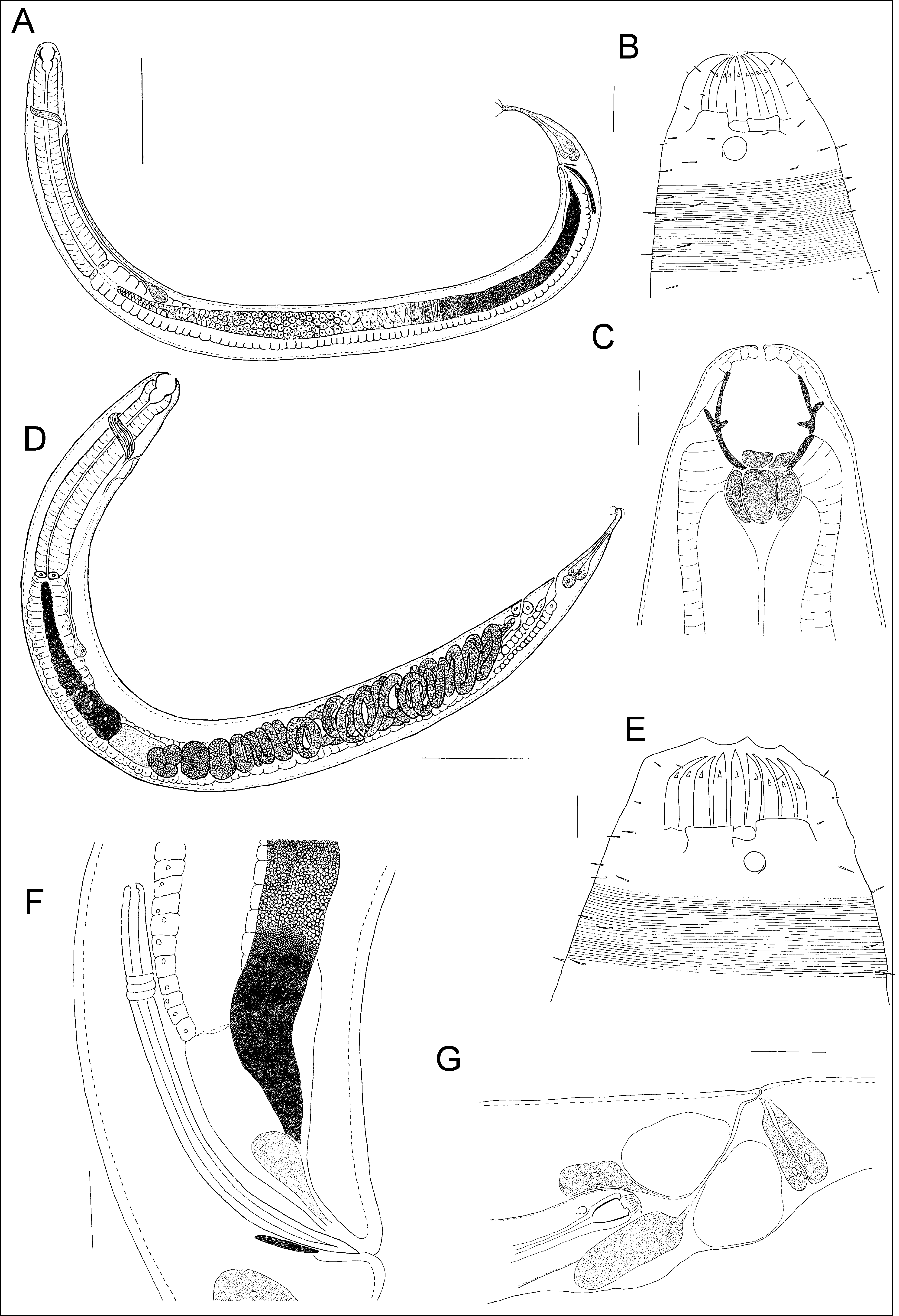
( Figures 1 View FIGURE 1 , 2 View FIGURE 2 and 3 View FIGURE3 ; Table 1)
Diagnosis of Parasphaerolaimus magdolnae sp. n. Large body attenuating towards posterior end. Stoma usually wider in female, surrounded by a ring of six non-sculptured plates. Amphids unispiral with distinct circular outline, situated posterior to the plates. Oesophagus cylindrical. Secretory-excretory pore at the level of nerve ring and ventral gland posterior to the cardia. Lateral alae present. Vulva at 80–90% of the body length. Spermatheca present. Spicules curved with a ventral indentation near the proximal end. Gubernaculum without apophysis. Tail conical-cylindrical with three terminal setae.
Etymology. The species is named in honor of Magdolna (Mrs. Lajosné Venekey), mother of the second author.
Holotype: male (slide MPEG.NEM 000111).
Paratypes: Seven paratype males (one male in slide MPEG.NEM 0 0 0 113 and six males in slides GENAQ- UFPA NEM 001–006) and ten paratype females (one female in slide MPEG.NEM 0 0 0 112, one female in slide MPEG.NEM 0 0 0 114 and eight females in slides GENAQ-UFPA NEM 007–014).
Type locality and Habitat : Holotype collected in Santa Cruz Channel (northeastern coast of Brazil) in February 2003, coordinates 7°39’S; 34°51’W, intertidal environment, surface sediment layer (0–20 cm), muddy sediment View Materials GoogleMaps . Paratypes collected in Santa Cruz Channel (northeastern coast of Brazil), 7°39’–43’S; 34°51–53’W, in February and June 2003, intertidal environment, surface sediment layer (0–20 cm), muddy sediment. View Materials
Description. Males ( Figures 1 View FIGURE 1 and 3 View FIGURE3 ). Large body (2.16–2.78 mm long) attenuating towards posterior end. Maximum diameter 96–180 µm (a=12.8–22.5). Cuticle striated, about 3 µm. Lateral alae visible from the cardia position till the end of the tail area. Inner and outer labial setae, both six in number, very short. Four cephalic setae 2–3 µm long. Subcephalic setae up to 4 µm. Cervical setae up to 8 µm. Buccal cavity large, divided into two sections. Cheilostom with longitudinal ribs. Anterior part of gymnostom with six non-sculptured plates (22–26 µm from the anterior end). Mouth bounded by thick, strong walls, continuous into the esophagus lumen. Anterior-most part of inner wall of the pharynx divided into six plates. Amphids unispiral with distinct circular outline, 8–11 µm in diameter (10.5–14.4% of the corresponding body diameter), situated posterior to the buccal cavity. Oesophagus cylindrical. Nerve ring at 26.8–34.4% of the oesophagus length. Single outstretched testis located on the right of the intestine, reaching almost to the base of pharynx. Spicules 126–234 µm long (1.5–2.1 of the corresponding body diameter), curved with a ventral indentation near the proximal end. Gubernaculum without apophysis. Tail conico-cylindrical with three terminal setae. Three caudal glands present.
Females ( Figures 1 View FIGURE 1 and 2 View FIGURE 2 ). Females quite similar to males, apart from minor morphometrical differences ( Table 1) in the range of body lengths. The head is wider than in males. Sexual dimorphism in amphids size (smaller than in males: 5–8 µm in diameter). One outstretched ovary situated to the right of intestine. Spermatheca present. Vulva situated far posterior, at about 80–90% from anterior end. Four vaginal glands observed. Tail conical-cylindrical with three terminal setae. Three caudal glands present.
Measures Nematode specimens Description of the reproductive strategy. In the female reproductive system of Parasphaerolaimus magdolnae sp. n. eggs and juveniles at different development stages could be found ( Figure 2D View FIGURE 2 ). Female gonad consisting of about 22 oocytes with granular cytoplasm and clear nucleus. The eggs are located after the spermatheca, followed by embryos and hatched juveniles. The juveniles are located in the posterior portion of the uterus up to the vulva region ( Figure 1G View FIGURE 1 ).
Relationships of P. magdolnae sp. n. with other Parasphaerolaimus species. The main characteristics that differentiate P. magdolnae sp. n. from the other Parasphaerolaimus species are the presence of only one testis, spicules morphology and size, and gubernaculum without dorsal apophysis.
From the ten species of Parasphaerolaimus considered as valid, only P. paradoxus , P. pentasetus , P. pilosus , and P. jintiani descriptions showed information about the number of testes ( Table 2). Among them two testes is attributed only to P. pentasetus . We have considered that the number of testes in those species of Parasphaerolaimus where there is not mention of the number of testes is the same as in family Sphaerolaimidae (two testes).
P. magdolnae sp. n. differs from the type species P. paradoxus by the spicules length relative to cloacal body diameter (1.5–2.1 vs 1.0, respectively) and tail shape (conico-cylindrical vs elongate conical, with a more or less filiform portion). Moreover, P. paradoxus cuticle is weakly striated while P. magdolnae sp. n. cuticle is striated with lateral alae. Besides that, de Man’s proportion “a” indicates that P. paradoxus is thinner than P. magdolnae sp. n. (37.0–49.0 vs 12.3–22.5) ( Table 2).
P. magdolnae sp. n. also differs from P. pilosus by the absence of gubernaculum with apophysis. Besides that, the males of P. pilosus are smaller than those of P. magdolnae sp. n. (1380–1545 vs 2160–2784 µm, respectively), have finely striated cuticle and short spicules (ca 1 cloacal body diameter, Table 2). Moreover, the position of the vulva in females of P. pilosus is closer to the middle of the body (55.5–67.1%) than in the new species (80–90%).
P. magdolnae sp. n. is similar to the recently described P. jintiani in the presence of only one testis, lateral alae and females with intra-uterine development of their offspring. Particularly the lateral alae in cuticle is also present in P. pilosus . The lateral alae in P. jintiani begins at about the middle of pharynx, meanwhile in P. magdolnae sp. n. it begins at the level of cardia. Moreover, several other characters and measurement also distinguish P. magdolnae sp. n. from P. jintiani . Particularly, the shape and size of spicules are different. The spicules of P. magdolnae sp. n. are longer than those of P. jintiani (1.5–2.1 corresponding body diameters vs. 1.3 corresponding body diameters, respectively) and curved with a ventral indentation near the proximal end; meanwhile, the spicules of P. jintiani are slightly arcuate, with swollen proximal end and pointed distal end. Although the females of both species have far posteriorly situated vagina, females of P. madolgnae sp. n. are larger than males, have larger maximum body diameter and they also have four vaginal glands, while females of P. jintiani are smaller than males and do not show vaginal glands. Moreover, P. magdolnae sp. n. showed a clear sexual dimorphism in amphids size.
Although P. lodosus female also possesses the vulva at the posterior end (around 80% of the total body); the only described female of this species is smaller (1870µm) than those of P. magdolnae sp. n. ( Table 2) and has a weakly striated cuticle. Moreover, the male specimens have smaller spicules than P. magdolnae sp. n. (60 µm vs. 126–234 µm, respectively).


No known copyright restrictions apply. See Agosti, D., Egloff, W., 2009. Taxonomic information exchange and copyright: the Plazi approach. BMC Research Notes 2009, 2:53 for further explanation.
|
Kingdom |
|
|
Phylum |
|
|
Class |
|
|
Order |
|
|
Family |
|
|
Genus |