
Atopocrinus ojii, Messing, 2020
|
publication ID |
https://doi.org/ 10.11646/zootaxa.4731.4.2 |
|
publication LSID |
lsid:zoobank.org:pub:85C4B527-6CAF-404B-87B9-B9FD5468213F |
|
DOI |
https://doi.org/10.5281/zenodo.3664747 |
|
persistent identifier |
https://treatment.plazi.org/id/03B89821-FF9A-E90C-7BAD-DEDDFC032B59 |
|
treatment provided by |
Plazi |
|
scientific name |
Atopocrinus ojii |
| status |
sp. nov. |
Atopocrinus ojii new species
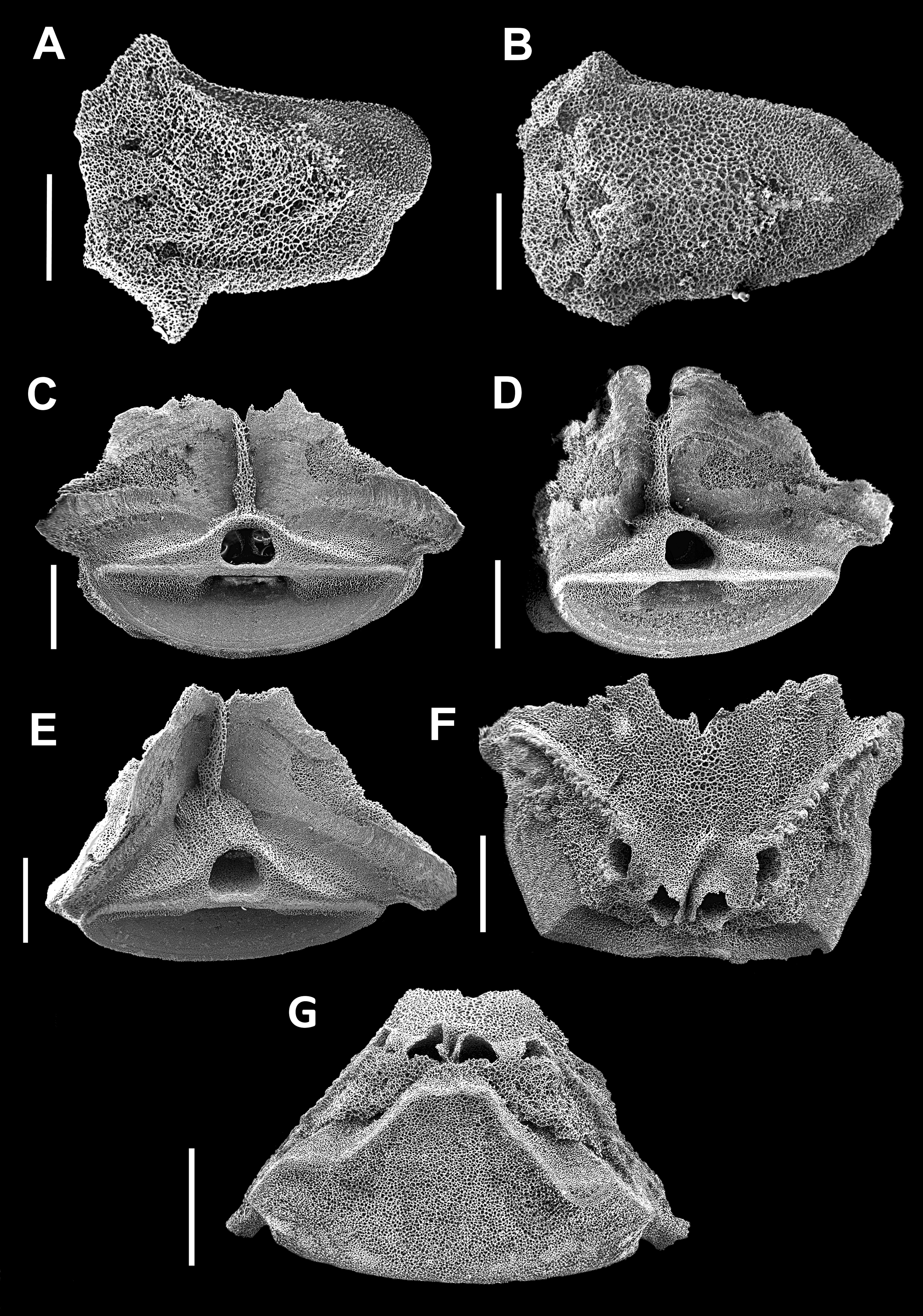
Table 1 View TABLE 1 , figures 4–11, 12B–F
Material examined. E of Miyakejima , Izu Is., Japan: OMNH cat. no. Iv2233, R / V Soyo, 34°31.8’N, 140°17.1’E, 1160 m, trawl, 14 Nov 1972, T. Okutani, coll. (holotype) GoogleMaps ; OMNH cat. no. Iv2448, R / V Soyo, 34°03.0’N, 140°02.0’E, 1210–1235 m, trawl, 5 Jul 1967, T. Okutani, coll. (1, paratype) GoogleMaps ; OMNH cat. no. Iv2980, R / V Soyo, 34°03.4’N, 140°04.7’E, 1270 m, trawl, 29 Jun 1969 (1, paratype) GoogleMaps . E of Chiba Prefecture, Japan: SAM cat. no. K2158, 35°09’N, 140°52’E, 522–537 m (Ext. no. C040), 24 Jun 2001, T. Oji, coll. (1, paratype) GoogleMaps . USNM cat. no. 1548291, R / V Tansei- Maru, Univ. Tokyo, cruise no. KT-01-8, sta. TK-1-5, 35°08.795’N, 140°51.061’E to 35°09.545’N, 140°51.933’E, 512–529 m, 3-m ORI beam trawl, 22 Jun 2001 (1, paratype, partly dissociated for SEM). GoogleMaps
Diagnosis. Atopocrinus with centrodorsal as tall as wide at base, or taller, and with small apical opening into centrodorsal cavity; HW 1.0–1.3. Cirrus sockets round or slightly oval, more deeply concave than in A. sibogae , in three columns per radial area proximally and two columns apically in large specimens; two columns per radial area in a smaller specimen. Deep “pore” between many or all fully developed sockets of a column; “pores” sometimes large enough so that sockets appear as short cylinders ‘attached’ to each other or the adjacent interradial ridge only at intervals around their circumferences. Interradial ridge sometimes crossed by distinct suture immediately aboral to suture between basal and centrodorsal as in A. sibogae , and often with 1 to 4 additional faint suture-like lines. Synarthrial tubercle on IBr2 moderately to strongly developed, with proximal angle in aboral view>90°; profile of arm base rounded or with distinct angle. Alternating articular tubercles on following proximal brachials weak to moderate.
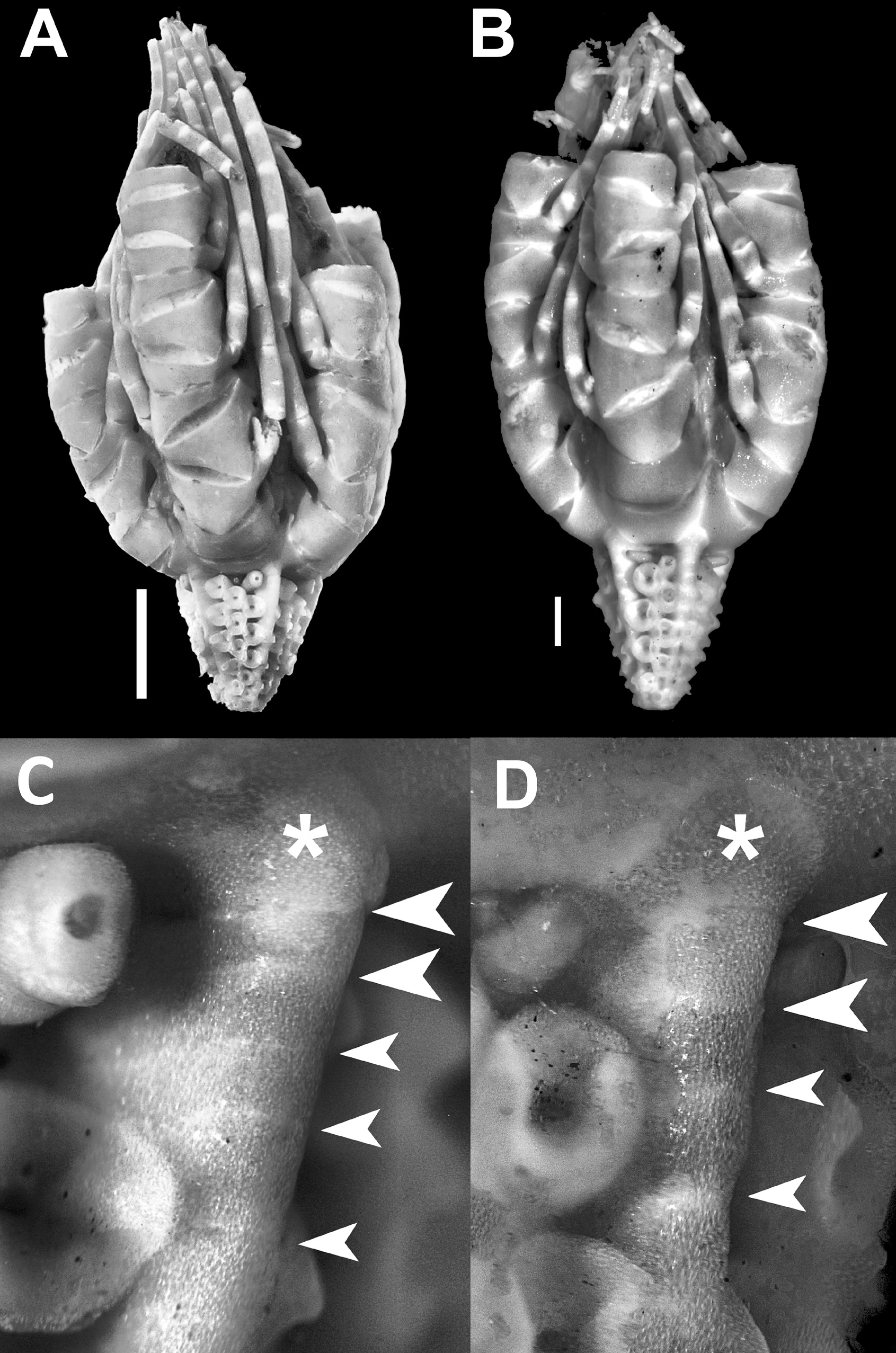
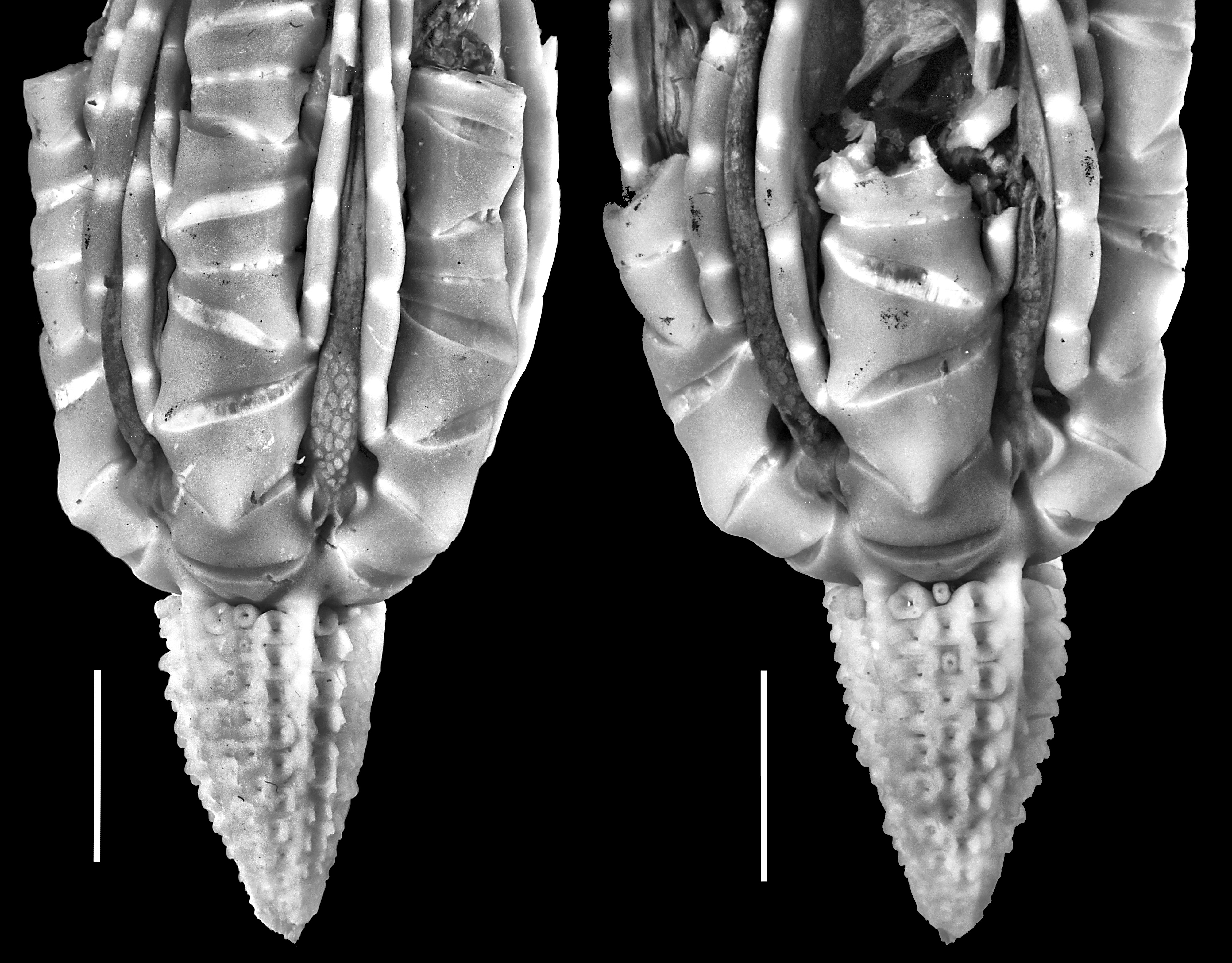
Description of holotype (OMNH Iv2233) ( Figures 4 View FIGURE 4 , 5 View FIGURE 5 , 12C View FIGURE 12 ).—Centrodorsal conical with straight sides. Interradial ridges along entire length of centrodorsal, stronger basally. Three of five ridges crossed by distinct suture immediately aboral to suture between basal and centrodorsal ( Figure 4 View FIGURE 4 B–D). Cirrus sockets in two lateral columns per radial area, of 6 sockets each; third, midradial column of 3 sockets (2 in one radial area) in basal half of each radial area; basalmost socket of several columns not fully developed; total number of functional sockets in Table 1 View TABLE 1 omitting one much smaller rudimentary cirrus at centrodorsal base in three radial areas; apicalmost socket in several columns obsolete.
Exterior ends of basals triangular or pentagonal, not of equal sizes; three crossed by fracture or suture-like line distinct from apparent suture with centrodorsal interradial ridge ( Figure 4D View FIGURE 4 ). Externally visible portion of radials extremely short and broadly U-shaped when specimen viewed from side ( Figure 4A View FIGURE 4 ), with diverging lateral margins and straight distal margin in aboral view; lateral margins of articulation with succeeding br1 parallel to oral-aboral axis ( Figure 4A, D View FIGURE 4 ).
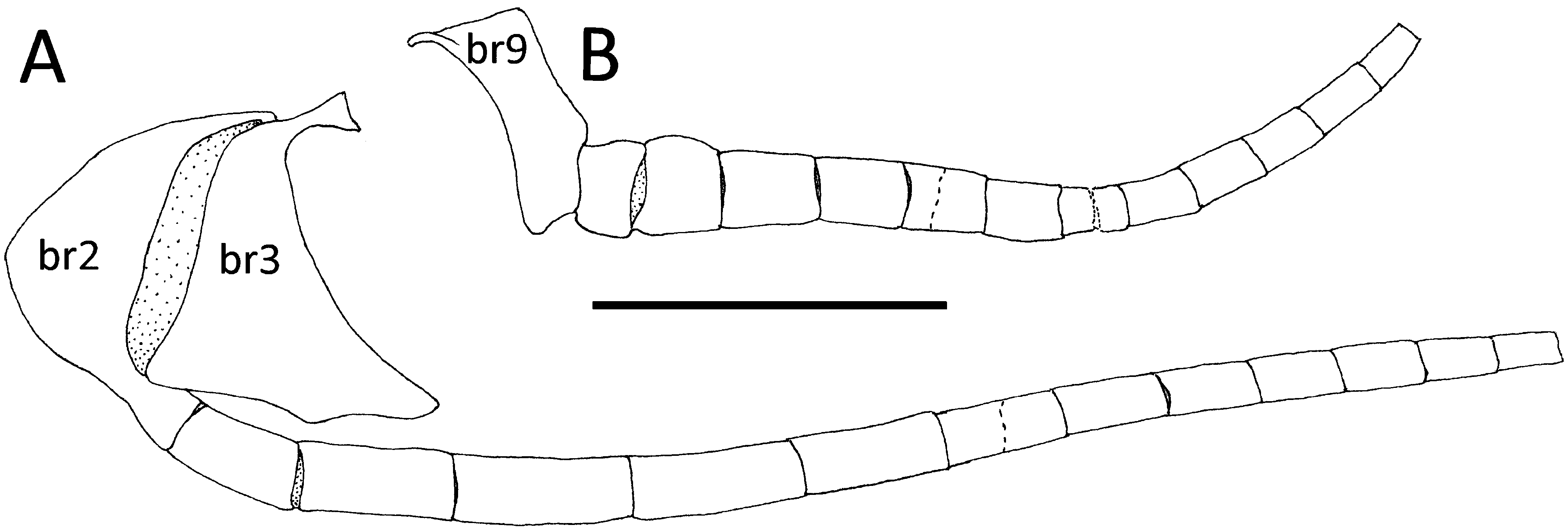
Arms retained to br5, br7, br10, br14, and br20. Br1 rectangular, not in contact laterally, with small lateral rounded triangular projections wider distally; distal margin straight or shallowly V-shaped. Br2 irregularly quadrate, with lateral margins diverging, one short and one long. Brr3 and 6 almost triangular. Brr4+5 and 7+8 strongly wedge-shaped, with long lateral margin on opposite side of arm from long lateral margin of preceding br. Brr9 and 10+11 slightly wedge-shaped. Brr7+8 and 10+11 with WL 1.7. Brr12 and 13 rectangular or slightly wedge-shaped, WL 2.5–2.8. Br14+15 and br16 wedge-shaped. Brr17–19 almost triangular. Brr16–19 with WL 2.0–2.2.
First three syzygies (brr4+5, 7+8 and 14+15) similar to those of A. sibogae , with only five narrow, widely separated radiating ridges. Succeeding syzygies (brr14+15, 20+21) with numerous fine, crowded ridges.
Pinnules arise from short lateral margin of brr2–9 (no pinnule on brr4 and 7—proximal ossicles of syzygial pairs), and from longer lateral margin on succeeding brachials ( Figure 5 View FIGURE 5 ). Pinnular articulations for P1–3 and Pa–b project at an angle from lateral margin of brachials (brr2–8) ( Figure 5A View FIGURE 5 ); articulations for succeeding pinnules flush with lateral margin of brachials ( Figure 5B View FIGURE 5 ). P1 on left side of br2 on 4 arms and on right side of 1 arm. P2 absent on br5 of one arm.
Remaining portion of P1 20 mm long, of 13 pinnulars (missing tip of unknown length); pinnulars longer than wide and smooth, increasing irregularly in length from P1 (1) (LW 1.4) through proximal third of pinnule length; LW of longest proximal pinnular 2.5–3.2; following pinnulars decreasing in length; distal pinnulars with LW 2.0; distalmost 3 remaining pinnulars shorter and with weak spines. Proximal pinnulars oval in cross section, attached to visceral mass to about P1 (5) and lacking ambulacral furrow; following pinnulars developing ambulacral furrow, becoming more pronounced on distal pinnulars. P2 similar, but P2 (1) with LW 1.0; pinnulars with greatest LW in proximal third. P3 broader basally than preceding; P3 (1) half-moon-shaped, wider than long; P3 (2) trapezoidal, narrower distally and as wide as long; middle pinnulars proportionately longest. Following pinnules all broken; second pinnular becoming wider than long; middle pinnulars weakly carinate, slightly expanded distally, with LW 1.0–1.3.
Disk mostly hidden within arm bases; anal papilla reaching level of br13; lateral surface of tegmen visible between adjacent arm bases with numerous small, more or less rhombic plates, forming pavement to level of brr2–3 and becoming more scattered and irregular, but submerged in tegmen tissue adorally, so that shapes are difficult to observe.
Description of large paratype (OMNH Iv 2448) ( Figures 6A, C View FIGURE 6 , 12D View FIGURE 12 ).—Similar to holotype but with centrodorsal as wide basally as tall, with midradial column of cirrus sockets reaching more than halfway to centrodorsal apex. One interradial ridge crossed by a distinct suture immediately aboral to suture between basal and centrodorsal (and a similar but partial suture in another ridge—both may be fractures in their respective basal ossicles). All five interradial ridges crossed by four additional faint suture-like lines ( Figure 6C View FIGURE 6 ). Cirrus sockets as in holotype but with 5–6 sockets in each lateral column and 2–4 in midradial column, again with one basalmost lateral socket not fully developed, and with one rudimentary cirrus at base of midradial column in each radial area. Aboral corners of radials swollen against ends of basals. Arms retained to br7 (3) and br10 (2). P1 on left side of br2 on 3 arms; on right side on 2 arms. All remaining pinnules incomplete (P1 retaining at most 11 segments).
Description of small paratype (OMNH Iv 2980) ( Figures 6B, D View FIGURE 6 , 12B View FIGURE 12 ).—Small specimen with 2 columns of 4 cirrus sockets plus one midradial rudimentary cirrus in each radial area; basalmost socket in one column in each radial area not fully developed (4 left, 1 right when viewed with apex downward). Interradial ridges of centrodorsal crossed by 3–5 faint suture-like lines aboral to centrodorsal/basal suture ( Figure 6D View FIGURE 6 ). (The basalmost of these may be equivalent to the supposed suture between A. H. Clark’s “reduced basal” and centrodorsal in A. sibogae .) Centrodorsal apex with round opening. External ends of basals pentagonal. Aboral corners of radials not swollen against ends of basals. Ray ossicles proportionately more elongated than in larger specimens, but still all wider than long. Arms retained to br10 (1) and br7 (4). P1 on left side of br2 on all arms. Most pinnules incomplete; longest pinnulars proportionally longer than in larger specimens; LW to 3.6. P3 missing only distal tip, of 10 segments, 5.6 mm long.
Description of paratype (SAM K2158) ( Figures 7 View FIGURE 7 , 12F View FIGURE 12 ).—Centrodorsal conical, taller than wide at base; sides straight, or slightly convex in proximal half; apical third slightly bent to one side. Interradial ridges along entire length of centrodorsal, stronger basally, each crossed by 1–2 faint apparent suture lines just aboral to suture between basal and centrodorsal. Tip of apex severed, exposing small cone of tissue. Cirrus sockets in two lateral columns per radial area, chiefly of 10 sockets each, with third, midradial column of 5–6 sockets reaching more than halfway to apex in each radial area. Pores between sockets fewer and smaller than those in other specimens. No fully developed cirri retained; small immature cirrus at base of four radial areas.
Exterior ends of basals roughly pentagonal, forming adoral extensions of centrodorsal interradial ridges. Sides of basals partly visible but disappearing into deep, low subradial clefts between centrodorsal margin and exterior surface of radials.
Externally visible portion of radials short, with diverging lateral margins; proximal corners slightly swollen over ends of basals; distal margin shallowly U-shaped in side view of specimen, almost straight in aboral view; distinct small “pore” between distolateral corners of adjacent radials.
Arms retained to br5 (2), br7 (2), and br10. Br1 rectangular, not in contact laterally; lateral margins straight and slightly projecting laterally in aboral view; lateral projections hemispherical in side view of arm; distal margin shallowly V-shaped. Br2 irregularly pentagonal, with lateral margins diverging; left short, right long. Synarthrial swelling between br1 and br2 well developed, midaboral, narrow. Br3 almost triangular, narrowing to right side. Br4+5 wedge-shaped, narrowest across syzygial articulation. Br6 short, wedge-shaped. Br7+8 not as strongly wedgeshaped as br4+5. Br9 almost oblong. Alternating articular swellings diminishing from brr3–4 through several following non-syzygial brachial pairs; negligible by brr8–9.
All remaining pinnules incomplete. P1 arising from left side of br2, longest remaining of 11 pinnulars, 19 mm long (missing tip); pinnulars compressed, longer than wide. LW of proximal pinnulars: P1 (1) 1.2, P1 (2) 2.3, P1 (3) 2.8, and P1 (4) 2.9; following pinnulars gradually shorter and more slender, but P1 (5) more elongated, LW 3.1. Abambulacral lateral margin of second through fourth pinnulars gently convex. P2 with pinnulars shorter and less elongated than in P1; LW of selected pinnulars: P2 (1) 1.6, P2 (2) 1.9, P2 (3) 1.8, P2 (7) 2.6, P2 (8) 2.5.
Sides of disk narrowly visible between proximal arms, with numerous small round calcareous plates. Oral surface of tegmen mostly mutilated. Proximal portions of each proximal pinnule connected to tegmen by thin sheet of tissue. Anal papilla with no visible plates.
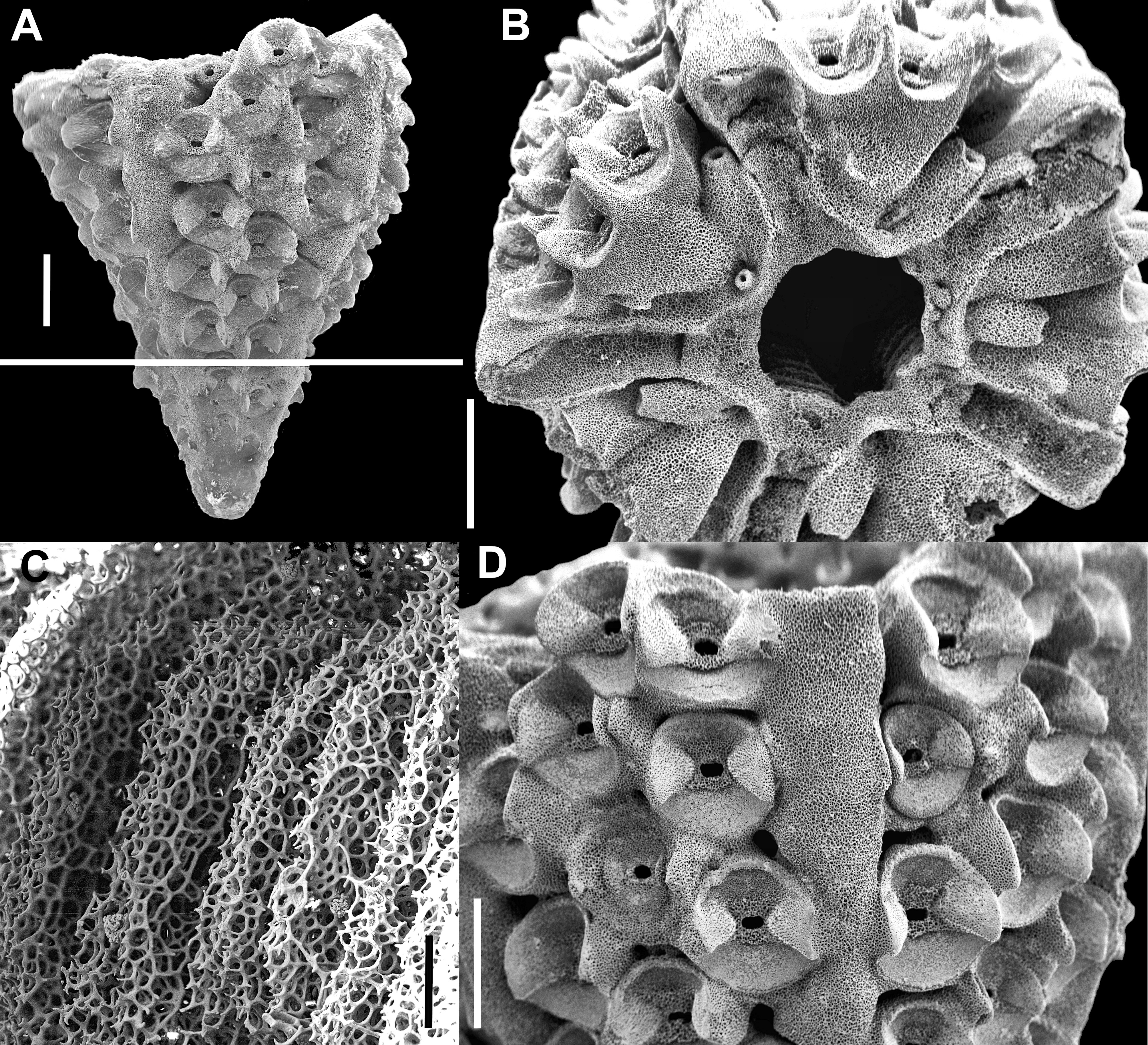
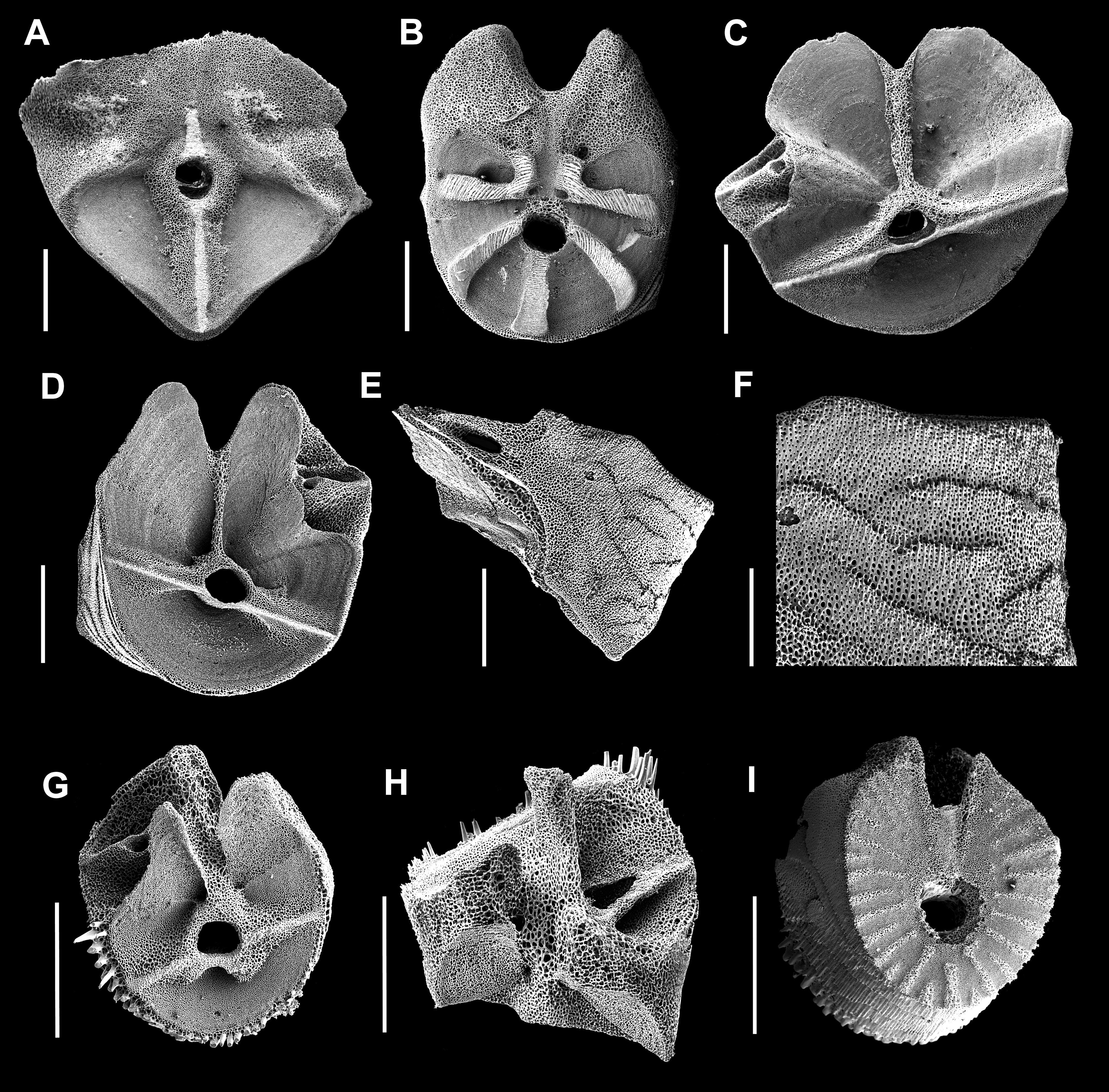
Paratype partly dissociated for scanning electron microscopy (USNM cat. no. 1548291) ( Figures 8 View FIGURE 8 –11, 10, D, F, G, 11E). Similar to holotype and large paratype but with sockets more crowded and numerous, inter-socket “pores” smaller, and no visible apical pore (after coating for SEM) ( Figure 8A, D View FIGURE 8 ). Adoral surface of centrodorsal with 5-rayed, stellate, thin-walled depression surrounding central cavity; depression rays narrow, interradial, reaching exterior, broader toward outer and inner ends; outer ends of rays truncated or rounded ( Figure 8B View FIGURE 8 ). One ray floored by three layers forming shallow steps rising toward central cavity (uppermost ray in Figure 8B View FIGURE 8 ). Each radial area of adoral centrodorsal surface with 1–2 tube-shaped rudimentary sockets not reaching exterior margin. Fully developed basalmost sockets also visible as tubes on adoral surface. Centrodorsal apex with one apparently obsolete socket in 2 radial areas. Interior of centrodorsal cavity with five broad interradial buttresses crossed by numerous regular fine ladder-like ridges and grooves ( Figure 8C View FIGURE 8 ); adoral opening ~0.25x basal diameter. No suture-like lines visible on interradial ridges ( Figure 8D View FIGURE 8 ). (Note: specimen not examined for this feature before dissociation for SEM.) Basals tongue-shaped, with no rosette; interior end of each basal wider, with two short, oblique, truncated projections, each abutting the projection of the neighboring basal; adoral surface convex; aboral surface flat or slightly concave, partially bordered by narrow ridge ( Figure 9 View FIGURE 9 A–B).
Radial articular facet deeply excavated, wider than tall ( Figure 9 View FIGURE 9 C–E). Aboral ligament fossa 4x wider than high, with wide, deep ligament pit. Central lumen surrounded by coarse stereom forming a tube with two low triangular “wings” extending laterally to ends of fulcral ridge. Muscle fossae parallel to oral-aboral axis, thin-walled, roughly triangular with rounded adoral and lateral corners (mostly broken), and separated medially by low narrow ridge of coarse stereom. Lateral ends of muscle fossae extending beyond ends of fulcral ridge. Weak ridge separating long narrow interarticular ligament fossae from muscle fossae, but with stereom similarly labyrinthic on both sides of ridge ( Figure 9C View FIGURE 9 ). Muscle fossae with surface layer of fine stereom with weak concentric lines, eroded in places in examined ossicles, revealing coarser underlying stereom lacking concentric features. Interior surface of radial ossicles with two pairs of canal openings ( Figure 9F View FIGURE 9 ). Lateral surfaces with a strong knobbed ridge running outward from just adoral of the outer canal openings to just short of the lateral ends of the muscle fossae ( Figure 9F View FIGURE 9 ). Aboral face flat; lateral-aboral margins excavated to accommodate basals ( Figure 9G View FIGURE 9 ).
Synarthry between br1 and br2 with adambulacral portion of fulcral ridge short, less than half length of abambulacral portion. Ligament fossae roughly triangular, restricted to abambulacral half of facet ( Figure 10A View FIGURE 10 ). Muscle fossae on proximal brachials large, roughly triangular, separated from each other by narrow ridge of coarse stereom, and separated from interarticular ligament fossae by weak ridge ( Figure 10C, D View FIGURE 10 ). Stereom of muscle fossae and interarticular ligament fossae similar. Middle brachials with distal border of spines ( Figure 10G, H View FIGURE 10 ). Pinnule articulations large, deeply excavated, with one muscle fossa ( Figure 10C, D, G, H View FIGURE 10 ).
Proximal three syzygies with five widely spaced ridges; two lateral ridges L- or ˩-shaped, each with the short arm oriented toward ambulacrum; ridge surfaces consisting of fine, closely crowded and often fused, concentrically-oriented solid stereom ridges; articular area of syzygy occupying about two-thirds of ossicle facet ( Figure 10B View FIGURE 10 ). Syzygies beyond 14+15 with numerous narrow ridges; several ridges very short and almost none reaching large central lumen; articular area occupying almost entire ossicle facet ( Figure 10I View FIGURE 10 ). Detached mid-arm fragment of 23 wedge-shaped to almost triangular brachials, wider distally with finely spinose distal margins, WL 1.4–1.6. Intersyzygial interval 3–5.
Exterior surface of brachials crossed by irregular, extremely fine “ledges”, each with a row of fine, distallydirected recumbent spines projecting slightly beyond the ledge edge, with rows of stereom pores between generally diminishing in size toward ledge ( Figure 10E, F View FIGURE 10 ).
Complete P1, 16 mm long, of 13 pinnulars, the distal 6 regenerating, short, with distal margins finely spinose.
Description of pinnule articulations dissociated for scanning electron microscopy (holotype OMNH Iv2233 and paratype USNM cat. no. 1548291) (Figure 11). P1 (1) with distal articulation roughly symmetrical with paired muscle fossae deeply excavated and almost fused (Figure 11A). P2 (1) with proximal articulation asymmetrical, with lumen and adjacent shallow ligament pit strongly offset from facet center; one arm of fulcral ridge much shorter than the other; single deep muscle fossa adjacent to lumen but recessed on adambulacral surface of ossicle (Figure 11B). Long proximal pinnulars of P2 with most of facet relatively flat with large abambulacral ligament fossa; distal facet with asymmetrical fulcral ridge and single deep muscle fossa slightly offset adjacent to lumen (Figure 11C); proximal facet with roughly symmetrical fulcral ridge and paired muscle fossae deeply recessed along adambulacral margin (Figure 11D, right end). Pinnulars hollow (Figure 11E). More distal pinnulars gradually developing deep V-shaped ambulacral groove (Figure 11F, H–I); lumen offset from facet center with short fulcral ridge restricted to side of facet opposite ambulacral groove; single deep muscle fossa on same side of groove as lumen and fulcral ridge (Figure 11H, I). Distal pinnulars with numerous long recumbent spines on one side and fine diagonal ridges similar to those of brachials ( Figure 10E, F View FIGURE 10 ) on the other (Figure 11G).
Etymology. Named for Tatsuo Oji, Professor, Department of Earth and Planetary Sciences, Nagoya University, Nagoya, Japan, for his many contributions to our understanding of both living and fossil Crinoidea .
Distribution. Known only off eastern Japan in 529 (possibly 512) to 1270 m.
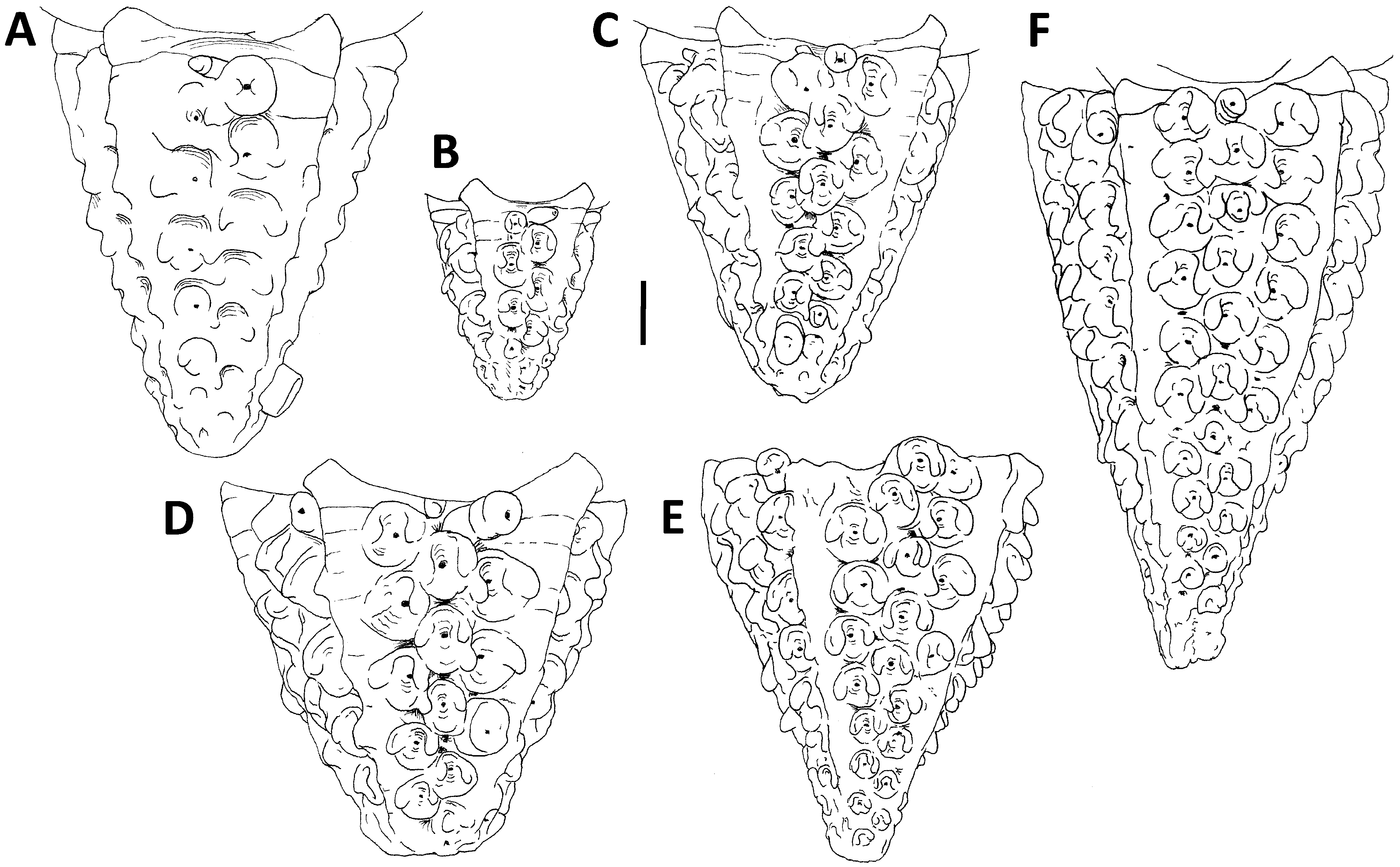
Remarks. The differences between similarly-sized specimens of A. sibogae and Atopocrinus ojii n. sp. appear significant and consistent enough to warrant recognition of the latter as a distinct species. Atopocrinus ojii n. sp. has three rather than two columns of sockets per radial area, round rather than squarish cirrus sockets, and centrodorsal with an apical pore, and pores between cirrus sockets. Figure 12 View FIGURE 12 illustrates the centrodorsals of all specimens of both species, and figure 13 illustrates how cirrus number relative to centrodorsal height distinguishes the two. Whether the two species differ or not in several other characters, e.g., presence or absence of a rosette, overall shape of basals, and structure of the adoral surface of the centrodorsal and its interior buttresses, cannot be determined at present, because A. sibogae remains known from a single specimen.
The inter-socket pores of Atopocrinus ojii n. sp. appear to be unique among extant crinoids. However, it could not be determined whether they communicate with the centrodorsal cavity or end blindly, possibly as pouches within the interradial buttresses inside the centrodorsal.
Four of the five specimens of A. ojii also have a pore in the centrodorsal apex. Gentle insertion of a fine needle in the two largest specimens met no resistance and suggests that the pore communicates with the centrodorsal cavity. A similar, although five-rayed, perforation corresponding to the central stalk canal remains at the aboral apex of the centrodorsal in feather stars following separation from the postlarval stalk ( Mortensen 1920, p. 30) but becomes plugged by stereom shortly thereafter. However, Mortensen (1918) noted that the pore remains in a half-grown specimen of Notocrinus virilis Mortensen, 1917 . Other species, including several fossil genera, retain a five-rayed depression at the aboral pole—the so-called dorsal star—although it is unknown how long any communication with the centrodorsal cavity remains after separation from the postlarval stalk in these taxa: e.g., Glenotremites , Loriolometra , Remesimetra (all Notocrinidae ), Kiimetra miocenica Shiibata & Oji, 2007 (Calometridae) ( Hess & Messing 2011).
In the single dissociated specimen of Atopocrinus ojii n. sp., the tongue-like basals lack a rosette. Messing & White (2001), described the zenometrid genera Zenometra and Sarametra as lacking a rosette as well. However, unlike Atopocrinus ojii n. sp., the inner end of each basal in both zenometrid genera ends abruptly and bears a pair of concavities, which appear to be partial versions of two openings in confamilial Psathyrometra . The openings in the latter are continuations of the aboral nerve canals of the overlying radials and correspond to the interradial and radial processes of the rosette of antedonids. The lack of similar structure in Zenometra and Sarametra may derive from poor preservation ( Messing & White 2001). Although a rosette as a discrete, centrally perforated, decagonal plate does not appear to exist in any of these genera, the inner ends of their basals exhibit structure not found in Atopocrinus ojii n. sp.
The centrodorsal of A. ojii n. sp. bears a striking resemblance to that of Spinimetra chesnieri Hess & Thuy, 2017 , from the Lower Jurassic (Toarcian, Tenuicostatum Zone). Both share the following features: conical centrodorsal with small apical opening and pores between cirrus sockets; aboral surface of centrodorsal with tube-shaped rudimentary sockets flanking narrow basals, and cirrus sockets with a pronounced horseshoe-shaped rim, each a blunt triangle projecting outward and downward. (Compare Figure 8D View FIGURE 8 with 14A, and 8C with 14B.) The combination exists in no other crinoids, extant or fossil, although two characters occur separately elsewhere: horseshoeshaped socket rims in Atelecrinidae and an apical pore in Notocrinidae , mentioned above. Spinimetra differs from Atopocrinus in lacking the interradial ridges on the centrodorsal, and in having basals with marginal crenulae. In addition, the centrodorsal of S. chesnieri appears to be constructed of multiple “nodals”; juvenile specimens of S. chesnieri suggested to Hess & Thuy (2017) that its centrodorsal was constructed of a stack of separate ossicles. It is tantalizing to consider the possibility that the faint suture-like lines that cross the interradial ridges in A. ojii n. sp. ( Figures 2 View FIGURE 2 , 4 View FIGURE 4 , 6 View FIGURE 6 ) might represent a vestige of this multi-ossicle construction (although the dissociated specimen of A. ojii exhibited no suture-like lines). As the cirrus sockets in this species develop as tubes adjacent to the centrodorsal base, it is not surprising that the faint suture-like lines do not cross them. Nevertheless, the characters that the two genera have in common may be convergences rather than homologies. As S. chesnieri is known only from centrodorsals, no further comparison with Atopocrinus is currently possible.
The video attributed to A. sibogae ( Figure 3 View FIGURE 3 ) shows that the distal half of each arm lacks pinnules. Other crinoids with distal arms similarly lacking pinnules include both feather stars—Atelecrinidae ( Messing 2013) and Sarametra (Zenometridae) ( Messing et al. 2018)—and stalked taxa: Porphyrocrinus (Phrynocrinidae) ( Messing 2016) and at least some Metacrininae (Isselicrinidae) (Tunnicliffe et al. 2015). Molecular data place two of these ( Atelecrinidae and Porphyrocrinus ) in a clade formerly treated as Bourgueticrinina ( Hess & Messing 2011) , but other members bear pinnules to arm tips (e.g., Democrinus, Bathycrinus ). It thus appears that distal arms lacking pinnules have arisen independently multiple times among extant crinoids.


No known copyright restrictions apply. See Agosti, D., Egloff, W., 2009. Taxonomic information exchange and copyright: the Plazi approach. BMC Research Notes 2009, 2:53 for further explanation.
|
Kingdom |
|
|
Phylum |
|
|
Class |
|
|
Order |
|
|
Family |
|
|
Genus |