
Atopocrinus sibogae A.H. Clark, 1912
|
publication ID |
https://doi.org/ 10.11646/zootaxa.4731.4.2 |
|
publication LSID |
lsid:zoobank.org:pub:85C4B527-6CAF-404B-87B9-B9FD5468213F |
|
DOI |
https://doi.org/10.5281/zenodo.3664751 |
|
persistent identifier |
https://treatment.plazi.org/id/03B89821-FF9E-E91B-7BAD-DF09FB1929E4 |
|
treatment provided by |
Plazi |
|
scientific name |
Atopocrinus sibogae A.H. Clark, 1912 |
| status |
|
Atopocrinus sibogae A.H. Clark, 1912
Table 1 View TABLE 1 , figures 1–3, 12A
Material examined. Laut Seram (formerly Ceram), Indonesia: NBC cat. no. V.ECH.C. 2097, Siboga sta. 177, 2°24’30”S, 129°38’30”E, 1300–1633 m, dead coral and stones, covered with manganese, 1 Sep 1899 (holotype). N of Pulau Sangihe, Indonesia: no specimen; video taken by ROV Little Hercules launched from NOAA R / V Okeanos Explorer , 4°14.894’N, 125°15.879’E, depth 1,151.1–1,151.7 m, 28 Jul 2010.
Diagnosis. Atopocrinus with centrodorsal taller than wide at base; HW 1.3. Cirrus sockets crowded in two columns per radial area, oblong or squarish (except apical and basalmost sockets round), surrounded by projecting rim with rounded triangular sides, and with no openings between sockets. Synarthrial tubercle on IBr2 strongly developed with proximal angle in aboral view <90° ( Figure 2A, B View FIGURE 2 ); profile of arm base with distinct angle at br1–2 articulation. Alternating articular tubercles on few following proximal brachials strongly developed.
Description of the holotype (NBC V.ECH.C. 2097). Centrodorsal elongated conical with straight sides and tip slightly blunted. Interradial ridges strong, each about as broad as adjacent cirrus socket column. Cirrus sockets recessed; lateral margins strongly rounded triangular, producing serrate profile; aboral margin of each socket produced as strong ridge across adoral border of adjacent socket. Immature proximal sockets projecting. Cirri lost, except for one tiny rudimentary cirrus in some radial areas. Deep subradial clefts half as tall as height of proximalmost mature cirrus sockets.
Ends of basals (=basal rays in A.H. Clark & A.M. Clark 1967) convex pentagonal, capping adoral ends of interradial ridges, extending inward to form sides and inner wall of subradial clefts. Exposed aboral surface of radials with WL ~2.0 (when viewed aborally) but projecting outward so that WL appears ~4.0 when specimen is viewed from the side.
Arms stout, all broken near bases; longest remaining portion 19 mm from subradial cleft to distal edge of br10. Br1 oblong in aboral view, with small deep notch in distal corners; distal margin V-shaped when specimen viewed from the side. Br2 irregularly quadrate, almost triangular; P1 arising from lateral projection on short lateral margin. Br1–2 with strong synarthrial tubercle. Br3 (on four arms) triangular, broader than long, with strongly concave sides. Br4+5 triangular; both br4 and br5 triangular, the former slightly longer. Following brachials similar to br3, becoming proportionally slightly longer with their shorter lateral margins slightly longer. Distalmost remaining brachial br10. Arm widths: 4 mm at br1; 3.5 mm at both br4+5 and br10.
Syzygies at br4+5, 7+8, and (where arm remains) 10+11, except 3+4, 6+7, and 9+10 on right posterior arm. Length from base of br1 to syzygy at br7+8 12.0 mm. Br7 distal syzygial facet with five radiating ridges restricted to abambulacral half of facet ( Figure 2E View FIGURE 2 ): two lateral ridges do not reach facet margin; three ridges arising from narrow ridge bordering abambulacral side of central canal—one midaboral, and one on either side between midaboral ridge and adjacent lateral ridge—all reach facet margin. Adambulacral half of facet dominated by pair of large shallow depressions separated by a narrow median groove; groove flanked by a pair of narrow, low flattened ridges that arise from the inner ends of the lateral ridges but lack the solid stereom of the latter. A pair of narrower, weak ridges lie parallel and adambulacral to the lateral ridges and do not reach the facet margin.
P1 arising from right side of br2 on four arms, moderately stout and somewhat compressed laterally. P1 (1) slightly trapezoidal, wider distally, with LW ~ 1.5 in lateral view; P1 (2) narrower distally, with LW 2.5–3.0, 2.3 mm long; P1 (3) narrower distally; following pinnulars similar but proportionately longer. P2 resembling P1. Following pinnules only with bases remaining; successive pinnules apparently becoming shorter and slenderer, with second pinnulars decreasing rapidly in length and distal width; Pc (2) (third pinnule, on br9—opposite side of arm from P1) shorter than proximal width and slightly trapezoidal; following pinnulars likely much more slender. P3 11.25 mm long, of 15 segments; greatest segment LW 1.2.
Disk concealed, somewhat mutilated, small and compact, similar to that of Atelecrinus , forming a high, rounded dome reaching level of br9, but beginning to curve inward at about level of br5. From here, ambulacra are supported on high narrow tissue bridges (as in Gephyrocrinus , Thalassocrinus , and Ptilocrinus ) and reach arms at ~br9. Pinnules connected to sides of tegmen by thin tissue sheets resembling the thicker bridges described above. Interradial sides of tegmen with thickened strip of tissue extending from base of disk to between sides of adjacent br1 and bearing cluster of ~12 rounded calcareous plates between adjacent br1 and br2. Deep oval pit between proximolateral corners of adjacent br1; similar larger pit adjacent to distolateral angles of br1, on both sides of each syzygy, and at each pinnule base (modified from A.H. Clark in A.H. Clark & A.M. Clark 1967, pp. 814–817).
Characters visible in video but not in holotype ( Figure 3 View FIGURE 3 ). — Pinnules developed along little more than half arm length; distal remainder of arm a long filament lacking pinnules (no scale recorded). Cirri of up to ~30 cirrals, straight for most of length, gently curved distally, about a third as long as pinnule-bearing portion of arm ( Figure 3B View FIGURE 3 ). Longest cirrals (~c6–7) with LW ~3.0; following cirrals gradually decreasing in length and width; middle cirrals with LW ~4.0; distalmost cirrals (except possibly penultimate) remaining longer than wide. Pinnules diminishing in length from P1; ~P6–P7 shortest, composed of pinnulars with LW no more than ~2.0; following pinnules increasing in length. Color: brachials maroon or orange-brown with yellow articulations; cirri pink with pale articulations; ambulacral tissue dark brown.
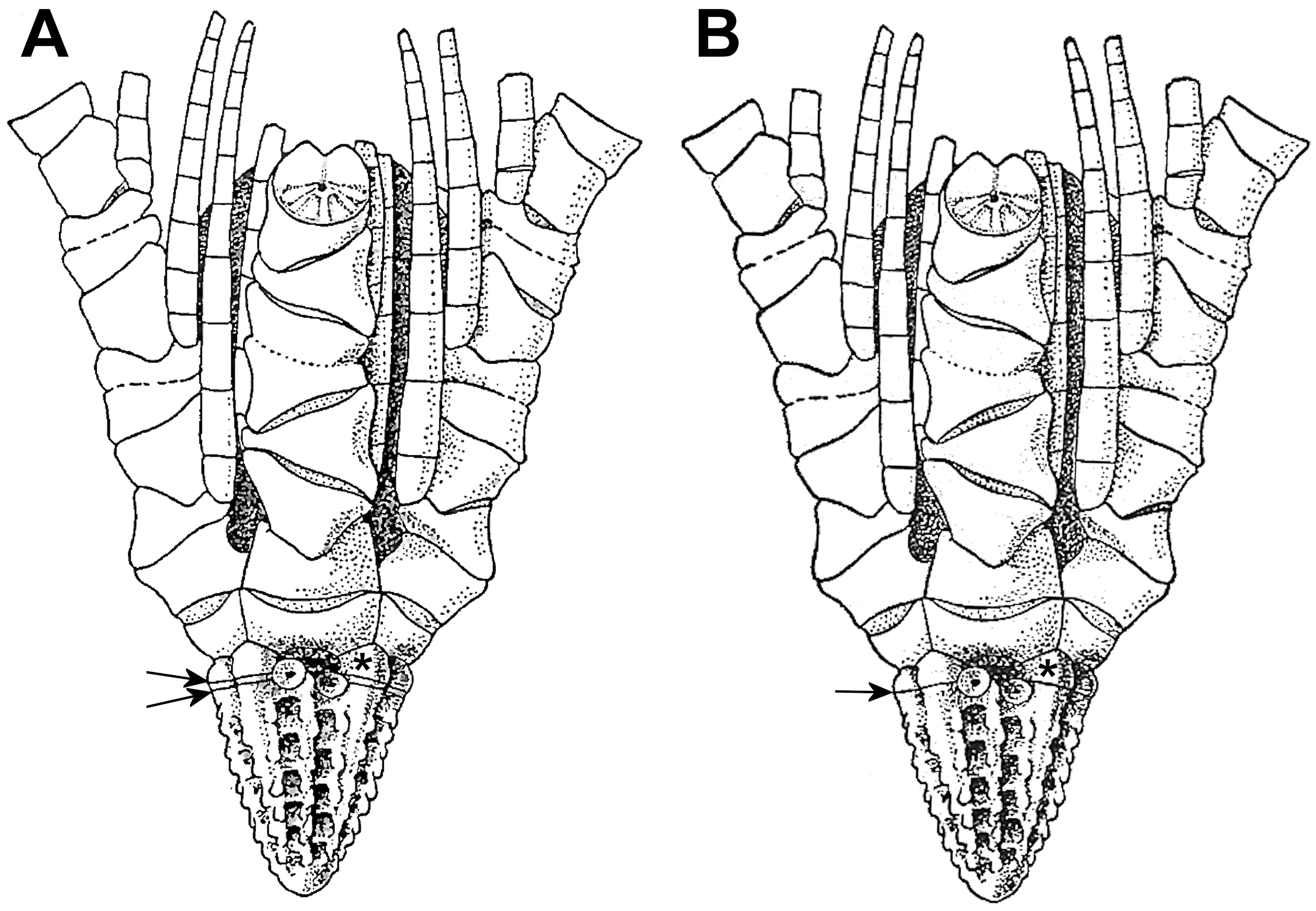
Remarks. A. H. Clark’s detailed descriptions (1918, and in A.H. Clark & A.M. Clark 1967) are basically accurate, although the number of functional cirrus sockets is closer to 60 than his 68, omitting apparently obsolete sockets near the apex and a rudimentary socket at the centrodorsal base in some radial areas. Also, in addition to the suture between the true basal (A. H. Clark’s basal ray) and centrodorsal (upper arrow in Figure 1A View FIGURE 1 and upper arrowhead in Figure 2C, D View FIGURE 2 ), close examination of the holotype reveals a fine line crossing the interradial ridges aboral to this suture (lower arrowhead in Figure 2C, D View FIGURE 2 ), corresponding to the suture between A.H. Clark’s supposed reduced basal (aboral to his basal ray) and centrodorsal (lower arrow in Figure 1A View FIGURE 1 ). This line lies at the level of the basalmost socket and disappears “behind” the socket rather than crossing it. Whether this is an actual suture or not is unclear (see discussion below). Two interradial ridges (not shown) each appear to have a faint second line aboral to the preceding, giving three apparent “sutures,” the most adoral of which is the true basal/centrodorsal suture.
The supposed “median ridge” in each radial area mentioned by A.H. Clark (in A.H. Clark & A.M. Clark 1967) appears instead to be just the midradial margins of successive alternating sockets from each of the two adjacent columns, augmented by their projecting fulcral tubercles, which form a serrated line along the middle of each radial area, rather than a separately elevated ridge. The fulcral ridges of each socket appear proportionally narrower than in atelecrinids with two columns per radial area, which creates in A. sibogae a relatively wider concavity surrounding each central socket lumen.
Although the pinnulars of A. sibogae have only been examined externally, their cylindrical structure and translucent appearance strongly suggest that, like those of Atopocrinus ojii n. sp., they are also hollow.
The in situ video (screen grab in Figure 3 View FIGURE 3 ) is attributed to A. sibogae based on a combination of location and depth ( Indonesia; 1,151 –1,152 m), tall centrodorsal with cirri in columns, and the five undivided arms. The characters of the distal portion of the arms lacking pinnules, the proximal pinnules and cirri visible in the video and screen grabs have not been included in the generic diagnosis pending recovery of complete specimens of the new species described below.
Figure 3A View FIGURE 3 shows the specimen in the video with four arms arranged in an almost monoplanar posture in response to near-bottom flow from the right rear, with the fifth arm (center foreground) parallel to the arm to its right but down current. All ambulacra face down current. Flow velocity is apparently too strong for the long primary podia to maintain a typical extended feeding posture, as the current forces them into both smooth and irregular down-current undulations (large arrows in Figure 3C, D View FIGURE 3 ) (note also the down-current deflection of the distal arm filaments in Figure 3A View FIGURE 3 , arrows). However, the shorter secondary podia are extended in straight lines characteristic of direct particle interception (small arrows in Figure 3C, D View FIGURE 3 ), as in some shallow-water feather stars, e.g., Antedon bifida ( LaHaye & Jangoux 1985) .
The tall centrodorsal and numerous long cirri that curve only slightly near their tips ( Figure 4A, B View FIGURE 4 ) are similar to those of deep-water Atelecrinidae ( Messing 2013) and similarly provide a broad-based platform that raises the calyx above the substrate, although not enough to permit arraying the arms in a parabolic posture as in some other feather stars with long cirri that typically cling to elevated perches, e.g., shallow-water Pontiometra andersoni ( Carpenter, 1889) ( Meyer & Macurda 1980) and Zygometra microdiscus ( Bell, 1882) ( Messing et al. 2006) .


| V |
Royal British Columbia Museum - Herbarium |
| NOAA |
National Oceanic and Atmospeheric Administration |
| R |
Departamento de Geologia, Universidad de Chile |
No known copyright restrictions apply. See Agosti, D., Egloff, W., 2009. Taxonomic information exchange and copyright: the Plazi approach. BMC Research Notes 2009, 2:53 for further explanation.