
Apophua sugaharai Momoi, 1978
|
publication ID |
https://doi.org/ 10.11646/zootaxa.3784.5.1 |
|
publication LSID |
lsid:zoobank.org:pub:6640D1B6-E304-4C6B-8E36-71F8FB2C347F |
|
DOI |
https://doi.org/10.5281/zenodo.6143605 |
|
persistent identifier |
https://treatment.plazi.org/id/03B8E80C-FFE0-F562-DA85-D89C25B0FB06 |
|
treatment provided by |
Plazi |
|
scientific name |
Apophua sugaharai Momoi, 1978 |
| status |
|
Apophua sugaharai Momoi, 1978 View in CoL
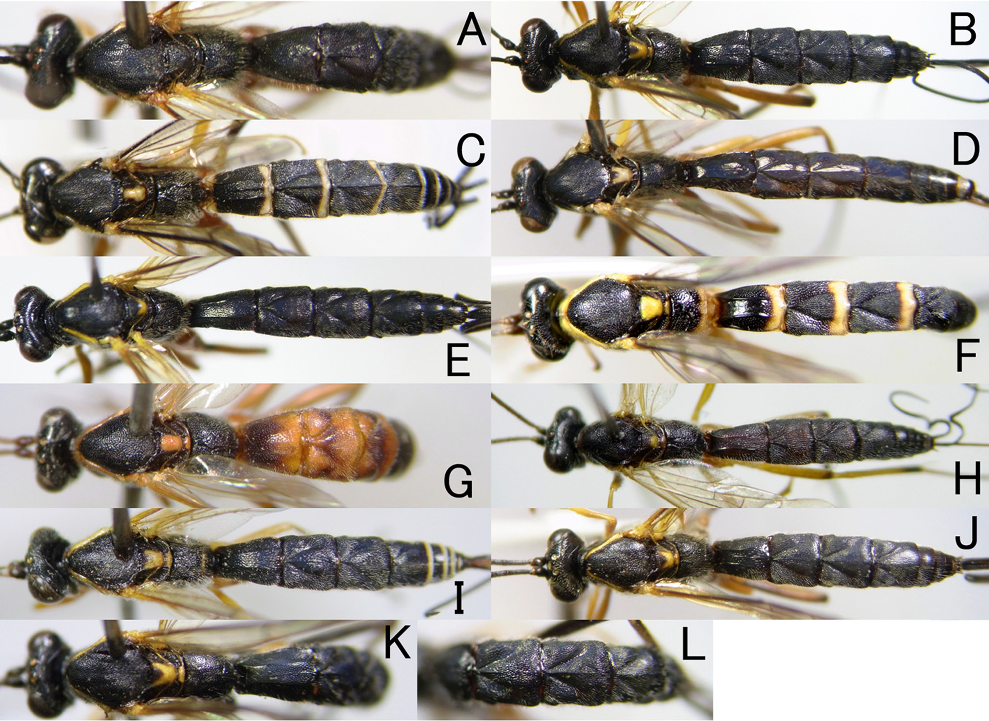
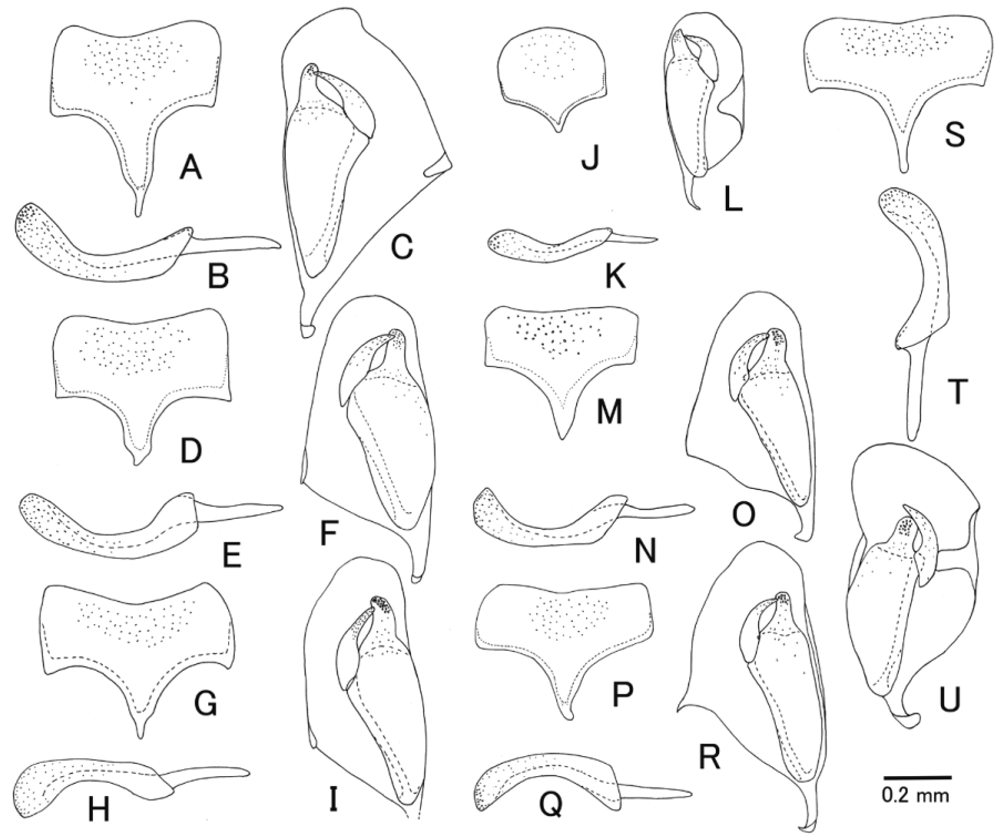
( Figs. 1 View FIGURE 1 I, 2 H, 4 J, 5 I, 6 I, 7 I, 8 M, N, 9 S–U)
Apophua sugaharai Momoi, 1978: 9 View in CoL .
Description. Female (n=4). Body: slender, its length 9.0–10.0 mm.
Head 0.6 times as long as wide. Clypeus 0.8 times as long as wide, its upper 0.6–0.7 punctate. Face 0.6–0.7 times as long as wide. MSL 0.7 times as long as BWM. OOD 1.6 times as long as OD. IOD 1.6 times as long as OD. Antenna with 44–47 flagellomeres. F1 2.0 times as long as F2.
Mesosoma. Lateral area of pronotum largely smooth ventral 0.5. Epicnemial carina present laterally. Mesopleural fovea smooth. All carinae of propodeum present but median section of lateromedian longitudinal carina and lateral longitudinal carina more or less obscured and restricted by weak ridge or sometimes absent. Fore wing length 7.5–8.5 mm, with vein 2 m-cu with two bullae. Hind wing with Cu 1 present, nebulous. Fore coxa without ridge ( Fig. 4 View FIGURE 4 J). Hind femur 6.4–6.5 times as long as maximum depth in lateral view. Hind TS1 2.5 times as long as TS2.
Metasoma punctate, shiny. Punctures on T1 to basal 0.5 of T4 slightly longitudinally striated by coalescent punctures. T1 1.4 times as long as maximum wide, its median dorsal carina nearly completely present, short median longitudinal keel sometimes present posteriorly ( Fig. 6 View FIGURE 6 I). T2 1.0 times as long as maximum width, short median longitudinal keel present only at base ( Figs. 6 View FIGURE 6 I, 7 I). T3 with short and weak median longitudinal at base ( Fig. 6 View FIGURE 6 I). Ovipositor sheath 2.2–2.3 times as long as hind tibia.
Coloration ( Figs. 1 View FIGURE 1 I, 2 H, 5 I, 7 I). Body rather strongly polished by dark blue reflection. Head black except for: clypeus, mandible excluding tip, malar space along mandible, narrow ventral spot on apex of pedicel, palpi yellow to yellowish-brown; antenna sometimes tinged with dark brown except for yellow area. Mesosoma black except for: upper margin of pronotum excluding median part, tegula, subtegular ridge, scutellum excluding basal spot, postscutellum, hind margins of each axilla, transverse stripe on propodeum along posterior transverse carina yellow. Wings hyaline with veins and stigma brown except for yellow wing base. Legs reddish-brown except for: fore and mid coxae, trochanters and trochantelli, dorsal part of hind coxa, hind trochanter yellow; mid TS5, ventral part of apex of hind coxa, hind trochantellus, base and apex of hind femur, subbasal band and apical 0.2 of hind tibia, hind tarsus including claw blackish-brown; hind tibia excluding brown area, hind tibial spur whitish-yellow. Metasoma black except for: posterior margin of T4–T7, membranous area of sternites, apex of subgenital plate whitish-yellow. Ovipositor reddish-brown. Yellow stripe on propodeum sometimes small in size.
Male (n=3). Similar to female. MSL 0.6; OOD 1.2–1.4 times as long as OD; IOD 1.4 times as long as OD. Subgenital plate with a long apodema sternalis, anterior margin slightly concave ( Fig. 9 View FIGURE 9 S). Inner margin of paramere strongly concave near basal inner angle (Fig. 8 M). Penis valve of aedeagus ca. 1.5 times as long as basal apodeme ( Fig. 9 View FIGURE 9 T). Ventral surface of scape and pedicel yellow.
Specimens examined. JAPAN [Hokkaido] 1M (paratype), Bibai, 1. ix. 1961, K. Kamijo leg. (MNHAH); 1F, Shimamatsu, 8. vii. 1965, K. Kusigemati leg. (KU). [Honshu] 1F (holotype), Iwate Pref., Wakare, 22. vi. 1968, T. Oku leg., (host: Ptycholoma circumclusana ) (MNHAH); 1M, Mt. Gassan, 2. ix. 1966, K. Kusigemati leg. (KU). [Shikoku] 1F, Tokushima Pref., Ichiu vil., Mt. Tsurugisan (1500m), 16. x. 1980, H. Takemoto leg. (NIAES); 1F, same data excluding T. Goto leg. (NIAES); 1F, Ehime Pref., Mt. Odamiyama, Mt. Mizuashiyama, 6. viii. 1994 (LT), E. Yamamoto leg. (NIAES).
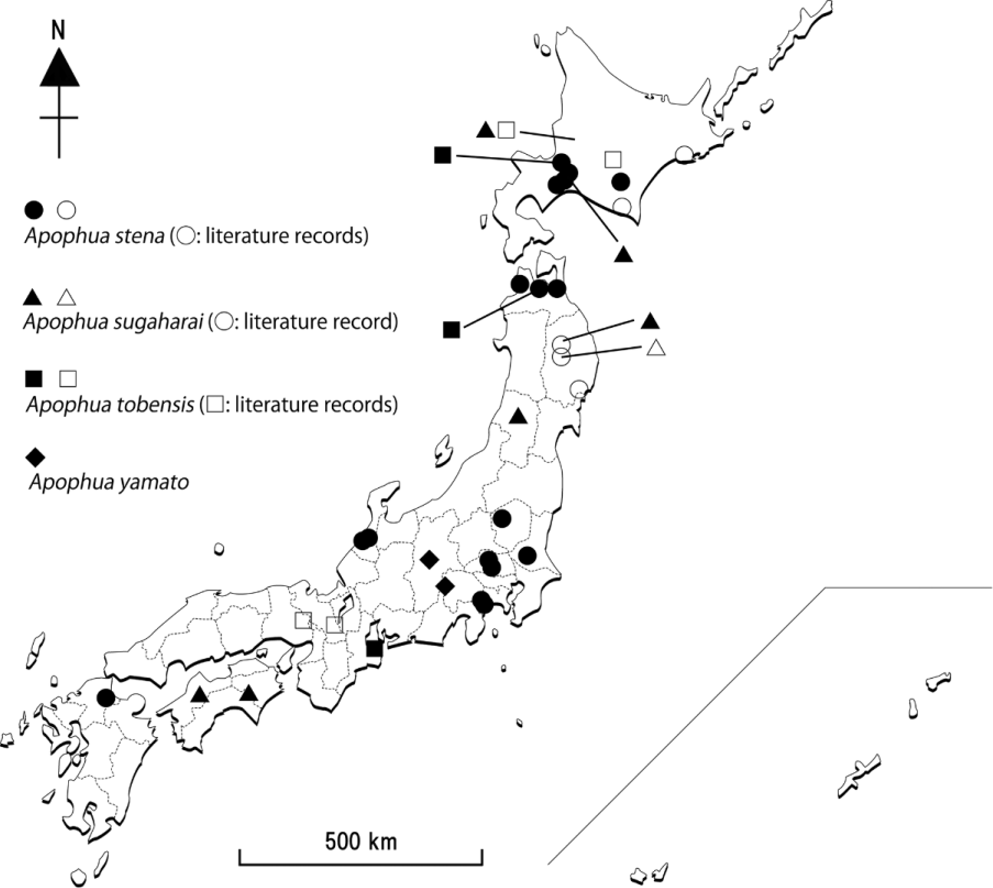
Distribution ( Fig. 13 View FIGURE 13 ). Japan (Hokkaido, Honshu and Shikoku*).
Biology. The following hosts are recorded in Japan: Ptycholoma lecheana circumclusana (Treitschke) and Pandemis heparana (Denis & Schiffermüller) ( Momoi, 1978) , Acleris alnivora Oku ( Nakaya, 2002) and Adoxophyes orana fasciata Walsingham ( Nakaya, 2009) .
Remarks. This species resembles A. honmai but it can be distinguished from the latter by the posterior margin of T4–T7 with conspicuous white band ( Figs. 1 View FIGURE 1 I, 2 H) (without white band in A. honmai ).


No known copyright restrictions apply. See Agosti, D., Egloff, W., 2009. Taxonomic information exchange and copyright: the Plazi approach. BMC Research Notes 2009, 2:53 for further explanation.
|
Kingdom |
|
|
Phylum |
|
|
Class |
|
|
Order |
|
|
Family |
|
|
Genus |
Apophua sugaharai Momoi, 1978
| Watanabe, Kyohei & Maeto, Kaoru 2014 |
Apophua sugaharai
| Momoi 1978: 9 |