
Apophua
|
publication ID |
https://doi.org/ 10.11646/zootaxa.3784.5.1 |
|
publication LSID |
lsid:zoobank.org:pub:6640D1B6-E304-4C6B-8E36-71F8FB2C347F |
|
DOI |
https://doi.org/10.5281/zenodo.6143583 |
|
persistent identifier |
https://treatment.plazi.org/id/03B8E80C-FFF7-F576-DA85-DDBE24A7F834 |
|
treatment provided by |
Plazi |
|
scientific name |
Apophua |
| status |
|
Key to Japanese species of the genus Apophua View in CoL View at ENA
(The males of aquilonia , elegans and yamato are unknown)
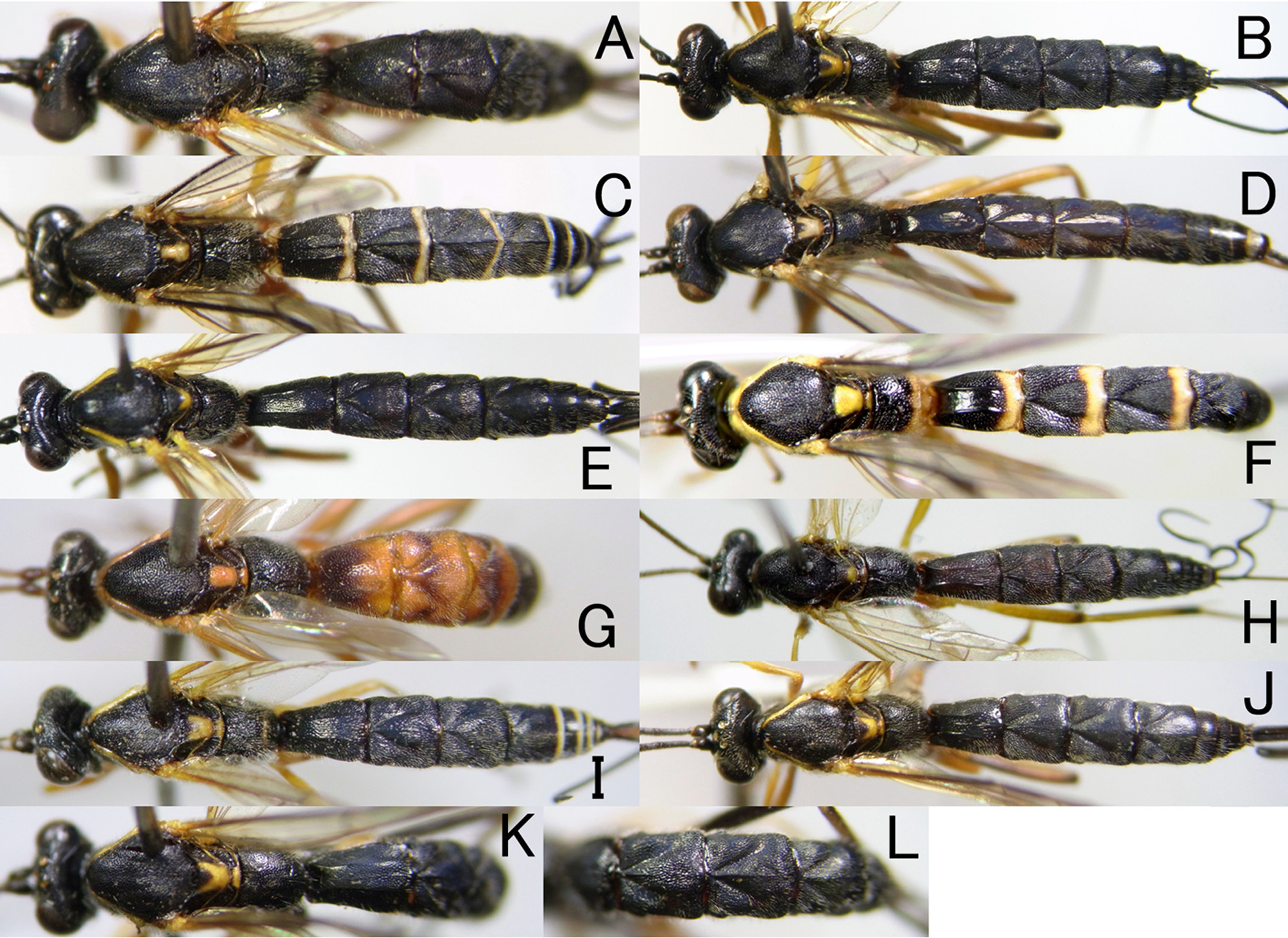
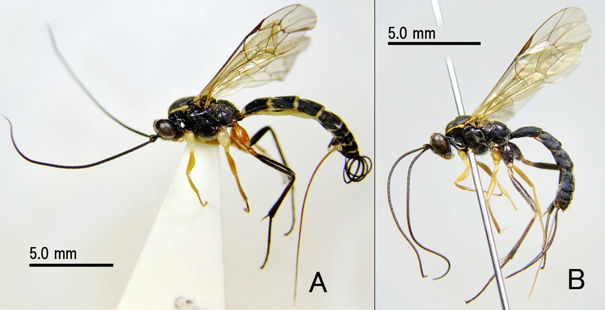
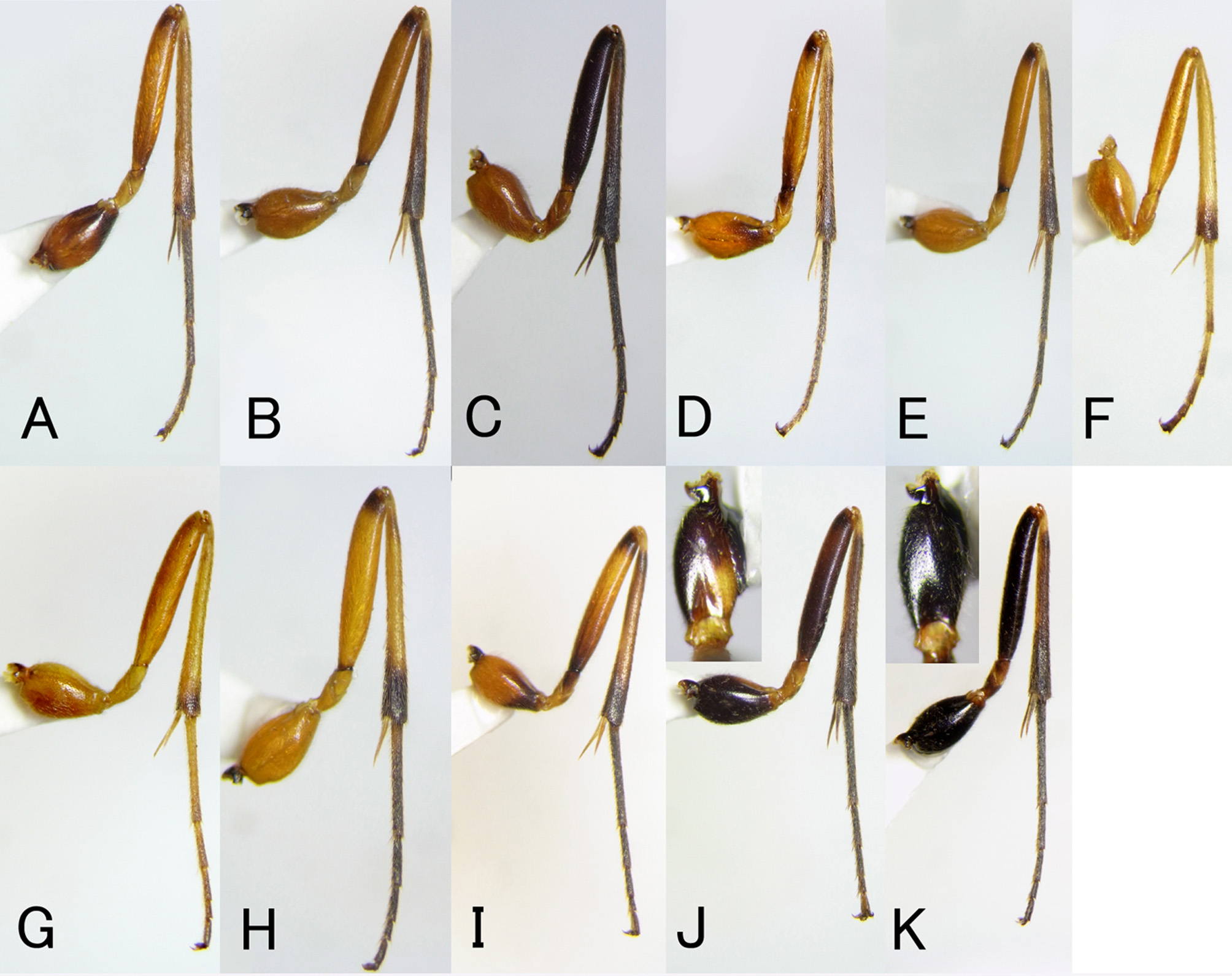
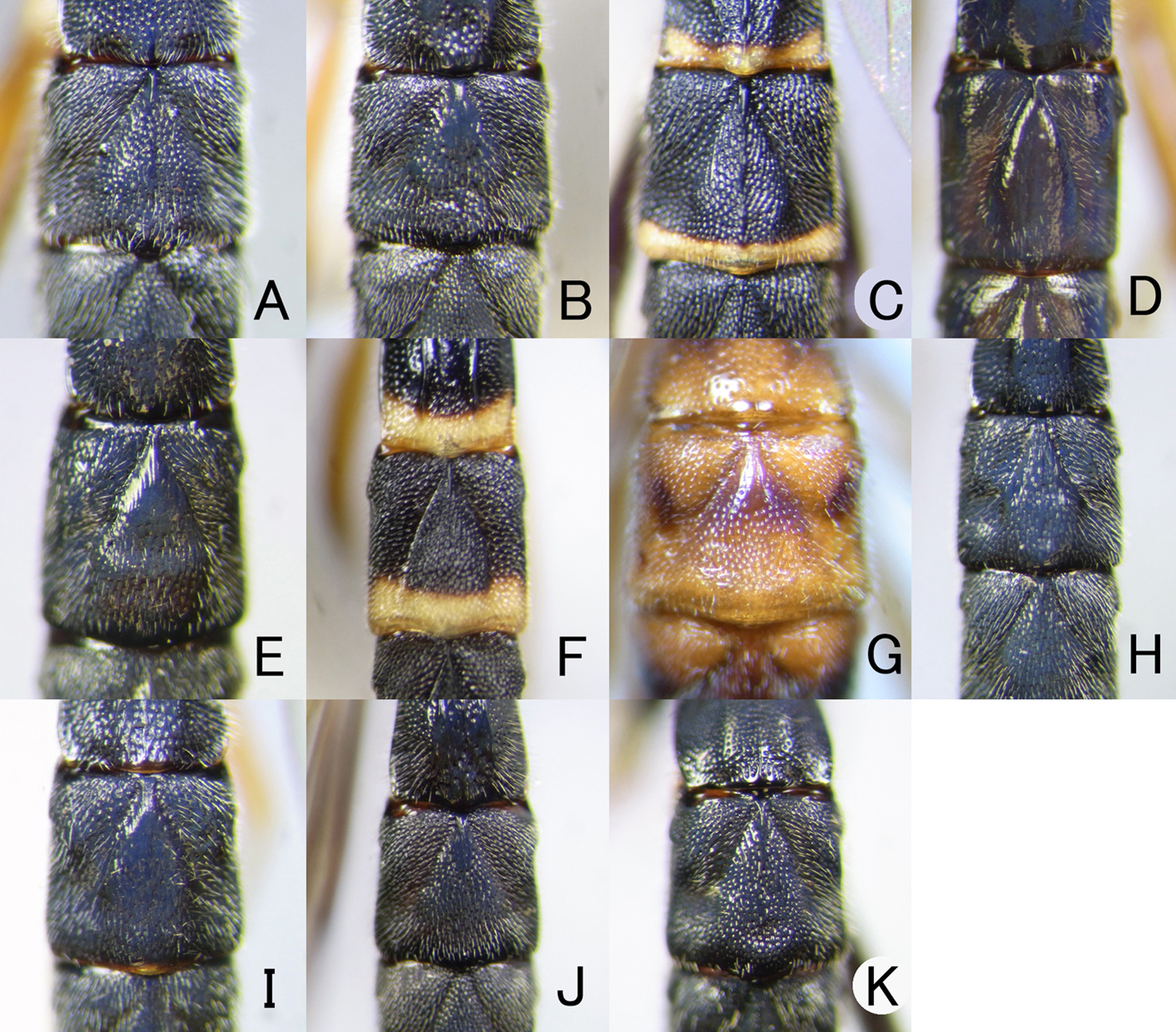
1. T1-T3 with strong median longitudinal carina ( Figs. 1 View FIGURE 1 C, 6 C, 7 C). Base and posterior margin of T1 and posterior margin of all other tergites yellow ( Figs. 1 View FIGURE 1 C, 3 A, 7 C). Face strongly transversely striated by coalescent punctures. Upper margin of pronotum with conspicuous yellow stripe ( Fig. 3 View FIGURE 3 A). Scutellum with yellow marking ( Fig. 1 View FIGURE 1 C). Hind coxa reddish-brown ( Figs. 3 View FIGURE 3 A, 5 C). Hind femur and tibia completely blackish-brown ( Figs. 3 View FIGURE 3 A, 5 C).............................. elegans sp. nov.
-. T1-T3 with weak and partly indistinct median longitudinal carina, or without it ( Figs. 1 View FIGURE 1 A, B, D–L, 6A, B, D–K, 7 A, B, D– K,). Face weakly transversely striated by coalescent punctures. Hind leg largely yellowish-brown ( Fig. 5 View FIGURE 5 A, B, D–I) or nearly entirely blackish-brown ( Figs. 3 View FIGURE 3 B, 5 J, K).................................................................. 2
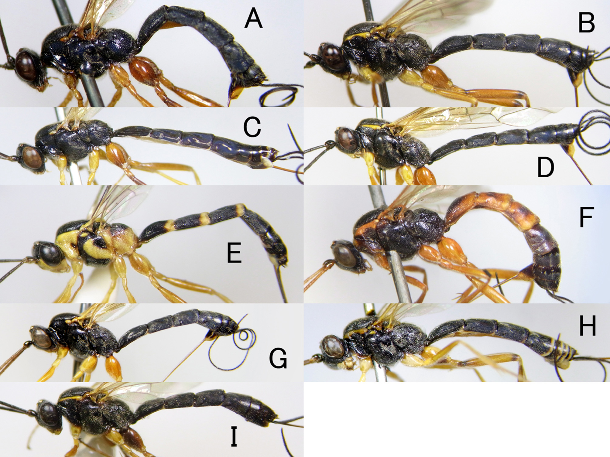
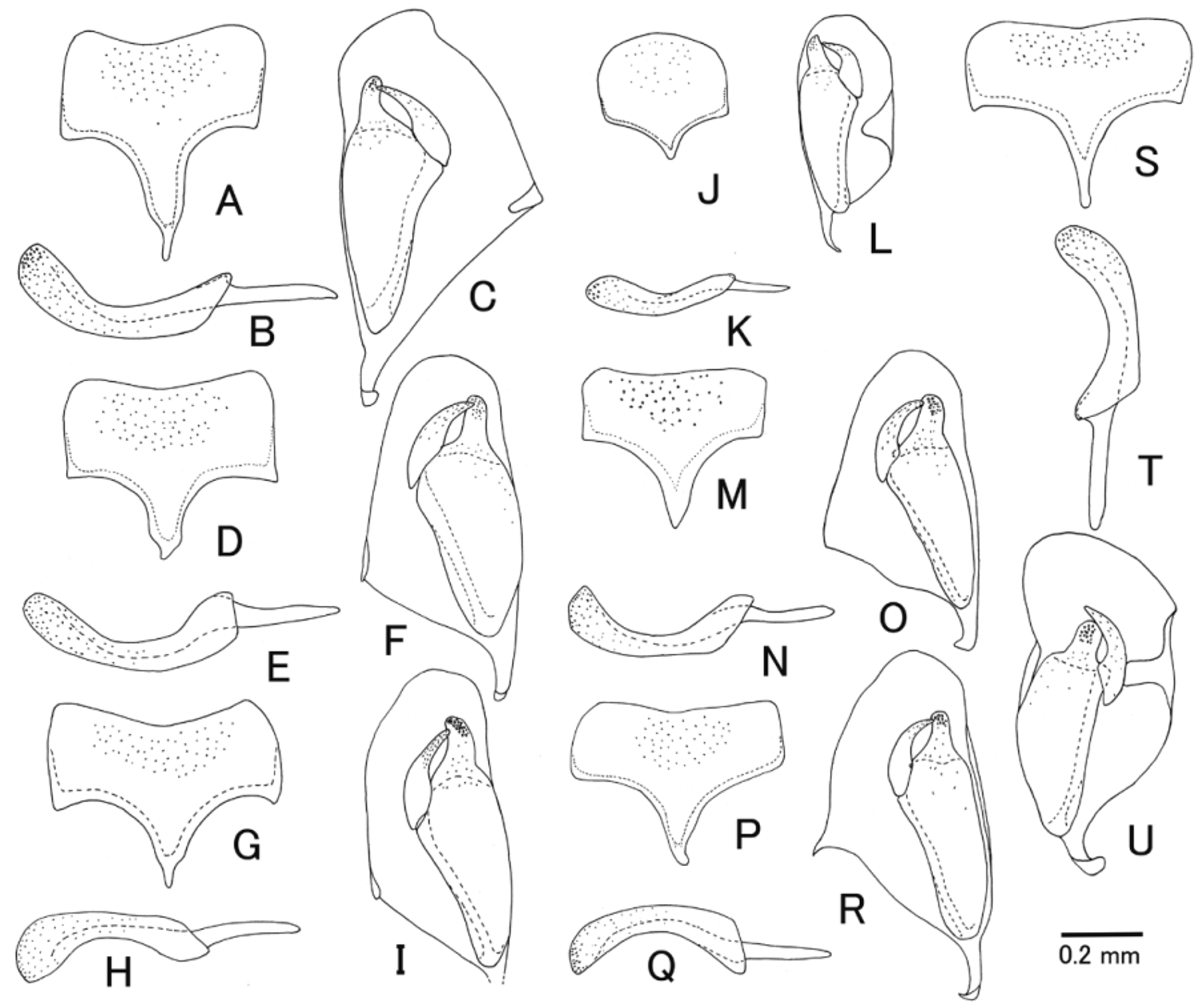
2. Pronotum largely yellow laterally ( Fig. 2 View FIGURE 2 E). Mesopleuron black with large yellowish marking ( Fig. 2 View FIGURE 2 E). T1–T3 with yellow posterior margin ( Figs. 1 View FIGURE 1 F, 2 E). Ovipositor very long, its sheath 3.0–3.7 times as long as hind tibia. Subgenital plate with a short apodema sternalis, anterior margin convex ( Fig. 9 View FIGURE 9 J). Inner margin of paramere slightly concave (Fig. 8 G).............................................................................................. kikuchii Uchida, 1928 View in CoL
-. Pronotum largely black laterally, yellow area at most present dorsally ( Fig. 2 View FIGURE 2 A–D, F–I). Mesopleuron without large yellow marking ( Fig. 2 View FIGURE 2 A–D, F–I). Ovipositor not very long, it sheath shorter than 2.5 times as long as hind tibia. Subgenital plate with a long apodema sternalis, anterior margin slightly concave ( Fig. 9 View FIGURE 9 A, D, G, M, P, S). Inner margin of paramere slightly concave (Fig. 8 A, C, E, I, K, M)................................................................................ 3
3. T2 longer than wide, its length 1.1–1.3 times as long as maximum width ( Figs. 1 View FIGURE 1 D, 6 D, 7 D). Metasomal tergites almost smooth (more or less punctate in male but sparser than other species), strongly shiny by strong blue reflection ( Fig. 7 View FIGURE 7. T 2 D). Epicnemial carina absent laterally. Propodeal carinae weak, usually absent (only restricted by slight convexity) except for posterior transverse carina and pleural carina. Yellow area on pronotum restricted near postero-lateral corner ( Fig. 2 View FIGURE 2 C). Median dorsal carina of T1 largely absent posteriorly ( Fig. 6 View FIGURE 6 D)...................................... evanescens ( Ratzeburg, 1848)
-. T2 shorter than 1.1 times as long as maximum width, usually square or transverse ( Figs. 1 View FIGURE 1 A, B, E–J, L, 6 A, B, E, G–K, 7 A, B, E–K). Metasomal tergites more or less punctate, weakly to rather strongly shiny ( Figs. 1 View FIGURE 1 A, B, E–J, L, 7 A, B, E–K). Epicnemial carina present laterally or absent in A. aquilonia View in CoL . Propodeal carinae except for posterior transverse carina and pleural carina absent to present. Median dorsal carina of T1 various, usually nearly complete ( Fig. 6 View FIGURE 6 A, B, E, G–K).............. 4
4. Epicnemial carina absent laterally. Fore coxa with ridge ( Fig. 4 View FIGURE 4 A, B). Scutellum black ( Fig. 1 View FIGURE 1 A). Median dorsal carina on T1 present only near base ( Fig. 6 View FIGURE 6 A)..................................................... aquilonia ( Momoi, 1963) View in CoL
-. Epicnemial carina present laterally. Scutellum usually with yellow marking ( Fig. 1 View FIGURE 1 B, E, G–K). Median dorsal carina on T1 well developed ( Fig. 6 View FIGURE 6 B, E, H–J), or if largely obsolete, T1–T3 reddish brown ( Figs. 1 View FIGURE 1 G, 2 F, 6 G, 7 G) or hind leg largely blackish-brown ( Fig. 5 View FIGURE 5 K)...............................................................................5
5. Hind femur and tibia blackish brown in lateral view ( Fig. 5 View FIGURE 5 J, K). Hind coxa largely blackish brown ( Fig. 5 View FIGURE 5 J, K). Upper margin of pronotum and scutellum yellow ( Figs. 2 View FIGURE 2 I, 3 B). Metasomal tergite black ( Fig. 1 View FIGURE 1 J–L).............................. 6
-. Hind femur and tibia reddish brown to brown in lateral view ( Fig. 5 View FIGURE 5 B, E, H, I). Hind coxa reddish brown ( Fig. 5 View FIGURE 5 B, E, H, I). Upper margin of pronotum and scutellum yellow ( Fig. 2 View FIGURE 2 B, D, H) or sometimes largely black ( Fig. 2 View FIGURE 2 G). Metasomal tergite sometimes partly tinged red ( Figs. 1 View FIGURE 1 G, 2 F)................................................................. 7
6. Ovipositor long, its sheath 2.9–3.0 times as long as hind tibia. Hind coxa with yellow marking dorsally ( Fig. 5 View FIGURE 5 J). Median dorsal carina of T1 at least present basal 0.6 ( Fig. 6 View FIGURE 6 J)......................................... tobensis ( Uchida,1928) View in CoL
-. Ovipositor short, its sheath 1.9–2.0 times as long as hind tibia. Hind coxa without yellow marking dorsally ( Fig. 5 View FIGURE 5 K). Median dorsal carina of T1 largely obsolete posteriorly, distinct area shorter than basal 0.5 ( Fig. 6 View FIGURE 6 K).............. yamato sp. nov.
7. Median dorsal carina of T1 shorter than 0.5 times of T1 ( Figs. 1 View FIGURE 1 G, 6 G). T1–T3 entirely (female) or partly (male: as posterior broad band) tinged with yellowish-red ( Figs. 1 View FIGURE 1 G, 2 F). Length of T2 distinctly shorter than wide ( Fig. 6 View FIGURE 6 G). Upper margin of pronotum with yellow stripe ( Fig. 2 View FIGURE 2 F)............. maetai Momoi, 1978 View in CoL (= genalis kasparyani Kuslitzky, 2007 View in CoL syn. nov.)
-. Median dorsal carina of T1 nearly complete, usually longer than 0.8 times of T1 (Fig. B, E, H, I). T1–T3 entirely black ( Fig. 1 View FIGURE 1 B, E, H, I), or in male of sugawarai with yellow posterior band, or if tinged reddish-brown, yellow area on upper margin of pronotum sometimes restricted to small spot before tegula ( Fig. 2 View FIGURE 2 G)............................................. 8
8. Posterior margin of all tergites (in male) or T4 and following all tergites (in female) with conspicuous yellow or white band ( Figs. 1 View FIGURE 1 I, 2 H). Propodeum usually with yellowish area posteriorly ( Fig. 1 View FIGURE 1 I). Upper margin of pronotum with yellow stripe ( Fig. 2 View FIGURE 2 H). Surface of tergites with blue reflection ( Figs. 1 View FIGURE 1 I, 2 H, 7 I).......................... sugaharai Momoi, 1978 View in CoL
-. Posterior margin of T4 and following tergites always without white or yellow band ( Figs. 1 View FIGURE 1 B, E, H, 2 B, D, G). Propodeum without yellow area ( Fig. 1 View FIGURE 1 B, E, H)....................................................................... 9
9. Small species, its body length 6.5–10.5 mm (usually less than 9.0 mm). Yellow area on upper margin of pronotum always restricted as small spot on posterior angle ( Fig. 2 View FIGURE 2 G). Anterior half of scutellum black ( Fig. 1 View FIGURE 1 H). T2 square, its length 1.0 times as long as maximum width ( Fig. 6 View FIGURE 6 H)....................................................... stena Momoi, 1963 View in CoL
-. Large species, its body length 8.5–11.0 mm. Yellow area of pronotum always large and broad, present along its upper margin ( Fig. 2 View FIGURE 2 B, D). Anterior half of scutellum usually yellow laterally ( Fig. 1 View FIGURE 1 B, E)..................................... 10
10. Robust species. T1–T3 usually with median longitudinal keel ( Fig. 6 View FIGURE 6 B). T1 1.2–1.4 times as long as maximum width ( Fig. 6 View FIGURE 6 B). T2 usually wide, its length 0.9–1.0 (both sexes) times as long as maximum width ( Fig. 6 View FIGURE 6 B). Surface on tergites without blue reflection or with slight blue reflection ( Fig. 7 View FIGURE 7. T 2 B)................................. bipunctoria ( Thunberg, 1822) View in CoL
-. Slender species. T1–T3 usually without median longitudinal keel except for bases of T2 and T3 ( Fig. 6 View FIGURE 6 E). T1 1.6–1.9 times as long as maximum width ( Fig. 6 View FIGURE 6 E). T2 usually slender, its length 1.0–1.1 (female) ( Fig. 6 View FIGURE 6 E) or 1.3 (male) times as long as maximum width. Surface of tergites with strong blue reflection ( Fig. 7 View FIGURE 7. T 2 E)........................ honmai Momoi, 1978 View in CoL


No known copyright restrictions apply. See Agosti, D., Egloff, W., 2009. Taxonomic information exchange and copyright: the Plazi approach. BMC Research Notes 2009, 2:53 for further explanation.