
Scyliorhinus duhamelii ( Garman, 1913 )
|
publication ID |
https://doi.org/ 10.11646/zootaxa.4601.1.1 |
|
publication LSID |
lsid:zoobank.org:pub:8A695352-8382-458F-A86A-17A198F780CA |
|
persistent identifier |
https://treatment.plazi.org/id/03B94378-D05B-0647-FF7D-FE2FFC2AA8A2 |
|
treatment provided by |
Plazi |
|
scientific name |
Scyliorhinus duhamelii ( Garman, 1913 ) |
| status |
|
Scyliorhinus duhamelii ( Garman, 1913) View in CoL
( Figs. 39–45 View FIGURE 39 View FIGURE 40 View FIGURE 41 View FIGURE 42 View FIGURE 43 View FIGURE 44 View FIGURE 45 ; Tabs. 3 View TABLE 3 , 5 View TABLE 5 , 11)
Common names: Duhamel’s catshark.
Catulus [sic] saxatilis: Duhamel, 1777: 304 , pl. 22, figs. 2–3 (catalogue, Mediterranean Sea).
Catulus duhamelii Garman, 1913: 73 –74 (original description, type locality: Adriatic Sea)
Scyliorhinus canicula: Springer, 1979: 130 View in CoL (only the part referring to the male specimen, MCZ 63–S).
Lectotype. MCZ 63 View Materials –S, male, 338.7 mm TL (Adriatic Sea) [designated herein].
Additional material examined. 22 specimens (see Appendix).
Diagnosis. Scyliorhinus duhamelii differs from all congeners by presenting a color pattern composed of scattered dark spots of varied sizes that also form aggregations (vs. dark spots absent in S. capensis , S. comoroensis , S. hesperius , S. meadi , S. torazame , and S. torrei ; reticulated pattern in S. retifer ; dark spots predominantly greater than spiracles in S. cervigoni , S. garmani , S. haeckelii , and S. stellaris ; spots with welldefined borders and not forming aggregations in S. boa , S. cabofriensis , S. canicula , and S. ugoi ); shallow nasoral grooves and posterior nasal flaps laterally situated (vs. grooves absent and posterior flaps situated on posterior border of excurrent apertures in other species, except S. canicula ); distance between nasal flaps 3.5–5 times smaller than width of the anterior nasal flap (vs. 6–7.5 times smaller in S. canicula ; two times in all other species); lower labial furrow 1.7–2.4 times smaller than mouth width (vs. more than 3 times in other species, except S. canicula ). The following combination of characters, although less conspicuous, also helps distinguish this species: dark spots scattered throughout the dorsolateral surface (vs. spots restricted to saddles in S. boa , S. cervigoni and, S. haeckelii ); anterior nasal flaps covering the upper lip (vs. flaps not reaching the upper lip in other species, except S. canicula , S. cervigoni , S. comoroensis , S. garmani and, S. stellaris ); interdorsal distance 0.6–1.0 times the anal base (vs. greater than the anal base in S. boa , S. cabofriensis , S. haeckelii , S. hesperius , S. meadi , S. retifer , S. torrei , and S. ugoi ); mandibular canal of lateral line system with 4 or 5 pores (vs. 6–7 in S. stellaris ); oral canal of lateral line system with 10–12 (vs. less than 10 pores in the other species, except S. torrei ); commissural teeth with two cusplets (vs. one cusplet in S. cervigoni , S. torazame and S. torrei ; four or more in S. boa , S. canicula and S. hesperius ); pelvic apron extending almost the entire length of pelvic inner margins (vs. extending to 2/3 of length in the other species, except in S. capensis , S. canicula , S. torazame , and S. torrei ); clasper with smooth terminal dermal cover (vs. rough in S. canicula and S. capensis ); terminal 3 cartilage absent (vs. present in S. boa , S. canicula , S. capensis , S. retifer , and S. torazame ); dorsal terminal 2 cartilage elongated and 1/4 length of dorsal terminal cartilage (vs. reduced and subtriangular in S. cabofriensis , S. capensis , S. cervigoni , S. haeckelii , and S. ugoi ; 1/3 of dorsal terminal cartilage in S. boa and S. comoroensis ; both cartilages the same length in S. torazame ); counts of monospondylous vertebrae 35–37 (vs. 44–46 in S. capensis ; 40–45 in S. cervigoni ; 48 in S. garmani ; 46–48 in S. meadi ; 43-47 in S. stellaris ; 32–35 in S. torrei ); upper tooth rows 41–45 (vs. 45–58 in S. cabofriensis ; 46–76 in S. capensis ; 48–54 in S. haeckelii ; 46–54 in S. meadi ; 47–56 in S. ugoi ); lower tooth rows 39–42 (vs. 44–50 in S. cabofriensis ; 43–53 in S. haeckelii ; 43–49 in S. meadi ; 43–53 in S. ugoi ); adult males at least 340 mm TL (vs. adult males greater than 450 mm TL in S. capensis , S. cervigoni , S. meadi , S. stellaris , and S. ugoi ; adult male 269 mm TL in S. torrei ).
Description. Morphometric and meristic data are given in Table 11, and neurocranial measurements in Table 5 View TABLE 5 .
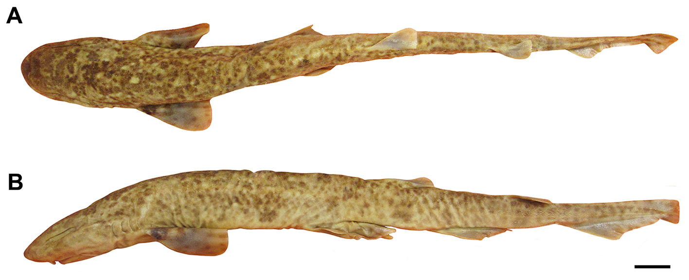
Body slender and cylindrical, tapering considerably posterior to cloaca ( Fig. 39 View FIGURE 39 ). Prepectoral length 0.4 times the prepelvic length. Trunk shorter than tail; snout-vent length 0.7–0.8 (0.8) times vent-caudal length. Pectoralpelvic space 1.3–1.7 (1.6) times the pelvic-anal space. Interdorsal space 1.9–3.1 (3.1) times the dorsal-caudal space ( Tab. 11). No interdorsal, postdorsal or postanal ridges; lateral crest on caudal peduncle absent.
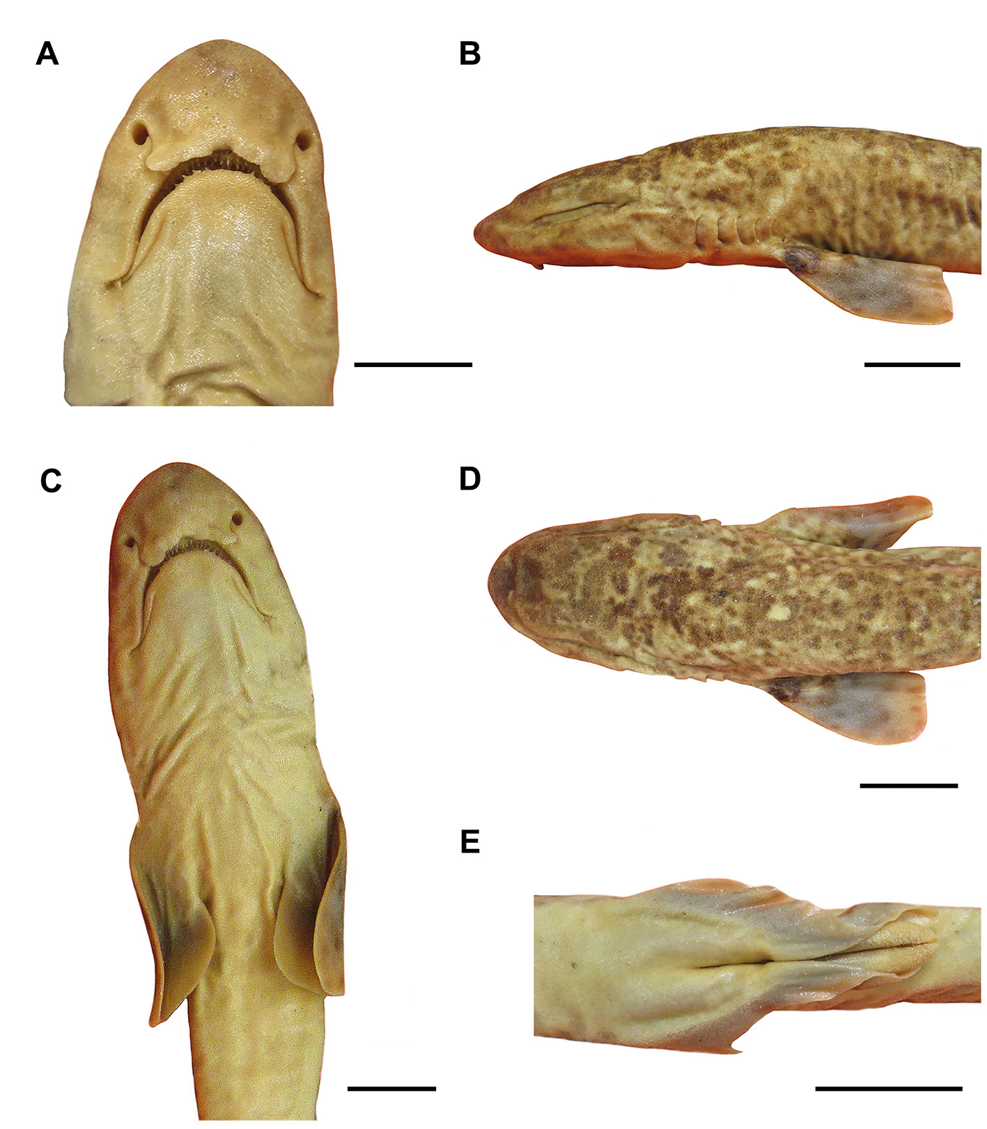
Head moderately broad and depressed; head length 1.8–2.3 (1.9) times head width ( Figs. 39 View FIGURE 39 , 40 View FIGURE 40 ). Snout relatively short, preoral length 0.6–0.7 (0.8) times mouth width and 0.7–0.8 (0.7) times smaller than preorbital length. Prenasal length 0.5–0.6 (0.5) times internarial space; preorbital length 0.9 (1.3) times interorbital space.
Eye large and slitlike, eye length 2.7–3.3 (3.3) times its height and 0.2–0.3 (0.2) times smaller than head length ( Fig. 39 View FIGURE 39 ). Eye dorsolateral on head, with lower edge medial to horizontal head rim in dorsal view; subocular ridge strong. Nictitating lower eyelid of rudimentary type, with shallow subocular pouch and secondary lower eyelid free from upper eyelid. Spiracle close behind but well separated from eyes, dorsolaterally on head and somewhat lower than level of eye notch. Spiracle diameter goes 3.5–5.5 (5.7) times in eye length and 4.5–9.5 (8.6) times in interorbital width.
First two gill openings about equally wide; first one twice as long as fifth. All gill openings slightly concave and not elevated on dorsolateral surface of head; gill filaments not visible externally.
Nostril with broad incurrent aperture, without nasal barbel, and small and oval excurrent aperture; shallow nasoral groove present. Anterior nasal flap large and triangular, covering the posterior nasal flap, excurrent aperture and upper lip; distance between anterior nasal flaps 3.7–5 (3.7) times smaller than width of anterior nasal flap ( Figs. 41 View FIGURE 41 A–B). Mesonarial ridge poorly developed and not exceeding the posterior border of the anterior nasal flap. Posterior nasal flap elongated and rodlike with a spatulate posterior edge, situated on the lateral edge of the excurrent aperture and corresponding to 1/3 of the anterior nasal flap. Mesonarial superior and inferior flaps conical and corresponding to 1/4 of anterior nasal flap. Internarial space 0.7–0.9 (1.2) times smaller than interorbital space.
Mouth arched, wider than long, its length goes 1.5–1.6 (1.5) times in mouth width ( Figs. 41 View FIGURE 41 A–B). Lower labial furrow long and narrow, 1.7–2.4 (2.1) times smaller than mouth width. Dorsal labial cartilage 1.3 times the ventral cartilage; anterior tip of dorsal labial cartilage reaching the orbital process of the palatoquadrate. Tongue flat and rounded, light-colored, with oral papillae hardly detectable.
Monognathic heterodonty gradual well developed; anterior teeth abruptly larger than the parasymphysial ones and lateral teeth smaller distally, with smaller and thicker cusps ( Fig. 42 View FIGURE 42 ). Sexual heterodonty not observed; only adult males examined. Tooth counts 19–22 19–23/ 17–20 1 17 –22 (20–21/ 20–1–18). Parasymphysial teeth with a principal cusp flanked by one cusplet on each side; cusplets 1/3 the height of the principal cusp and half of its width. Protuberances on medial portion of the crown base and striae restricted to the crown base on the labial surface. Anterior teeth similar in shape but greater than the parasymphysial ones; principal cusp three times greater than the cusplets. Anterior upper teeth with a principal cusp slightly oblique and medial portion of root with a more pronounced concavity. Protuberances and striae restricted to the crown base. Lateral teeth with three cusplets, two at the mesial edge and one at the distal edge. Mesial proximal and distal cusplets corresponding to half the height of the principal cusp and mesial marginal poorly developed; principal cusp slightly oblique on both jaws. Protuberances less prominent on crown base and striae running from the crown base until half the height of principal cusp. Commissural teeth similar to anterior ones in number of cusplets; principal cusp stouter and slightly oblique. Protuberances well prominent on the crown base and striae from the crown base until half the height of principal cusp. Ectodermal pits present in lateral and commissural teeth, restricted to the crown base.
Lateral trunk denticles with flat, elongated teardrop-shaped crowns, 1.9–2.1 times as long as wide, anterior part covered with ectodermal pits ( Tab. 3 View TABLE 3 ). Crown with a strong medial ridge extending its entire length onto long principal cusp. Dermal denticles above the pectoral fin presenting well-developed cusplets, 0.4 times the principal cusp; distinct lateral ridges. Cusplets poorly developed in posterior regions with short or reduced lateral ridges ( Fig. 43 View FIGURE 43 ).
Pectoral base 0.8–1.0 (0.8) times mouth width ( Fig. 41C View FIGURE 41 ). Pectoral anterior margin 2.1–2.5 (2.0) times its base and 1.5–2.3 (1.7) times the posterior margin. Pectoral fin skeleton aplesodic with radials mostly divided into three segments. Propterygium and mesopterygium trapezoidal; former smaller than latter. Propterygium with one proximal segment; mesopterygium with 3–4 proximal segments fused proximally. Metapterygium with 8 segments. Metapterygial axis rectangular and corresponding to 1/5 of metapterygium.
Pelvic fin triangular ( Fig. 41E View FIGURE 41 ); pelvic anterior margin 0.9–1.1 (0.8) times the posterior margin and 0.9 times the pelvic base. Pelvic inner margins of males fused through almost all of their extension, with sharp edges; claspers of juveniles covered by the pelvic apron and evident only with lifted apron.
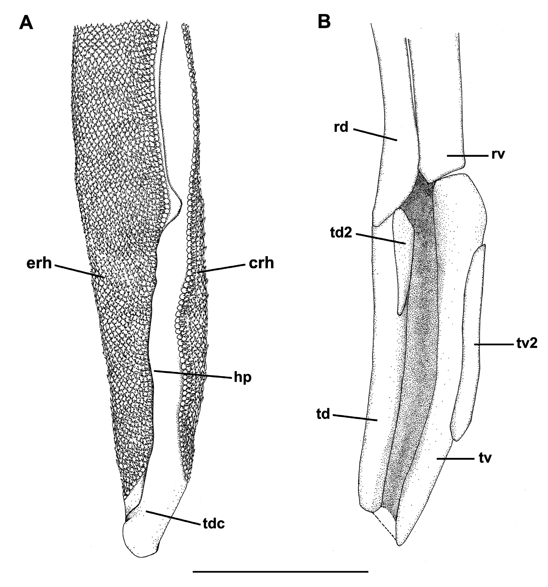
Clasper short and cylindrical ( Fig. 41 View FIGURE 41 ), sometimes extending beyond free rear tips of pelvic fins; clasper inner length 1.3–1.4 (1.4) times the pelvic anterior margin, 1.5–2.1 (1.5) times clasper outer length and 3.7–5.5 (6.0) times the clasper base. Most of clasper surface except dorsomedial surface of glans, medial border of cover rhipidion, rhipidion, and terminal dermal cover, covered by dermal denticles with anteriorly directed crowns ( Fig. 44A View FIGURE 44 ). Clasper hooks absent. Rhipidion poorly developed and extending to the hindquarter of clasper; insertion of rhipidion at the anterior portion of dorsal terminal 2 cartilage. Cover rhipidion expanded medially reaching the exorhipidion and sometimes covered by this anteriorly; both cover rhipidion and exorhipidion covering the clasper groove. Envelope inconspicuous; pseudosiphon poorly developed, visible only internally. Terminal dermal cover smooth, extending for 1/4 of the ventral terminal cartilage, contacting the exorhipidion, and covering the cover rhipidion.
Clasper skeleton relatively simple ( Fig. 44B View FIGURE 44 ). Ventral marginal cartilage shorter than dorsal one; ventral terminal beginning anteriorly, but ending together with the dorsal terminal. Terminal 3 cartilage absent. Dorsal terminal 2 cartilage elongated and rod-like, medially positioned on the dorsal terminal cartilage; this cartilage supports the rhipidion and corresponds to 1/3 of the length of ventral terminal cartilage. Ventral terminal 2 cartilage slender, on the ventral terminal cartilage and corresponding to midlength of this; ventral terminal 2 cartilage beginning at the 1/3 anterior of the dorsal terminal 2.
First dorsal fin triangular, with nearly straight anterior margin, rounded apex and angular free rear tip ( Fig. 39 View FIGURE 39 ). First dorsal fin origin opposite and slightly anterior to the pelvic fin insertion. First dorsal fin insertion opposite to the anterior 2/5 of pelvic-anal distance. Anterior margin 1.3–1.4 (1.3) times first dorsal fin base; first dorsal fin height 0.6–0.7 (0.6) times its base.
Second dorsal fin smaller than the first and triangular, sometimes subrectangular ( Fig. 39 View FIGURE 39 ). Second dorsal fin origin opposite to the 1/5 posterior of the anal fin in females and slightly anterior to the anal in insertion in males. First dorsal fin insertion opposite or posterior to the posterior tip of the anal fin. Anterior margin 1.2 (1.3) times base of second dorsal fin; second dorsal base 1.5–2.0 (2.2) times its height and 1.1–2.2 (2.4) times the dorsalcaudal distance. First dorsal fin 1.3–1.5 (1.4) times larger than the second dorsal fin.
Anal fin triangular, apically narrow and not falcate ( Fig. 39 View FIGURE 39 ); anal fin base 1.7 (1.8) times the second dorsal fin base. Anal fin anterior margin nearly straight, apex narrowly rounded, free rear tip acutely pointed, and inner margin straight. Anal fin base 0.6–0.7 (1.0) times the interdorsal distance and 1.3–1.4 (2.1) times the dorsal-caudal distance. Anal anterior margin 1.5–2.3 (2.1) times the posterior margin; anal fin height 0.5–0.6 (0.3) times its base.
Caudal fin narrow-lobed and asymmetrical ( Fig. 39 View FIGURE 39 ). Dorsal caudal lobe 1.8–2.2 (2.9) times larger than preventral lobe; subterminal caudal margin 1.2 (1.6) times the terminal margin. Caudal crest of enlarged denticles absent on caudal fin margins.
Neurocranium broad and somewhat flattened, corresponding to 7.5–9.2% TL. Rostrum length similar to the distance between lateral rostral cartilages. Nasal capsule wider than long, oval-shaped and expanded laterally; width 1.1–1.2 times its length. Anterior fontanelle broad and subrectangular in males (females not available for dissection); epiphyseal notch very prominent. Orbital region corresponding to 2.2 times in the nasobasal length. Otic capsule short, its length 4.9–5.1 times in nasobasal length and width 3.1–3.9 times otic capsule length. Width across postorbital processes 1.5 times the preorbital processes width ( Tab. 5 View TABLE 5 ).
Coloration in alcohol. Body beige with dark brown spots with poorly defined borders, forming aggregations and rosettes, predominantly greater than the spiracles; distributed along the dorsolateral surfaces and fins ( Figs. 39 View FIGURE 39 , 40 View FIGURE 40 ). Saddles and longitudinal dark stripe absent. Spots darker on the dorsal surfaces than on lateral surfaces. Light spots, when present, diffuse, scattered and larger than spiracles. Spots less evident from the pelvic fins to caudal tip; distal edge of fins not pinkish. Belly and ventral surface of paired fins without spots, cream in color.
Distribution. Examined specimens distributed along Adriatic and Mediterranean Seas, along the continental shelves of Croatia, Greece, Tunisia and Argelia ( Fig. 45 View FIGURE 45 ).
Biological data. Adult male at 340 mm TL; largest male examined 436 mm TL. Largest female examined 420 mm TL; no data on sexual maturity of females. This species is a benthic dweller in waters of depths 43– 75 m.
Etymology. The specific name ‘duhamelii’ was dedicated to Duhamel du Monceau, author that first noted the differences between this species and S. canicula .
Remarks. Garman (1913) described Catulus duhamelii (= Scyliorhinus duhamelii ) on the basis of differences in color pattern, dorsal fin positions and distance between anterior nasal flaps, distinguishing it from S. canicula and S. stellaris . Springer (1979) considered S. duhamelii a junior synonym of S. canicula , arguing that no differences between the syntypes of S. duhamelii and other specimens of S. canicula could be found. Specimen MCZ 63–S (male, 338.7 mm TL, Adriatic Sea) mentioned by Garman (1913) is here designated the lectotype of S. duhamelii , to be treated as the only specimen of the type series corresponding to the original description of this species. Specimen MCZ 60–S (female, 410.6 mm TL; Nice, France) also mentioned by Garman (1913) is reidentified here as S. canicula . Garman (1913) reported that the capture locality of the female specimen would be the Adriatic Sea, whereas the male would be from Nice, France. However, Hartel & Dingerkus (1997) pointed out that Garman (1913) had inverted the collection data in his publication, based on the labels inside the jar and information in the specimen catalog of the Museum of Comparative Zoology.


No known copyright restrictions apply. See Agosti, D., Egloff, W., 2009. Taxonomic information exchange and copyright: the Plazi approach. BMC Research Notes 2009, 2:53 for further explanation.
|
Kingdom |
|
|
Phylum |
|
|
Class |
|
|
Order |
|
|
Family |
|
|
Genus |
Scyliorhinus duhamelii ( Garman, 1913 )
| Soares, Karla D. A. & De, Marcelo R. 2019 |
Scyliorhinus canicula: Springer, 1979 : 130
| Springer, S. 1979: 130 |
Catulus duhamelii
| Garman, S. 1913: 73 |