
Scyliorhinus boa ( Goode & Bean, 1896 )
|
publication ID |
https://doi.org/ 10.11646/zootaxa.4601.1.1 |
|
publication LSID |
lsid:zoobank.org:pub:8A695352-8382-458F-A86A-17A198F780CA |
|
persistent identifier |
https://treatment.plazi.org/id/03B94378-D064-0616-FF7D-F905FAB6ACFB |
|
treatment provided by |
Plazi |
|
scientific name |
Scyliorhinus boa ( Goode & Bean, 1896 ) |
| status |
|
Scyliorhinus boa ( Goode & Bean, 1896) View in CoL
( Figs. 3–9 View FIGURE 3 View FIGURE 4 View FIGURE 5 View FIGURE 6 View FIGURE 7 View FIGURE 8 View FIGURE 9 ; Tabs. 1–3 View TABLE 1 View TABLE 2 View TABLE 3 )
Common names: boa catshark ( United States), rousette boa ( France), Alitán boa ( Spain).
Scylliorhinus retifer var. boa Goode & Bean, 1896: 17 View in CoL (original description, type locality: Northwestern Atlantic). Regan, 1908: 457 (only the part referring to the holotype of S. boa View in CoL ).
Catulus boa: Garman, 1913: 77 (except the part referring to the holotype of S. haeckelii View in CoL ); White, 1937: 117 (systematics, listed).
Scyliorhinus boa: Bigelow & Schroeder, 1948: 204 View in CoL –207 (except the part referring to the holotype of S. haeckelii View in CoL ); Springer, 1966: 601 –602, fig. 15a (taxonomic review, except the part referring to the holotype of S. haeckelii View in CoL ); Springer, 1979: 128 –129, figs. 81, 82 (taxonomic review); Uyeno et al., 1983: 51 (catalogue, French Guiana and Suriname); Compagno 1984: 357 –358 (FAO catalogue); Cervigón & Alcalá, 1999: 94 –96, fig. 41 (catalogue, Venezuela); Compagno, 1999: 480 (listed); Kiraly et al., 2003: 15 (catalogue, Atlantic coast of the United States); Compagno et al., 2005: 247, pl. 42 (compilation); Mejía-Falla et al., 2007: 139 (listed); Castro, 2011: 336, fig. 86a (catalogue, North America); Kyne et al., 2012: 58 (listed, conservation status for North America, Central America and Caribe); Ebert et al., 2013a: 374, 378, pl. 52 (compilation); Oliveira et al., 2015: 31 (catalogue, Rio Grande do Norte, Brazil); Weigmann, 2016: 43 (listed).
Scyliorhinus fernandezi Weibezahn, 1953: 3 View in CoL –7, fig. 1 (original description, type locality: La Guaira, Venezuela); Cervigón, 1966: 60 –61 [synonym of S. boa View in CoL ]; Lasso et al., 1998: 4 (catalogue, MHNLS).
Scyliorhinus retifer boa Springer & Sadowsky, 1970: 90 View in CoL –91 (only part referring to the type specimen and specimens of island platforms from Bonaire to Hispaniola).
Scyliorhinus retifer haeckelii: Cadenat & Blache, 1981: 183 View in CoL -184, 125b (catalogue, compared with species of Mediterranean and western coast of Africa).
Scyliorhinus haeckelii: Springer, 1979: 136 View in CoL (incorrect identification, USNM 181695 and USNM 188061).
Holotype. MCZ 1335 View Materials , male, 149.4 mm TL ( Lesser Antillean , Barbados, 365 m depth).
Additional material examined. 41 specimens (see Appendix).
Diagnosis. Scyliorhinus boa differs from all congeners by presenting a color pattern composed of black spots of varied sizes (but predominantly smaller than spiracle) bordering saddles and not present within these (vs. no dark spots in S. capensis , S. comoroensis , S. hesperius , S. meadi , S. torazame , and S. torrei ; reticulated pattern in S. retifer ; spots predominantly within saddles and with approximate bilateral symmetry in S. haeckelii and S. ugoi ; spots predominantly larger than the spiracle in S. cervigoni , S. garmani and S. stellaris ; spots spread around the body not bordering saddles in S. cabofriensis S. canicula , and S. duhamelii ). The following combination of characters, although less conspicuous, also helps distinguish this species: dark spots spiracle-sized and forming a distinct row on the lateral line (vs. row absent in S. cabofriensis , S. haeckelii and S. ugoi ); anterior nasal flap not reaching the upper lip (vs. flaps reaching the lip, and sometimes covering it, in S. canicula , S. cervigoni , S. comoroensis , S. duhamelii , S. garmani , and S. stellaris ); oral canal of lateral line system with 8–10 pores (vs. 5–6 pores in S. hesperius ; 10–12 in S. duhamelii ; 9–13 in S. torrei ); commissural teeth presenting three cusplets (vs. two or less in the other species, except in S. canicula , S. capensis and S. hesperius ); internarial space 4.3–7.1% TL (vs. 1.8–2.7% TL in S. cabofriensis ; 2–4% in S. haeckelii ; 1.4–2.4% in S. ugoi ); interdorsal space larger than the anal fin base (vs. 0.6–1.0 times in S. capensis , S. cervigoni , S. comoroensis , S. duhamelii , S. garmani , S. stellaris , and S. torazame ); clasper with smooth terminal dermal cover (vs. rough in S. canicula and S. capensis ); envelope medially expanded (vs. poorly developed or absent in other species, except in S. hesperius and S. retifer ); cover rhipidion well-developed (vs. poorly developed in S. canicula and S. duhamelii ); cover rhipidion with no dermal denticles (vs. present in the others, except in S. cervigoni , S. hesperius and S. retifer ); terminal 3 cartilage present (vs. absent in the other species, except in S. canicula , S. capensis , S. retifer , and S. torazame ); dorsal terminal 2 cartilage rod-like and corresponding to one third of dorsal terminal cartilage (vs. reduced and subtriangular in S. cabofriensis , S. capensis , S. cervigoni , S. haeckelii , and S. ugoi ; 1/4 of dorsal terminal cartilage in S. canicula , S. duhamelii , S. retifer , S. stellaris , and S. torrei ; same length in S. torazame ); neurocranium with basal plate width corresponding to 40.7–43.8% of nasobasal length (vs. 63.3% NL in S. meadi ; 48.3–50.6% in S. stellaris ; 48.7–53.2% in S. torrei ); distance between nasal apertures 12.8–15.6 % NL (vs. 27.8–37.6% in S. canicula ; 20.1–26.9% in S. capensis ; 20.5% in S. cervigoni ; 20.1% in S. duhamelii ; 17.9–21.6 % NL in S. hesperius ; 27.6–29.8% in S. stellaris ; 11.6% in S. meadi ; 17.2–28.1% in S. torazame ); counts of monospondylous vertebrae 39–42 (vs. 44–46 in S. capensis ; 35–37 in S. duhamelii ; 48 in S. garmani ; 46–48 in S. meadi ; 43–47 in S. stellaris ; 32–37 in S. torazame ; 30–35 in S. torrei ); somewhat medium sized adult males, at about 350 mm TL, and adult females at least 400 mm TL (vs. sizes greater than 450 mm for males and females in S. capensis , S. cervigoni , S. meadi , S. stellaris , and S. ugoi ; 269 mm TL in S. torrei ).
Description. Morphometric and meristic data are given in Table 1 View TABLE 1 , and neurocranial measurements in Table 2 View TABLE 2 .
Body slender and cylindrical, tapering considerably posterior to cloaca ( Figs. 3 View FIGURE 3 , 4 View FIGURE 4 ). Prepectoral length 0.4 times the prepelvic length. Trunk shorter than tail; snout-vent length 0.7–0.8 (0.7) times vent-caudal length. Pectoral-pelvic space 1.3–1.7 (1.9) times the pelvic-anal space. Interdorsal space 1.9–2.2 (2.1) times the dorsalcaudal space ( Tab. 1 View TABLE 1 ). No interdorsal, postdorsal or postanal ridges; lateral crest on caudal peduncle absent.
Head moderately broad and depressed; head length 1.4–1.6 (1.7) times head width ( Figs. 3 View FIGURE 3 , 4 View FIGURE 4 ). Snout relatively short, preoral length 0.6–0.7 (0.6) times mouth width and 0.8–1.0 (0.7) times smaller than preorbital length. Prenasal length 0.5–0.7 (0.6) times internarial space; preorbital length 0.6–1.0 (0.9) times interorbital space.
Eye large and slitlike, eye length 1.9–2.8 (3.7) times its height and 0.2–0.3 (0.3) times smaller than head length ( Figs. 4 View FIGURE 4 , 5 View FIGURE 5 C–D). Eye dorsolateral on head, with lower edge medial to horizontal head rim in dorsal view; subocular ridge strong. Nictitating lower eyelid of rudimentary type, with shallow subocular pouch and secondary lower eyelid free from upper eyelid. Spiracle close behind but well separated from eye, dorsolaterally on head and somewhat lower than level of eye notch. Spiracle diameter goes 3.8–6.2 (5.8) times in eye length and 5.7–11.8 (8.8) times in interorbital width.
First two gill openings about equally wide; first one twice as long as fifth. All gill openings slightly concave and not elevated on dorsolateral surface of head; gill filaments not visible externally.
Nostril with broad incurrent aperture, without nasoral groove or nasal barbel, and small and oval excurrent aperture. Anterior nasal flap large, triangular, and covering posterior nasal flap and excurrent aperture, extending just anterior to mouth, close to the upper lip but not touching it ( Figs. 5 View FIGURE 5 A–B). Mesonarial ridge distinct but not exceeding the posterior edge of the anterior nasal flap. Posterior nasal flap rectangular, situated on the posterior edge of the excurrent aperture. Mesonarial superior and inferior flaps conical and corresponding to 1/4 of anterior nasal flap. Internarial distance 1.1–1.4 (1.4) times smaller than interorbital distance.
Mouth arched, wider than long, its length goes 1.6–2.0 (1.5) times in mouth width ( Fig. 5 View FIGURE 5 A–B). Lower labial furrow short and narrow, 3.3–3.9 (4.9) times smaller than mouth width. Dorsal labial cartilage 1.3 times the ventral cartilage; anterior tip of dorsal labial cartilage reaching the orbital process of the palatoquadrate. Tongue flat and rounded, light-colored, with oral papillae hardly detectable.
Monognathic heterodonty gradual well developed; anterior teeth abruptly greater than the parasymphysial ones and lateral teeth smaller distally, with smaller and thicker principal cusps ( Fig. 6 View FIGURE 6 ). Sexual heterodonty not observed; only adult males examined. Tooth counts 19–25 20–24/ 17–22 1 19 –24 (19–20/ 19–1–20). Parasymphysial teeth with a principal cusp flanked by one cusplet at each side; cusplets 1/3 height of principal cusp. Protuberances on the crown base or striae absent along the labial surface. Anterior teeth larger than the parasymphysial one and principal cusp less stout. Anterior teeth with a principal cusp and two or three cusplets. Anterior upper teeth with two cusplets on the mesial edge and a poorly developed cusplet on the distal edge; one cusplet poorly developed present on each side of the principal cusp in the lower teeth. Protuberances and striae restricted to the crown base. Lateral teeth in both jaws smaller than the anterior ones, with one principal cusp and three cusplets; principal cusp oblique in upper teeth and straight in lower ones. Mesial proximal cusplet half the height of the principal cusp, 1/3 larger than the distal cusplet and two times the mesial marginal cusplet. Protuberances on the crown base and striae running from the crown base to one half the height of the principal cusp on labial surface. Commissural teeth similar to the anterior ones in number of cusplets. Principal cusp stronger, inclined toward the commissure in upper teeth and nearly straight in lower ones; two cusplets at the mesial edge and one at the distal edge. Mesial proximal and distal cusplets 2/3 the height of the principal cusp and half of its width. Mesial marginal cusplet poorly developed and corresponding to half the height of the mesial proximal cusplet. Protuberances present and striae running from the base toward the apex of the crown on the labial surface. Ectodermal pits present on lateral and commissural teeth, restricted to the crown base.
Lateral trunk denticles with flat, elongated teardrop-shaped crowns, 1.7–1.9 times longer than wide ( Tab. 3 View TABLE 3 ); anterior part covered with ectodermal pits. Crown with a strong medial ridge extending its entire length onto long principal cusp. Dermal denticles above the pectoral fin presenting well-developed cusplets, 0.4 times the principal cusp; prominent lateral ridges. Cusplets poorly developed in posterior regions, with short or reduced lateral ridges ( Fig. 7 View FIGURE 7 ).
*Values for dermal denticles of Scyliorhinus cabofriensis , S. haeckelii and S. ugoi can be found in Soares et al. (2015, 2016).
**Skin samples of S. comoroensis and S. garmani were not available.
Pectoral base 0.7–1.0 (0.6) times mouth width. Pectoral anterior margin 1.7–2.4 (2.6) times its base and 1.6–2.0 (1.9) times the posterior margin. Pectoral fin skeleton aplesodic with radials mostly divided into three segments. Propterygium and mesopterygium trapezoidal; the former smaller than the latter. Propterygium with one proximal segment; mesopterygium with 3–4 proximal segments fused proximally. Metapterygium with 9 segments. Metapterygial axis rectangular and corresponding to 1/5 of metapterygium.
Pelvic fin triangular ( Fig. 5E View FIGURE 5 ); pelvic anterior margin 1.1–1.4 (1.1) times the posterior margin and 0.9–1.2 (1.0) times the pelvic base. Pelvic inner margins of males fused by 2/3 of their extension; claspers of juveniles evident without lifting the pelvic apron.
Clasper short and cylindrical ( Fig. 5E View FIGURE 5 ), sometimes extending beyond free rear tips of pelvic fins; clasper inner length 0.2–0.9 (0.5) times the pelvic anterior margin, 1.0–1.9 (2.5) times the outer length and 2.7–5.6 (3.7) times the clasper base. Most of clasper surface except dorsomedial surface of glans, envelope, cover rhipidion, rhipidion, and terminal dermal cover, covered by dermal denticles with anteriorly directed crowns ( Fig. 8A View FIGURE 8 ). Clasper hooks absent. Rhipidion well-developed, partly covered medially by a prominent exorhipidion and anteriorly by the cover rhipidion; insertion of rhipidion at anterior portion of dorsal terminal 2 cartilage and extending to the end of glans. Cover rhipidion expanded medially reaching the exorhipidion, sometimes covered by it anteriorly, and covering the clasper groove. Envelope expanded medially and covering the anterior border of the cover rhipidion; pseudosiphon poorly developed, visible only internally. Terminal dermal cover extending for 1/3 of the ventral terminal cartilage, contacting the exorhipidion, and covering the cover rhipidion.
Clasper skeleton relatively simple ( Fig. 8B View FIGURE 8 ). Ventral terminal cartilage beginning anteriorly, but ending together with the dorsal one. Terminal 3 cartilage present and medially situated like a conical and subtriangular anterior projection of ventral terminal cartilage. Dorsal terminal 2 cartilage elongated and rod-like, medially positioned on the dorsal terminal cartilage; this cartilage supports the rhipidion and corresponds to 1/3 of the length of ventral terminal cartilage. Ventral terminal 2 cartilage slender above the ventral terminal cartilage corresponding to one half its length.
First dorsal fin triangular, square-tipped in some juveniles, with nearly straight anterior margin, rounded apex and angular free rear tip ( Figs. 3 View FIGURE 3 , 4 View FIGURE 4 ). First dorsal fin origin posterior to the insertion and above half of inner margin of pelvic fin; males presenting first dorsal fins slightly posterior. First dorsal fin insertion opposite to the anterior 2/ 5 of pelvic-anal distance. Anterior margin 1.3–1.4 (1.4) times first dorsal fin base; first dorsal fin height 0.6–0.7 (0.6) times its base.
Second dorsal fin smaller than the first and triangular, sometimes subrectangular ( Figs. 3 View FIGURE 3 , 4 View FIGURE 4 ). Second dorsal fin origin slightly behind the anal midbase and insertion opposite to the posterior end of the anal fin. Anterior margin 1.2–1.3 (1.3) times base of second dorsal fin; second dorsal base 1.2–1.3 (2.0) times its height and 1.0–1.1 (1.4) times the dorsal-caudal distance. First dorsal fin 1.0–1.3 (1.2) times larger than the second dorsal fin.
Anal fin low, apically narrow, not falcate and similar to second dorsal fin ( Figs. 3 View FIGURE 3 , 4 View FIGURE 4 ); anal fin base 1.2 (1.5) times the second dorsal fin base. Anal fin anterior margin nearly straight, apex narrowly rounded, free rear tip acutely pointed, and inner margin straight. Anal fin base 0.6–0.7 (1.0) times the interdorsal distance and 1.3–1.4 (2.1) times the dorsal-caudal distance. Anal anterior margin 1.5–2.3 (2.1) times the posterior margin; anal fin height 0.5–0.6 (0.3) times its base.
Caudal fin narrow-lobed and asymmetrical ( Figs. 3 View FIGURE 3 , 4 View FIGURE 4 ). Dorsal caudal lobe 1.8–2.2 (2.9) times larger than preventral lobe; subterminal caudal margin 1.2 (1.6) times the terminal margin. Caudal crest of enlarged denticles absent on caudal fin margins.
Neurocranium broad and somewhat flattened, corresponding to 7.5–9.2% TL. Rostrum length similar to the distance between lateral rostral cartilages. Nasal capsule wider than long, oval-shaped and expanded laterally; width 1.1–1.2 times its length. Anterior fontanelle broad and subrectangular in males (females not available for dissection); epiphyseal notch very prominent. Basal plate flat with narrow borders, its width goes 2.3–2.4 times in nasobasal length. Orbital region 2.2 times smaller than the nasobasal length. Otic capsule short, its length goes 4.9–5.1 times in nasobasal length and width 3.1–3.9 times otic capsule length. Width across postorbital processes 1.5 times the preorbital processes width ( Tab. 2 View TABLE 2 ).
Coloration in alcohol. Background color light beige or cream with seven or eight saddles along the body, almost inconspicuous in relation to the background color ( Figs. 3 View FIGURE 3 , 4 View FIGURE 4 ). Black spots well defined, of varied sizes, predominantly smaller than the spiracle and bordering saddles, not present within these. Black spots spiracle-sized forming a distinct row above the lateral line; below it, spots more numerous. Light spots smaller than the spiracle and poorly defined on lateral surfaces; indistinct or absent posteriorly to first dorsal fin. Males slightly more pigmented than females with more black spots below the lateral line and more prominent saddles. Juveniles with black spots spiracle-sized, bordering the saddles and sometimes inside them. Belly, ventral surface of paired and anal fins without spots, cream in color.
Distribution. This species is endemic to the Caribbean Sea in the Western Central Atlantic, and known to occur in the Caribbean slope of Barbados, Lesser Antilles, Hispaniola ( Haiti and the Dominican Republic), Jamaica, and the continental slope of Nicaragua, Colombia, Venezuela, Guianas and Brazil, from Amapá to Rio Grande do Norte. No records exist from Panama and Costa Rica ( Fig. 9 View FIGURE 9 ).
Biological data. Adult male known at 350 mm TL; largest male examined was 500 mm TL. Adult female approximately 400 mm TL; largest female examined, 516 mm TL. Maximum length reported in literature, 540 mm TL ( Compagno 2002; Kiraly et al. 2003). Little is known about the reproduction and biology of the species. This species is a benthic dweller in water depths of 36–677 m, usually caught in deepwater trawls ( Kiraly et al. 2003). Conservation status ‘Least Concern’ ( Burgess et al. 2009).
Remarks. Weibezahn (1953) described S. fernandezi based on a specimen collected from north of Porto de La Guaira, Venezuela, at 36 m in depth. In the original description, the specimen was identified as MHNLS 7717, receiving posteriorly a new catalogue number, MHNLS 3052. Scyliorhinus fernandezi was considered a synonym of S. boa by Cervigón (1966, 1975) and Cervigón & Alcalá (1999), while Springer (1966, 1979) presented different proposals on the taxonomic status of S. fernandezi , considering it a synonym of S. boa ( Springer 1966) and, later, a synonym of S. haeckelii ( Springer & Sadowsky 1970; Springer 1979). Springer (1979) revalidated S. haeckelii and included S. fernandezi as a synonym of the former, but no explanations were provided. This last proposal was followed by Soares et al. (2016), but the authors did not examine specimens of S. boa .
Although the holotype of S. fernandezi was not examined in this study, the information obtained from the literature on the species ( Weibezahn 1953; Cervigón 1966, 1975; Cervigón & Alcalá 1999; Springer 1966, 1979) as well as the examination of specimens of S. boa and S. haeckelii collected in different locations agree with the proposal presented by Cervigón (1966, 1975) and Cervigón & Alcalá (1999). Scyliorhinus fernandezi is considered here a synonym of S. boa , rejecting the proposal presented by Springer (1979) and Soares et al. (2016).
The identification of specimens USNM 181695 and USNM 188061 as S. haeckelii cited and examined by Springer (1979) is corrected here. The reidentification of these and other specimens (see Appendix) contribute significantly to update geographic range of S. boa , restricting it from the continental shelf of Nicaragua to the Potiguar Bay, Rio Grande do Norte, Brazil, and with records for Barbados, Porto Rico and the Virgin Islands.

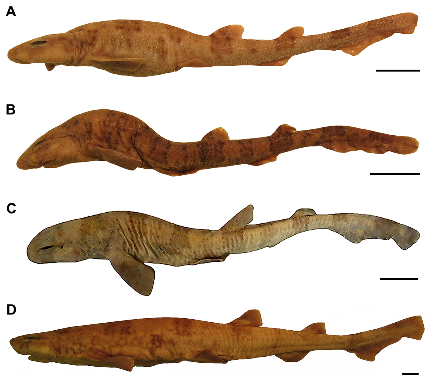


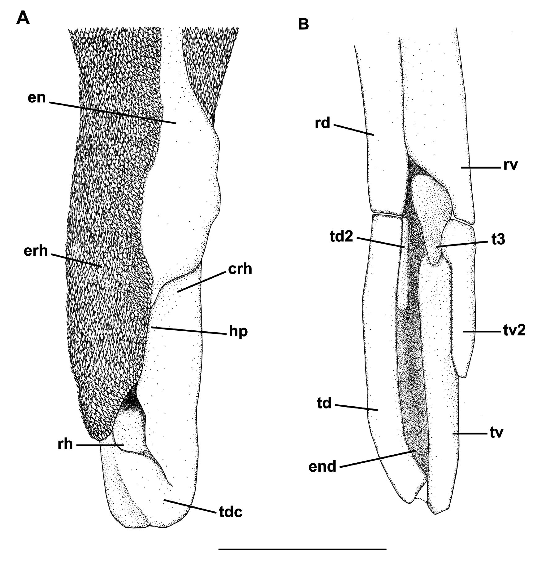

TABLE 1. Morphometric and meristic data of Scyliorhinus boa. SD, standard deviation; n, number of examined specimens. Total length (TL) in mm, other measurements as percentages of TL.
| Characters | Holotype | n | Range | Mean | SD |
|---|---|---|---|---|---|
| Total length (TL) | 149.4 | 42 | 97.6–4881.1 | 261.8 | 97.3 |
| Precaudal length | 70.7 | 42 | 70.7–78.8 | 74.3 | 2.1 |
| Eye-spiracle length | 0.9 | 42 | 0.6–1.5 | 1.0 | 0.2 |
| Prenasal length | 3.5 | 42 | 2.4–5.2 | 3.4 | 0.6 |
| Preoral length | 5.3 | 42 | 3.8–6.7 | 5.3 | 0.8 |
| Preorbital length | 7.2 | 42 | 3.7–8.1 | 6.9 | 1.3 |
| Prespiracular length | 10.3 | 42 | 9.2–12.2 | 11.0 | 1.6 |
| Prebranchial length | 15.3 | 42 | 13.3–17.7 | 16.0 | 1.7 |
| Head length | 19.7 | 42 | 17.2–22.1 | 20.7 | 1.7 |
| Prepectoral length | 15.8 | 42 | 15.1–20.2 | 18.7 | 1.8 |
| Prepelvic length | 38.5 | 42 | 34.3–44.7 | 39.8 | 2.1 |
| Snout-vent length | 42.4 | 42 | 35.8–44.6 | 42.5 | 2.1 |
| Vent-caudal length | 60.0 | 42 | 49.5–60.1 | 56.6 | 2.6 |
| Pre-first dorsal length | 46.1 | 42 | 42.3–52.9 | 47.6 | 2.7 |
| Interdorsal distance | 10.5 | 42 | 9.3–13.4 | 10.9 | 1.0 |
| Dorsal-caudal distance | 5.0 | 42 | 4.1–7.2 | 4.8 | 0.9 |
| Pectoral-pelvic distance | 19.0 | 42 | 12.8–19.0 | 15.6 | 1.5 |
| Pelvic-anal distance | 10.2 | 42 | 7.4–14.9 | 11.1 | 1.5 |
| Anal-caudal distance | 7.6 | 42 | 6.3–11.5 | 7.6 | 1.1 |
| Interorbital distance | 7.9 | 42 | 5.9–8.0 | 7.8 | 0.9 |
| Internarial distance | 5.4 | 42 | 4.3–7.1 | 5.6 | 0.5 |
| Mouth length | 5.6 | 42 | 3.4–6.1 | 5.0 | 0.8 |
| Mouth width | 8.2 | 42 | 6.7–9.8 | 8.3 | 0.9 |
| Lower labial furrow length | 1.0 | 42 | 1.7–3.0 | 2.2 | 0.3 |
| Eye length | 5.2 | 42 | 3.1–5.4 | 4.4 | 0.6 |
| Eye height | 1.4 | 42 | 1.1–2.8 | 1.8 | 0.4 |
| Spiracle length | 0.9 | 42 | 0.5–1.4 | 1.1 | 0.3 |
| First gill slit height | 2.7 | 42 | 2.0–4.1 | 2.6 | 0.4 |
| Fifth gill slit height | 1.1 | 42 | 0.9–2.4 | 1.4 | 0.2 |
| Pectoral length | 11.4 | 42 | 11.2–15.4 | 13.1 | 1.2 |
| Pectoral anterior margin | 13.0 | 42 | 12.0–16.5 | 14.6 | 1.2 |
| Pectoral base | 4.9 | 42 | 4.9–9.5 | 6.9 | 1.1 |
| Pectoral posterior margin | 7.0 | 42 | 5.9–10.5 | 8.4 | 1.3 |
| Pectoral inner margin | 5.8 | 42 | 5.7–8.5 | 6.6 | 0.7 |
| Pelvic length | 10.6 | 42 | 8.4–12.1 | 10.7 | 1.1 |
| Pelvic anterior margin | 6.4 | 42 | 5.9–8.2 | 7.1 | 0.8 |
| Pelvic posterior margin | 5.8 | 42 | 4.3–7.6 | 6.0 | 0.9 |
| Pelvic base | 6.5 | 42 | 5.0–9.5 | 7.2 | 1.0 |
| Pelvic inner length | 4.1 | 42 | 2.8–5.9 | 4.0 | 1.0 |
......continued on the next page
No known copyright restrictions apply. See Agosti, D., Egloff, W., 2009. Taxonomic information exchange and copyright: the Plazi approach. BMC Research Notes 2009, 2:53 for further explanation.
|
Kingdom |
|
|
Phylum |
|
|
Class |
|
|
Order |
|
|
Family |
|
|
Genus |
Scyliorhinus boa ( Goode & Bean, 1896 )
| Soares, Karla D. A. & De, Marcelo R. 2019 |
Scyliorhinus retifer haeckelii:
| Cadenat, J. & Blache, J. 1981: 183 |
Scyliorhinus haeckelii: Springer, 1979 : 136
| Springer, S. 1979: 136 |
Scyliorhinus retifer boa Springer & Sadowsky, 1970 : 90
| Springer, S. & Sadowsky, V. 1970: 90 |
Scyliorhinus fernandezi
| Lasso, C. A. & Lasso-Alcala, O. M. & Capelo, C. J. 1998: 4 |
| Cervigon, F. 1966: 60 |
| Weibezahn, F. H. 1953: 3 |
Scyliorhinus boa: Bigelow & Schroeder, 1948 : 204
| Weigmann, S. 2016: 43 |
| Oliveira, J. E. L. & Nobrega, M. F. & Garcia, J. & Sampaio, C. & Di Dario, F. & Fischer, L. G. & Mincarone, M. M. 2015: 31 |
| Kyne, P. M. & Carlson, J. K. & Ebert, D. A. & Fordham, S. V. & Bizzarro, J. J. & Graham, R. T. & Kulka, D. W. & Tewes, E. E. & Harrison, L. R. & Dulvy, N. K. 2012: 58 |
| Castro, J. I. 2011: 336 |
| Mejia-Falla, P. A. & Navia, A. F. & Mejia-Ladino, L. M. & Acero, A. P. & Rubio, E. A. 2007: 139 |
| Compagno, L. J. V. & Dando, M. & Fowler, S. 2005: 247 |
| Kiraly, S. J. & Moore, J. A. & Jasinsky, P. H. 2003: 15 |
| Cervigon, F. & Alcala, A. 1999: 94 |
| Compagno, L. J. V. 1999: 480 |
| Compagno, L. J. V. 1984: 357 |
| Uyeno, T. & Matsuura, K. & Fujii, E. 1983: 51 |
| Springer, S. 1979: 128 |
| Springer, S. 1966: 601 |
| Bigelow, H. B. & Schroeder W. C. 1948: 204 |
Catulus boa: Garman, 1913 : 77
| White, E. G. 1937: 117 |
| Garman, S. 1913: 77 |
Scylliorhinus retifer var. boa
| Regan, C. T. 1908: 457 |
| Goode, S. B. & Bean, T. H. 1896: 17 |