
Pseudosinella aggtelekiensis Stach, 1929
|
publication ID |
https://doi.org/ 10.5281/zenodo.213677 |
|
DOI |
https://doi.org/10.5281/zenodo.6166544 |
|
persistent identifier |
https://treatment.plazi.org/id/03B98133-FFF3-6A1F-FF46-CDAC6C9285A9 |
|
treatment provided by |
Plazi |
|
scientific name |
Pseudosinella aggtelekiensis Stach, 1929 |
| status |
|
Pseudosinella aggtelekiensis Stach, 1929
Figs 1–9 View FIGURES 1 – 4 View FIGURE 9
Lepidocyrtus (Pseudosinella) aggtelekiensis Stach, 1929: 296 .
Diagnosis. Eyes and pigmentation absent. Labium with M1M2reL1L2 basal setae, r strongly reduced. Dorsal macrosetae: R111/32/0201+3. On Abd.IV proximal macroseta in position A4. Setal pattern of abdominal tergite II: pABq1q2. Abd.IV with supplementary microseta s in front of anterior trichobothrium and 3+3 smooth mesosetae. Antennal segments with conical microsetae cm. Apical half of Ant.III segment without additional leaf-like setae, Ant.II apically with 1 such modified seta. Foot complex as in Fig. 8. Unguiculus without any tooth on outer lamella. Tibiotarsal tenent hair pointed. Trochanteral organ with 18–22 smooth setae.
Type material. Hungary, Domica-Baradla cave system, Baradla Cave, 18–20.viii.1924, leg. E. Dudich & E. Bokor; ibid. 1.xi. and 5.xii.1928, leg. E. Dudich (not examined, number of specimens and place of deposition unknown).
Examined material from type locality. Hungary, Slovak-Aggtelek Karst, Domica-Baradla cave system, Baradla Cave, 5 specimens mounted on a permanent slide, 19.xii.1929, leg. Dudich. ISEA, Polish Academy of Sciences Kraków, Poland. Slovakia, Slovak-Aggtelek Karst, Domica-Baradla cave system, Čertova diera Cave, “Dóm netopierov” Hall and “Vstupná sieň” Hall, 5 specimens (3 females, 2 males), collected by pitfall traps, 23.x.–9. xii.1997, leg. Ľ. Kováč; ibid. Líščia diera Cave, “Veľká sieň” Hall, 1 specimen (female), 1.xii.2000, leg. P. Ľuptáčik. 6 specimens saved in collection of the Muséum Nationalle d´Histoire Naturelle ( MNHN) in Paris.
, 8; side
internal
,
III leg
of
trochanter
, 7;
side
left). 8,
– dorsally (7 Figs, III. m ̝ Abd) 50,, 6 6 – side; 5 Figs left, (m dorsally 200:: ̝ segment, bars Scale II. Abd., 5 enlarged: aggtelekiensis , macroseta Pseudosinella of a—tip internal., 8 II – 5 leg FIGURES of unguis
Other examined material. Slovakia, Slovak-Aggtelek Karst, Ardovská Cave, “Zrútený dóm” Dome, 2 specimens, pitfall trap, 29.iv.–13.vi.1997, leg. Ľ. Kováč; ibid., main cave passage in upper level, 6 specimens, pitfall traps, 4.iv.–30.x.1997, leg. Ľ. Kováč; ibid., “Rozprávková sieň” Hall, 11 specimens, collected on bat guano, 31.iii.2011, leg. Ľ. Kováč; ibid., “Zrútený dóm” Dome, 2 specimens, collected on rotten wood, 10.x.2008, leg. P. Ľuptáčik; Gombasecká Cave, “Blatistá chodba” Passage, 7 specimens, 22.x.1999, hand collecting on surface of water pool and on rotten wood, leg. Ľ. Kováč; ibid., 4 specimens, collected on rotten wood, 8.x.2008, leg. P.
Ľuptáčik; Hačavská Cave, 3 specimens, pitfall trap, 24.v.–23.vi.1996, leg. Ľ. Kováč; Majkova Cave, 4 specimens, pitfall trap, 5.iii.–15.v.1998, leg. Ľ. Kováč and A. Mock; Milada Cave, “Dóm Vysokých Tatier” Dome, 5 specimens, collected on bait, 8.ix.2010. leg. P. Ľuptáčik. Other material kept in the Institute of Biology & Ecology, Faculty of Science, P. J. Šafárik University ( IBE FS UPJŠ) Košice.
Redescription. Body 2.1–2.4 mm long. White, without traces of pigmentation. Scales on antennae and legs absent; ventral side of manubrium with scales.
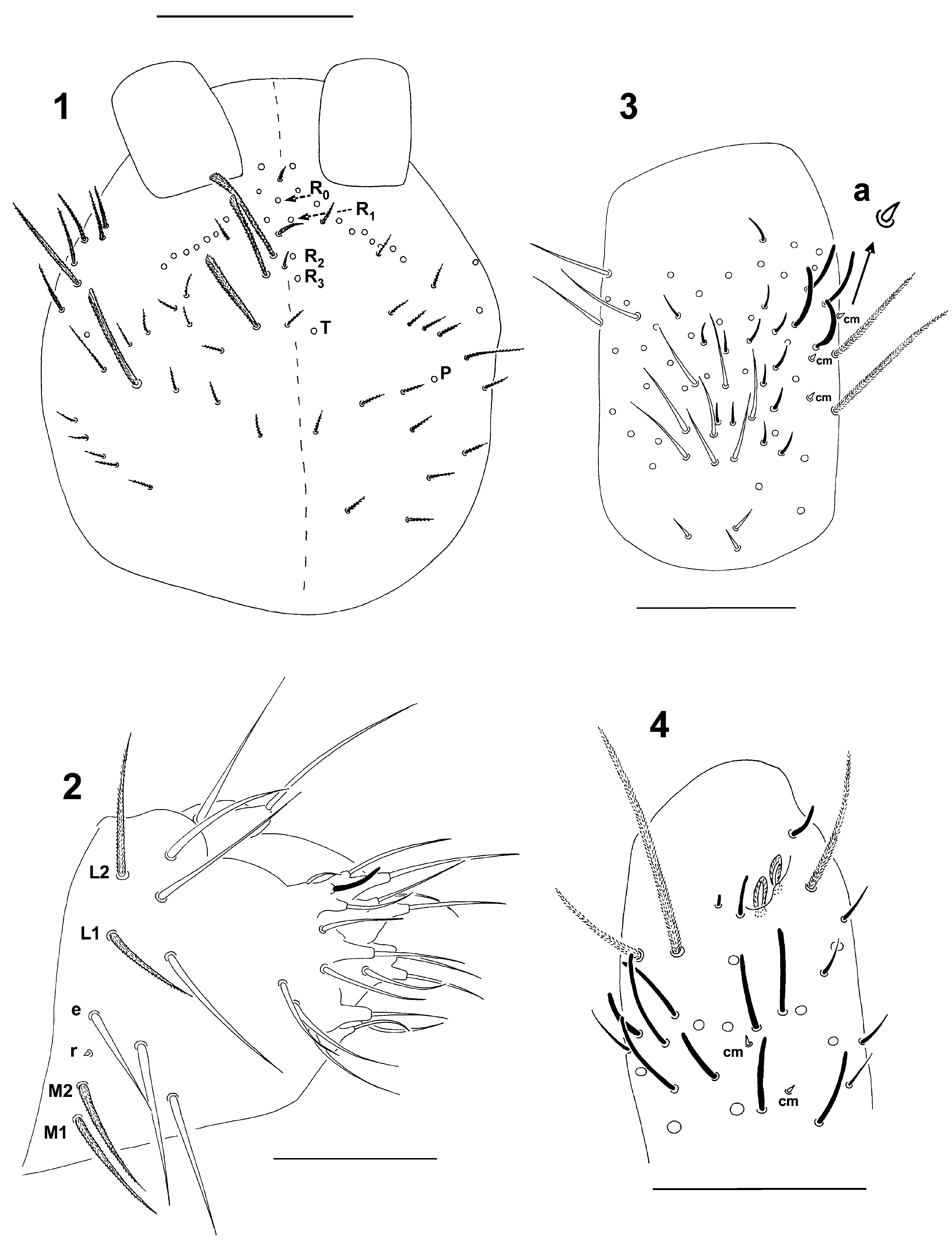
Head. Eyes absent. Dorsal macrosetae R111 or R (R0 R1 R2) + R3 T P. Macrosetae ciliated (65–85 μm) with blunt apex (dorsal ones) or sharply pointed (lateral ones); mesosetae finely ciliated (15–40 μm, Fig. 1 View FIGURES 1 – 4 ). Posterior row with finely ciliated and sharply pointed mesosetae (55 μm). Short trichobothrium (35 μm) situated laterally to ocular macroseta. Praelabral setae ciliated, labral setae smooth. Setal pattern of labrum: 4/554. Labium in adults with M1M2reL1L2 basal setae; M1, M2, L1 and L2 finely ciliated, seta r strongly reduced, smooth ( Fig. 2 View FIGURES 1 – 4 ); in juveniles and subadults seta e finely ciliated (i.e. E) as others. Frontal row of labial setae smooth.
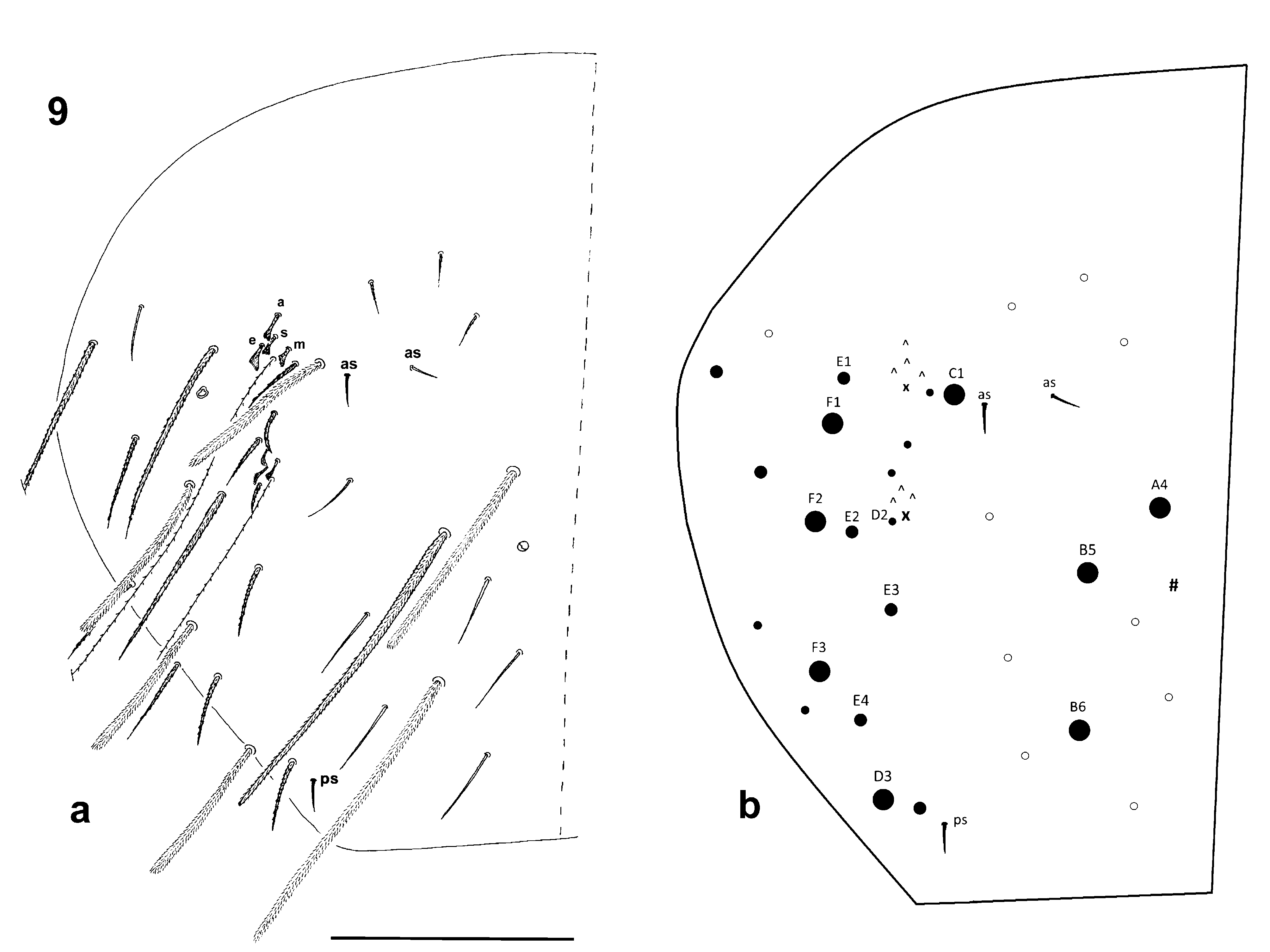
Thorax and abdomen (Figs 5, 6 and 9). Dorsal macrosetae: /32/0201+3. Microsensillar formula 10/10100, microsensilla (ms) strong and placed laterally (5 μm), on Th.II and Abd.I anteriorly, on Abd.III posteriorly. Formula of smooth mesosetae 11/01133, mesosetae (s) progressively elongated from Th.II (10 μm) to Abd.V (17 μm). Smooth mesoseta on Th.II in anterior position placed laterally to ms. Abd.IV with 3 smooth mesosetae, 2 anterior as (medial and lateral) and 1 posterior ps. Setal pattern of abdominal tergite II: pABq1q2 (Fig. 5); macroseta A 0.63% of the length of macroseta B (90 and 145 μm, respectively). Abd.IV with 4 supplementary microsetae (blunt, ciliated) in front of anterior trichobothrium (microseta s present) and 3 such microsetae in front of posterior trichobothrium ( Fig. 9 View FIGURE 9 a, b). Medial macrosetae of Abd.IV: anterior A4 broad with blunt apex (190 μm), medial B5 and posterior B6 more slender, equally long (300 μm) with apex sharply pointed devoid of ciliation. Entire setal pattern of Abd.IV shown in Figs 9 View FIGURE 9 a and 9b.
Appendages. Antennae longer than head (1280: 600 μm). Antennal segments I: II: III: IV as 140: 300: 340: 500 (µm); densely covered with ciliated meso- and macrosetae (25–70 µm), numerous curved sensilla (15–20 µm) and thin microsensilla (10–12 µm). Ventral side of segments with groups of conical microsetae cm (2.5 µm; Figs 3 and 4 View FIGURES 1 – 4 ). Apical bulb on Ant.IV absent, subapical organite not seen. Apical part of Ant. III with antennal organ consisting of 2 wrinkled, leaf-like sensory setae (10 µm) partly hidden behind cuticular folds, 2 guard sensilla (10 µm) and short rod (4 µm; Fig. 4 View FIGURES 1 – 4 ). Ant. III without additional leaf-like setae; several modified sensilla with thickened basal part (15 µm): 3 ventral and 3–4 internal situated in vertical rows, and 5–6 dorsal grouped together. Apical part of Ant.II with 1 dorso-external leaf-like seta (10 µm); segment externally with 4–5 sensilla with thickened base (15 µm), internal side with 1 such seta. Ant.I with 3 dorsal and 3 ventral basal microsetae (8–10 µm); ventrally with group of 8–14 thin microsensilla (10 µm) and 8–10 smooth setae (20–25 µm); 4–6 external sensilla (18–20 µm), 1 or 2 of which thicker and shorter (14 µm); ( Fig. 3 View FIGURES 1 – 4 ). Conical microsetae cm (2.5 µm) present on ventral side of Ant.II–IV, on Ant. I 3–4 such microsetae situated externally ( Figs 3 and 4 View FIGURES 1 – 4 ).
Unguis of legs I, II and III as 64, 62 and 60 μm; tibiotarsi 25 μm wide. Unguis with 2 short proximal (basal) teeth in 16% length and 1 short internal tooth in 26 % length of unguis (positions in % measured on leg I); 1 short external tooth (12 μm); apical and lateral teeth on unguis absent (Fig. 8). Unguiculus 46 µm long, external tooth absent. Tibiotarsal tenent hair pointed, 30 µm long, inner macrosetae of tibiotarsi differentiated (except of proximal setae whorl)—thick, apically smooth, obliquely cut and sharply pointed (Fig. 8). Metatibiotarsus (leg III) with 1 differentiated internal seta placed in first whorl, smooth, pointed, rather long (50 μm). Trochanteral organ (leg III) consists of 18–22 smooth setae (12–30 μm; Fig. 7). Ventral tubus with 14–16 ciliated setae on lateral flap. Manubrial plaque on each side with 2 pseudopores, 2 internal and 2 external ciliated setae. Manubrium: dens: mucro as 410: 530: 30 (µm). Apical part of dens (0.1 of the length) not crenulated. Mucro elongated with apical teeth apparently longer than anteapical one, 1 short basal seta reaching anteapical tooth.
Both sexes known.
Discussion. There are another two species of the genus Pseudosinella without eyes and identical macrosetae formula R111/32/0101+3, P. antennata Bonet, 1929 and P. unguilonginea Jordana & Beruete, 1983 . Both represent cave adapted species distributed in karst of the Navarra province in Spain. They also share other characters with P. aggtelekiensis , i.e. supplementary microseta s on Abd.IV segment present, tenent seta on tibiotarsus pointed, and external tooth on unguiculus absent. However, P. aggtelekiensis has different pattern of head macrosetae (R0 R1 R2 R3 T P) compared to P. antennata and P. unguilonginea (both with R0 R1 R2 S T P). Other difference between P. aggtelekiensis and both species from Navarra is in pattern of basal labial setae (M1M2reL1L 2 in P. aggtelekiensis ; m 1m 2rel1l 2 in P. antennata and P. unguilonginea ). P. aggtelekiensis and P. antennata have very similar shape of unguis and unguiculus (unguis with short teeth - 2 proximal, 1 internal and 1 lateral). P. unguilonginea has, in the contrary, apparently elongated and narrowed unguis with very short proximal teeth, and minute internal and lateral teeth. Presence of spiniform, smooth ventral seta on tibiotarsus of the third leg is common character for P. aggtelekiensis and P. unguilonginea , its status in P. antennata being unknown.
Pseudosinella aggtelekiensis has several other characters unique among species of the genus. For example 3–4 external conical microsetae, resembling rests of broken scales, are present on Ant.I segment. In the contrary to remnants of scales, these microsetae have regular sharp apex and apparent basal circle. Abd.IV tergum has 3 blunt and supplementary microsetae in front of posterior trichobothrium (instead of usually 2) and 2 anterior smooth mesosetae as, medial and lateral (instead of only medial as). Moreover, of medial macroseta the anterior one is placed in longitudinal row A (position A4). Medial macrosetae B5 and B6 are rather long with apex unusually sharply pointed and devoid of ciliation.
Distribution. The species was described by Stach (1929) from the Baradla Cave, a part of the Domica-Baradla cave system (approx. length 25 km) situated in the Slovak-Aggtelek Karst in the border region of Slovakia and Hungary. It was later detected also in the Szabadság and Béke caves in the same region ( Loksa 1961, Dányi 2011). Strouhal and Vornatscher (1975, p. 512) listed P. aggtelekiensis in the catalogue of cave fauna of Austria. The species was considered to inhabit the Bärenhöhle Cave in NE Alps (Hartelsgraben near Hieflau, Austria). However, Christian (1987) stressed that conspecifity of Styrian taxon with P. aggtelekiensis is unprobable. We also had possibility to study slide labelled as ”Kärnten, Hartelsgrabenhöhle in Gesäure cca 1200 m, 1940, leg. H. Franz, Pseudosinella aggtelekiensis “ deposited in the Institute of Systematics and Evolution of Animals, Polish Academy of Science, Kraków. Unfortunately the slide contains only legs and antennae of several Pseudosinella specimens. However, the shape and composition of unguis is different from that of P. aggtelekiensis (more slender shape, proximal teeth very short, internal and external teeth minute). Two slides of the same collection are labelled as ” Austria, Gasslhöhle bei Ebensee an Traun, det. Stach, Lepidocyrtus (Pseudosinella) aggtelekiensis “ with one specimen each. However, the status of the specimens does not allow observation of principal taxonomic characters, except for the shape and arrangement of unguis that is apparently different from that of P. aggtelekiensis (more slender shape and ungual teeth as in previous case). Finally, the species has been reported also from Romania as a member of soil communities ( Fiera 2007).
Pseudosinella aggtelekiensis represents an obligate cave species with endemic distribution restricted to the Slovak-Aggtelek Karst region located between Slovakia and Hungary. The species has been found to inhabit the Domica-Baradla cave system (type locality) and caves Ardovská, Drienka, Krásnohorská, Majkova, Milada, Szabadság and the Obrovská Abyss ( Stach 1929, Dudich 1932, Loksa 1961, Kováč et al. 2005a, b, Papáč et al. 2006, 2007). Literature data referring the occurrence of Pseudosinella aggtelekiensis in caves or soils of other regions are un-probable and should be verified.
Troglobionts P. unguilonginea and P. antennata , very similar by their morphology, inhabit caves of the Navarra region in northern Spain. Their distribution ranges are vicariant, the first species being restricted to Aralar ridge karstic area, the second one to adjacent Urbasa–South area, respectively. P. aggtelekiensis , P. antennata and P. unguilonginea are example of parallelisms in evolution of subterranean species in different ” Pseudosinella “ lineages ( Christiansen 1961). Pseudosinella aggtelekiensis may be considered as a form derived from older fauna of Tertiary origin based on its distribution restricted to caves, small (endemic) distribution range, level of troglomorphy and several conspicuous morphological characters.
Remarks to biology. Mixture of clay particles and fungal hyphae usually prevails in content of the gut. Moreover, we observed characteristic spores of micromycete genera Alternaria , Fusarium , Ulocladium and Tetracoccosporium in the gut of several specimens (A. Nováková, det.).
No known copyright restrictions apply. See Agosti, D., Egloff, W., 2009. Taxonomic information exchange and copyright: the Plazi approach. BMC Research Notes 2009, 2:53 for further explanation.
|
Kingdom |
|
|
Phylum |
|
|
Class |
|
|
Order |
|
|
Family |
|
|
Genus |
Pseudosinella aggtelekiensis Stach, 1929
| Kováč, Ľubomír & Rusek, Josef 2012 |
Lepidocyrtus (Pseudosinella) aggtelekiensis
| Stach 1929: 296 |