
Pleuronema elegans, Pan & Huang & Fan & Ma & Al-Rasheid & Miao & Gao, 2015
|
publication ID |
https://doi.org/10.4467/16890027AP.15.003.2190 |
|
DOI |
https://doi.org/10.5281/zenodo.8377645 |
|
persistent identifier |
https://treatment.plazi.org/id/03B9BA0A-FFB1-FFF1-FCC0-3C45FAB31C14 |
|
treatment provided by |
Felipe |
|
scientific name |
Pleuronema elegans |
| status |
sp. nov. |
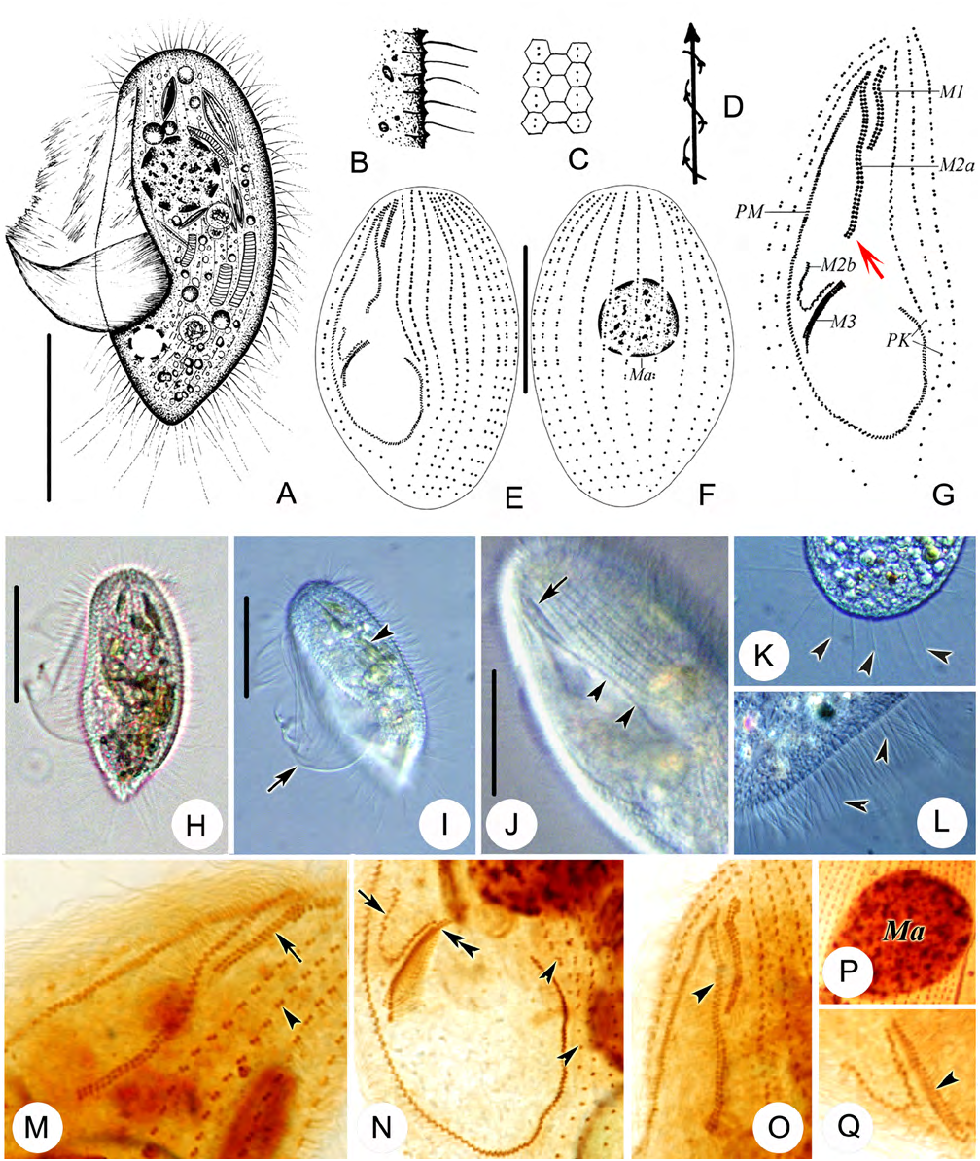
Pleuronema elegans spec. nov. ( Fig. 2 View Fig , Table 1 View Table 1 )
Diagnosis: Size in vivo 90–115 × 45–60 µm with a distinctly pointed posterior end; contractile vacuole located dorsally near posterior end; about 10 prolonged caudal cilia; consistently two preoral and 18 or 19 somatic kineties; membranelle 1 with a length about 50% that of the anterior part of membranelle 2 which is double-rowed with its posterior end straight but not hook-shaped; marine habitat.
Type locality: Swimming beach, Qingdao, northern China ( 36°06′N; 120°32′E) GoogleMaps .
Type slides: The holotype slide (registration number: PXM-20120515) and one paratype slide (registration number: NHMUK 2013.8.15.1) with protargol stained-specimens are deposited in the Laboratory of Protozoology , Ocean University of China ( OUC) and the Natural History Museum, London, respectively .
Etymology: This new form named ‘ elegans ’ refers to its elegant body shape.
Description: The body is about 100 × 50 µm in vivo, slender oval in outline, with a distinctly pointed posterior end ( Figs 2A, H, I View Fig ). Buccal field cavity is about 70% of body length with a conspicuous, saillike, undulating membrane ( Fig. 2I View Fig ). Pellicle is rigid and slightly notched with closely arranged extrusomes, which is about 3 µm long ( Fig. 2B View Fig ). Cytoplasm is colourless to slightly grayish, packed with large amounts of green ingested algae and shining globules of varying size, food vacuoles which are usually large and filled with bacteria, and blue irregularly-shaped crystals (<6 μm in diameter) ( Figs 2A, H, I View Fig ). One spherical ma- cronucleus, about 32 × 32 µm, located in anterior half of cell. No micronucleus is observed ( Fig. 2P View Fig ). Single contractile vacuole is about 10 µm in diameter, located slightly dorsally near posterior end of cell ( Fig. 2A View Fig ). Somatic cilia is about 12 µm long ( Figs 2A, L View Fig ). There are about ten prolonged caudal cilia, each is about 30 µm in length ( Fig. 2K View Fig ).
The cell swims moderately fast while rotating about main body axis, sometimes lying motionless along substrate such as bottom of Petri dish or detritus ( Fig. 2D View Fig ).
There are eighteen or 19 somatic kineties, which are composed of dikinetids in anterior 60% of body and monokinetids in posterior third, extending almost the entire length of the cell, terminating anteriorly at a small glabrous apical plate ( Figs 2E, F View Fig ). There are consistently two preoral kineties to the left of the buccal field ( Figs 2G, N View Fig ).
Oral apparatus is typical for genus: M1 comprises two longitudinal rows of basal bodies, the length of which is about 50% that of the anterior part of M2a ( Figs 2G, M, O View Fig ). M2a is double-rowed with its posterior end straight; posterior part of M2b is V-shaped, and is distinctly separated from M2a ( Figs 2G, N View Fig ). M3 is three-rowed with a similar length to that of M1 ( Figs 2G, Q View Fig ). Length of paroral membrane is about 70% of body length. Silverline system is typical for the genus with a near-hexagonal honeycomb pattern ( Fig. 2C View Fig ).
SSU rRNA gene sequence: The SSU rRNA gene sequence of Pleuronema elegans spec. nov. has been deposited in the GenBank database with the accession number, length and G+C content as follows: KF840518 View Materials , 1661 bp, 42.75%.
Remarks and comparison: Based on its conspicuously pointed posterior end and marine habitat, Pleuronema elegans spec. nov. most resembles two nominal species: P. czapikae Wang et al., 2008 and P. tardum Czapik & Jordan, 1977 and can be readily separated from other congeners.
Pleuronema elegans spec. nov. can be clearly distinguished from P. czapikae Wang et al., 2008 through its different body shape (slender oval in outline, with a distinctly pointed posterior end in P. elegans vs. elongate-elliptical in outline, almost parallel-sided with both ends slightly pointed in P. czapikae ), fewer somatic kineties (18–19 vs. 29–35 in P. czapikae ) and M2a double-rowed with its posterior end straight (vs. mostly two-rowed but with a short section that is single-rowed, posterior end invariably hook-shaped in P. czapikae ) ( Wang et al. 2008b).
Pleuronema elegans spec. nov. differs from P. tardum in having more preoral kineties (two vs. one in P. tardum ), less somatic kineties (18–19 vs. 40–50) and a different ratio of M1 and M3 to the anterior part of M2a (50% vs. M1 and M3 very short and M2a extreme- ly long in P. tardum ) ( Czapik and Jordan 1977).
| NHMUK |
Natural History Museum, London |
No known copyright restrictions apply. See Agosti, D., Egloff, W., 2009. Taxonomic information exchange and copyright: the Plazi approach. BMC Research Notes 2009, 2:53 for further explanation.
|
Kingdom |
|
|
Phylum |
|
|
Class |
|
|
SubClass |
Scuticociliatia |
|
Order |
|
|
Family |
|
|
Genus |