
Schizopelex huettingeri Malicky 1974
|
publication ID |
https://doi.org/ 10.11646/zootaxa.4311.2.8 |
|
publication LSID |
lsid:zoobank.org:pub:A700167F-C2DD-432E-8286-96DBCB643203 |
|
DOI |
https://doi.org/10.5281/zenodo.6049577 |
|
persistent identifier |
https://treatment.plazi.org/id/03B9E056-AA6A-C01C-FF50-8F4BFCDDD18B |
|
treatment provided by |
Plazi |
|
scientific name |
Schizopelex huettingeri Malicky 1974 |
| status |
|
Description of the final instar larva of Schizopelex huettingeri Malicky 1974 View in CoL
Biometry. Body length ranging from 11.4 to 11.9 mm, head width from 1.59 to 1.75 mm (n = 3).
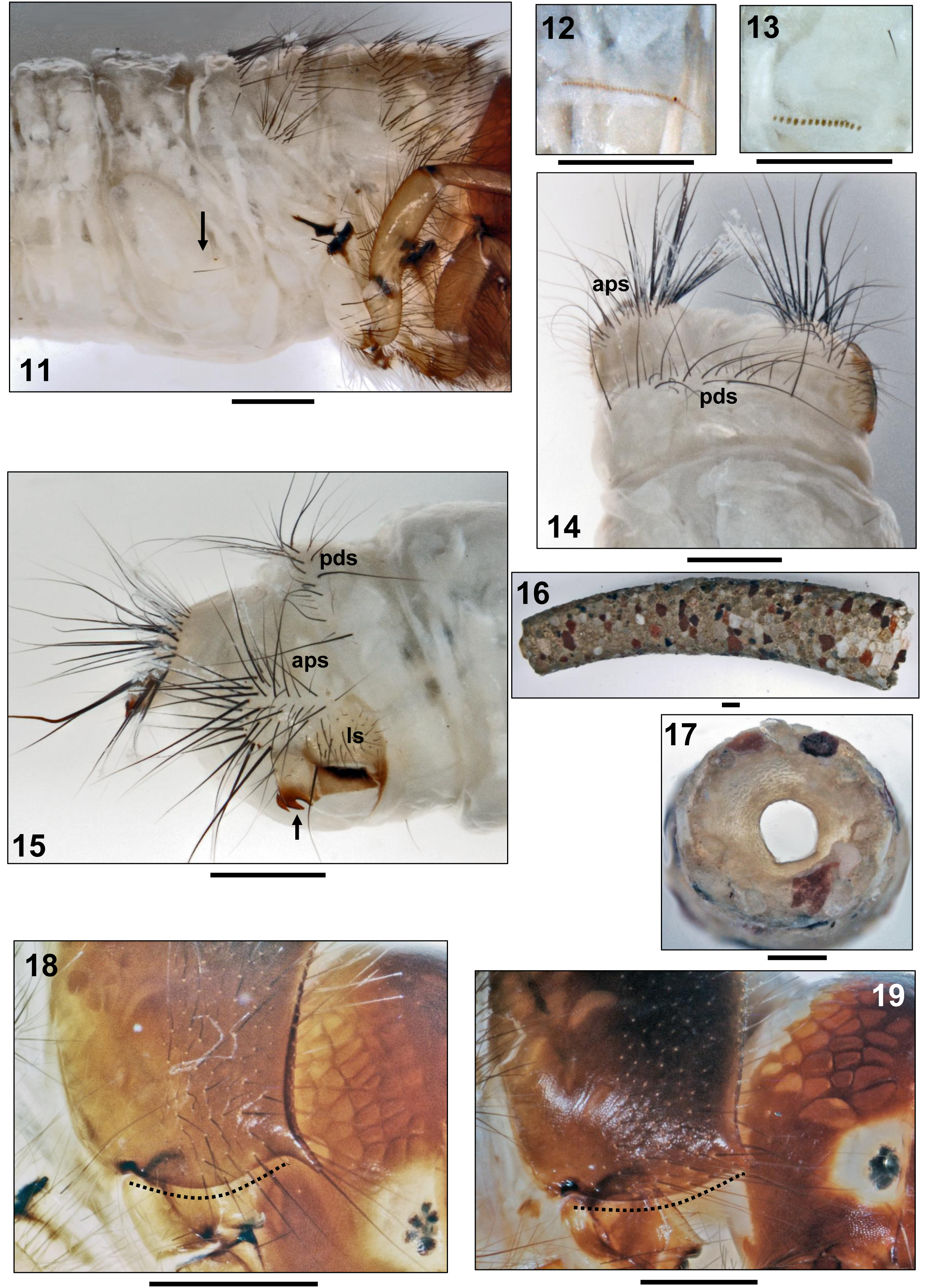
Head. Head capsule nearly round, dorsally medium to chestnut brown, posterolaterally and ventrally yellowish white, with smooth surface and large, pale muscle attachment spots ( Figs. 1–3 View FIGURES 1 – 4 ). Distinct white semicircle beneath each eye ( Figs. 1, 3 View FIGURES 1 – 4 ). Dorsolateral ridge extending from posterodorsal corner of white semicircular ring around each eye ( Fig. 3 View FIGURES 1 – 4 ) to anterior parietal margin where ridge creating inward-bent conical projection bearing antenna ( Fig. 1 View FIGURES 1 – 4 , arrow). Frontoclypeus narrow and elongated, with shallow central constriction and subapical bulge ( Fig. 1 View FIGURES 1 – 4 ). Head capsule with complete set of 18 pairs of primary setae: 10 dorsal and 2 ventral primary setae on each parietal, 6 pairs of primary setae on frontoclypeus. Labrum medium brown, narrowly rectangular, with 3 pairs of primary setae. Submentum broadly triangular, light brown, with dark brown anterior transverse band ( Fig. 2 View FIGURES 1 – 4 ). Mandible bases yellowish brown, distal sections black, of shredder type, right mandible with terminal tooth and up to 3 teeth along mesal edge, left mandible slightly serrate ( Fig. 1 View FIGURES 1 – 4 ).
Thorax. Anterior section of pronotum medium to dark brown, posterior section yellowish brown to medium brown, with pale muscle attachment spots. Pronotum without transverse ridge (present in other caddisfly taxa; e.g., Limnephilinae ), heavily sclerotized, with anterolateral corners elongated and pointed; pair of plates meeting mesally in narrow, straight suture; surface smooth ( Figs. 3 View FIGURES 1 – 4 , 5 View FIGURES 5 – 10 ). Ventrolateral pronotal margin straight ( Fig. 3 View FIGURES 1 – 4 , dotted line). Each pronotal half covered by 160–180 setae concentrated on anterior, darker pronotal section; setal type 1 pale, flexible and procumbent, setal type 2 long, straight and medium to dark brown. Anterior pronotal margin with single row of setae of both types ( Figs. 3 View FIGURES 1 – 4 , 5 View FIGURES 5 – 10 ). Anterior part of coxopleurite long and corniform ( Figs. 3 View FIGURES 1 – 4 , arrow, 4). Prosternal sclerites and prosternal horn lacking.
Mesodorsum covered by 4 sclerotized plates (2 large central, 2 small lateral), whitish, with pale yellow muscle attachment spots, distinct anterior margin, less-distinct posterior margin ( Figs. 5 View FIGURES 5 – 10 ms, 6cs, 6ls); suture between central and lateral plates inconspicuous ( Fig. 6 View FIGURES 5 – 10 , arrows). Setal counts per central sclerite are 75–85 in anterior group and 25–30 in posterior group ( Fig. 5 View FIGURES 5 – 10 ms); lateral sclerite with 45–50 black setae ( Fig. 6 View FIGURES 5 – 10 ls).
Metadorsum covered by colourless and barely visible, weak sclerites arranged in 2 parallel transverse bands. Setal counts per sclerite: 44–48 setae in anterior group, 20–25 setae in posterior group ( Fig. 5 View FIGURES 5 – 10 mt).
Legs medium to light brown ( Figs. 7–9 View FIGURES 5 – 10 ). Forelegs short and stout, each with proximal section of femur enlarged and flattened, strongly narrowing apically, thereby creating edge interacting with tibia when bent inwards ( Fig. 7 View FIGURES 5 – 10 ). Coxa with ventral group of long black setae, trochanter with dense ventral brush of pale, flexible setae. Dorsal edge of femur with large groups of proximodorsal and distodorsal black setae. Tibia with groups of long black dorsal and ventral setae and with pale apical spine. Strong tarsal claw sickle-shaped, with stout pale basal spine. Midlegs much more slender, coxae weakly sclerotized, femora not enlarged. Hind legs even more slender, tarsal claws elongated, setation less than on other legs ( Figs. 7–9 View FIGURES 5 – 10 ).
Abdomen. Abdominal segment I with pair of flat, oblique lateral and 1 low, inconspicuous dorsal protuberances ( Fig. 11 View FIGURES 11 – 19 ); setation consisting of 1 pair of ventral sa 1 setae ( Fig. 10 View FIGURES 5 – 10 , arrows) and 1 lateral protuberance seta per side ( Fig. 10 View FIGURES 5 – 10 lp). Gills consisting of tiny single filaments and in presegmental position only. Dorsal gills present at most on abdominal segments I to IV, ventral gills on segments III to VII and lateral gills on II to III. Lateral fringe lacking; however, with lateral row of tiny serrate lamellae on each side of abdominal segments III to VII ( Fig. 12 View FIGURES 11 – 19 ), and with row of forked lamellae on each side of segment VIII ( Fig. 13 View FIGURES 11 – 19 ).
Dorsal sclerite of abdominal segment IX lacking, soft cuticle with 18–41 black setae of almost equal length on posterodorsal border ( Figs. 14, 15 View FIGURES 11 – 19 pds). Dorsum of each anal proleg with cluster of 30–40 black setae ( Figs. 14, 15 View FIGURES 11 – 19 aps). Weakly sclerotized lateral sclerite of each anal proleg with 22–25 black setae of varying length ( Fig. 15 View FIGURES 11 – 19 ls). Anal proleg claws each with sharply angled crook and dorsal accessory hook ( Fig. 15 View FIGURES 11 – 19 , arrow).
Larval case. Cylindrical, tapering, strongly curved, made of flat sand grains of uniform size arranged in puzzle-like pattern, thereby creating rather smooth surface ( Fig. 16 View FIGURES 11 – 19 ). Case length 11.5 to 13.1 mm, anterior width 2.7 to 2.8 mm, posterior width 1.8 to 1.9 mm (n = 3). Posterior foramen partly closed by slightly conical, silken membrane with round central hole 0.55 mm in diameter ( Fig. 17 View FIGURES 11 – 19 ).


No known copyright restrictions apply. See Agosti, D., Egloff, W., 2009. Taxonomic information exchange and copyright: the Plazi approach. BMC Research Notes 2009, 2:53 for further explanation.