
Spilomena Shuckard, 1838
|
publication ID |
https://doi.org/10.11646/zootaxa.5068.2.6 |
|
publication LSID |
lsid:zoobank.org:pub:6385D6D2-9442-4626-BA3C-65504B22CD24 |
|
DOI |
https://doi.org/10.5281/zenodo.5705420 |
|
persistent identifier |
https://treatment.plazi.org/id/03BA4074-F366-FF9E-FF16-8CC5B68DE396 |
|
treatment provided by |
Plazi |
|
scientific name |
Spilomena Shuckard |
| status |
|
Key to the Indian species of Spilomena Shuckard View in CoL
1. Inner mandibular tooth broad and blunt.................................................................... 2
— Inner mandibular tooth pointed............. S. keralaensis Tessy, Sureshan et Girish Kumar ( Fig. 2 View FIGURES 1–8 of Rajan et al. 2018)
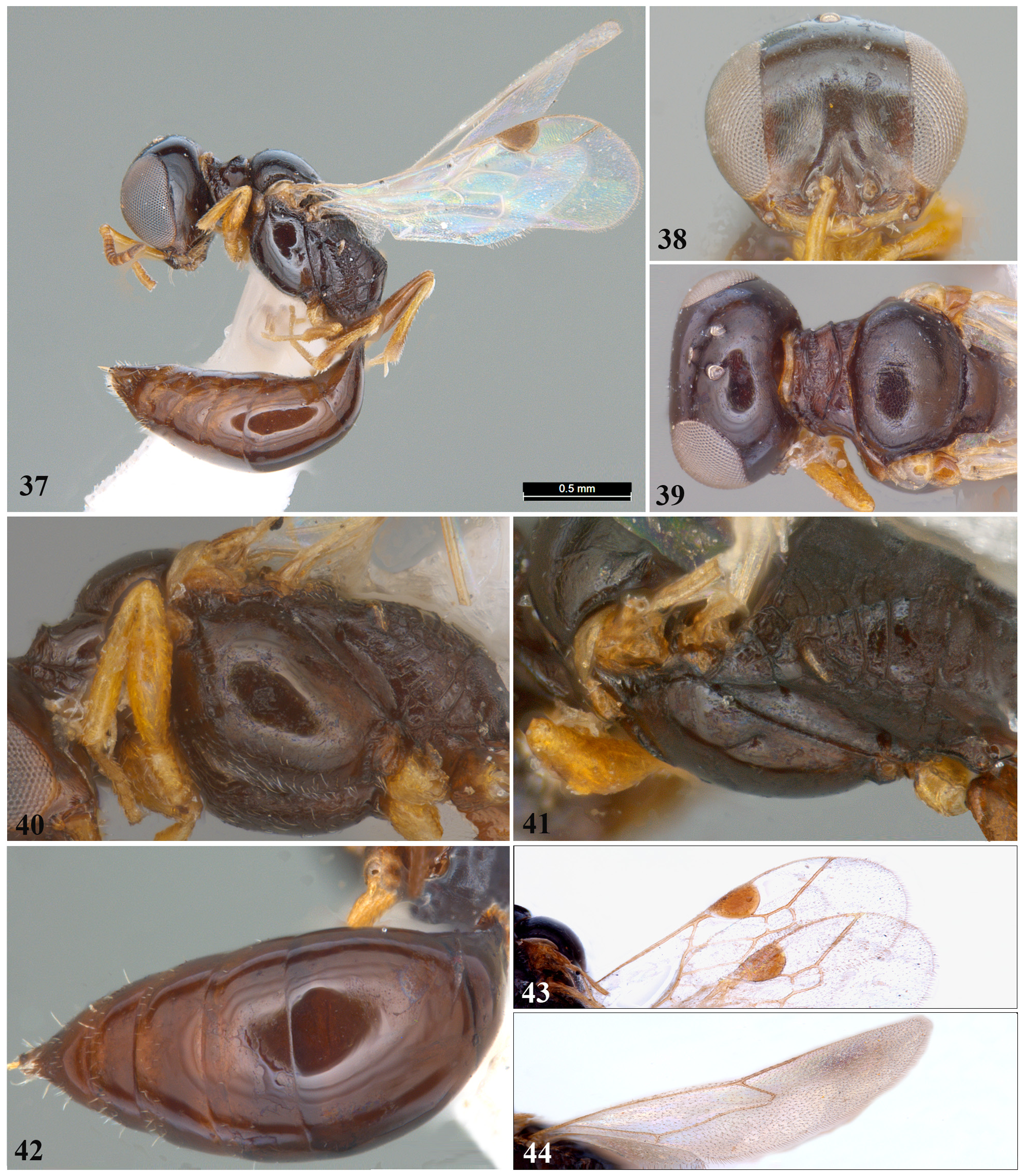
2. Interantennal tubercle not distinctly pointed ( Figs 2 View FIGURES 1–8 , 10 View FIGURES 9–16 , 18 View FIGURES 17–20 , 22 View FIGURES 21–28 , 30 View FIGURES 29–36 & 38 View FIGURES 37–44 ); mesosoma black or brownish black ( Figs 1 View FIGURES 1–8 , 9 View FIGURES 9–16 , 17 View FIGURES 17–20 , 21 View FIGURES 21–28 , 29 View FIGURES 29–36 & 37 View FIGURES 37–44 ); body length 2.5 – 4.6 mm....................................................................... 3
— Interantennal tubercle acutely pointed ( Fig. 46 View FIGURES 45–52 ); mesosoma brownish yellow with few black patches ( Fig. 49 View FIGURES 45–52 ); body length 5.6 mm.................................................. S. tuberculata Tessy, Sureshan et Girish Kumar , sp. nov.
3. Posterior surface of propodeum without median carina ( Figs 5 View FIGURES 1–8 , 13 View FIGURES 9–16 , 20 View FIGURES 17–20 & 37 View FIGURES 37–44 ); recurrent vein of fore wing interstitial ( Fig. 15 View FIGURES 9–16 ) or nearly so ( Figs 7 View FIGURES 1–8 & 31 View FIGURES 29–36 ) with first transverse cubital vein.................................................... 4
— Posterior surface of propodeum with median carina ( Figs 25 View FIGURES 21–28 & 41 View FIGURES 37–44 ); recurrent vein of fore wing received well within first submarginal cell ( Figs 27 View FIGURES 21–28 & 43 View FIGURES 37–44 )............................................................................. 7
4. Recurrent vein of fore wing interstitial with first transverse cubital vein ( Fig. 15 View FIGURES 9–16 ).................................. 5
— Recurrent vein of fore wing nearly interstitial with first transverse cubital vein ( Fig. 7 View FIGURES 1–8 & 35 View FIGURES 29–36 ).......................... 6
5. Body black ( Fig. 17 View FIGURES 17–20 ); propodeum rugose ( Fig. 20 View FIGURES 17–20 ).......................................... S. indostana Turner View in CoL
— Mesosoma black with mesoscutum laterally and mesopleuron yellowish brown ( Fig. 9 View FIGURES 9–16 ); propodeum entirely transversely striate ( Fig. 12 View FIGURES 9–16 ).................................................. S. fulvopleuris Tessy, Sureshan et Binoy , sp. nov.
6. Interantennal tubercle extending into long median carina reaching up to mid frons ( Fig. 2 View FIGURES 1–8 ); anterior carina of pronotal collar deeply emarginate ( Fig. 3 View FIGURES 1–8 ); inner eye margin slightly concave ( Fig. 2 View FIGURES 1–8 ); upper frons and vertex without setigerous punctures ( Fig. 2 View FIGURES 1–8 ); propodeal dorsum transversely striate, with two median longitudinal carinae; posterior surface of propodeum with irregular transverse ridges; lateral surface of propodeum finely aciculate ( Figs 5 & 4 View FIGURES 1–8 ); body length 3.5 mm.......................................................................... S. attenboroughi Tessy, Sureshan et Binoy , sp. nov.
— Interantennal tubercle extending into short median carina, at most reaching lower frons ( Fig. 30 View FIGURES 29–36 ); anterior carina of pronotal collar straight ( Fig. 31 View FIGURES 29–36 ); inner eye margin straight ( Fig. 30 View FIGURES 29–36 ); upper frons and vertex with setigerous punctures ( Fig. 30 View FIGURES 29–36 ); propodeal dorsum and posterior surface of propodeum rugose and rugulose, lateral surface of propodeum transversely striate ( Figs 33 & 32 View FIGURES 29–36 ); body length 4.6 mm........................... S. sahyadriensis Tessy, Sureshan et Girish Kumar , sp. nov.
7. Mesopleuron finely reticulate, with several well-defined punctures ( Fig. 24 View FIGURES 21–28 ); gena imbricate; frons reticulate; anterior carina of pronotal collar slightly emarginate ( Fig. 23 View FIGURES 21–28 ); stigma elongate ( Fig. 27 View FIGURES 21–28 ); propodeal enclosure with delimiting furrow; lateral surface of propodeum with transverse ridges ( Figs 25 & 24 View FIGURES 21–28 ).............. S. reticularis Tessy, Sureshan et Binoy , sp. nov.
— Mesopleuron finely aciculate, with ill-defined punctures ( Fig. 40 View FIGURES 37–44 ); gena finely aciculate; frons imbricate; anterior carina of pronotal collar deeply emarginate ( Fig. 39 View FIGURES 37–44 ); stigma not elongate ( Fig. 43 View FIGURES 37–44 ); propodeal enclosure without delimiting furrow; lateral surface of propodeum finely aciculate ( Figs 41 & 40 View FIGURES 37–44 ) .............. S. tsunekii Tessy, Sureshan et Girish Kumar , sp. nov.


No known copyright restrictions apply. See Agosti, D., Egloff, W., 2009. Taxonomic information exchange and copyright: the Plazi approach. BMC Research Notes 2009, 2:53 for further explanation.
|
Kingdom |
|
|
Phylum |
|
|
Class |
|
|
Order |
|
|
Family |