
Phyllopodopsyllus setouchiensis Kitazima, 1981
|
publication ID |
https://doi.org/10.11646/zootaxa.3718.6.1 |
|
publication LSID |
lsid:zoobank.org:pub:4ED0B842-AFD9-486F-A01F-01B633972B4A |
|
DOI |
https://doi.org/10.5281/zenodo.6157992 |
|
persistent identifier |
https://treatment.plazi.org/id/03BA87BB-FFC3-FFA9-35C7-AACBFC5AFC03 |
|
treatment provided by |
Plazi |
|
scientific name |
Phyllopodopsyllus setouchiensis Kitazima, 1981 |
| status |
|
Phyllopodopsyllus setouchiensis Kitazima, 1981
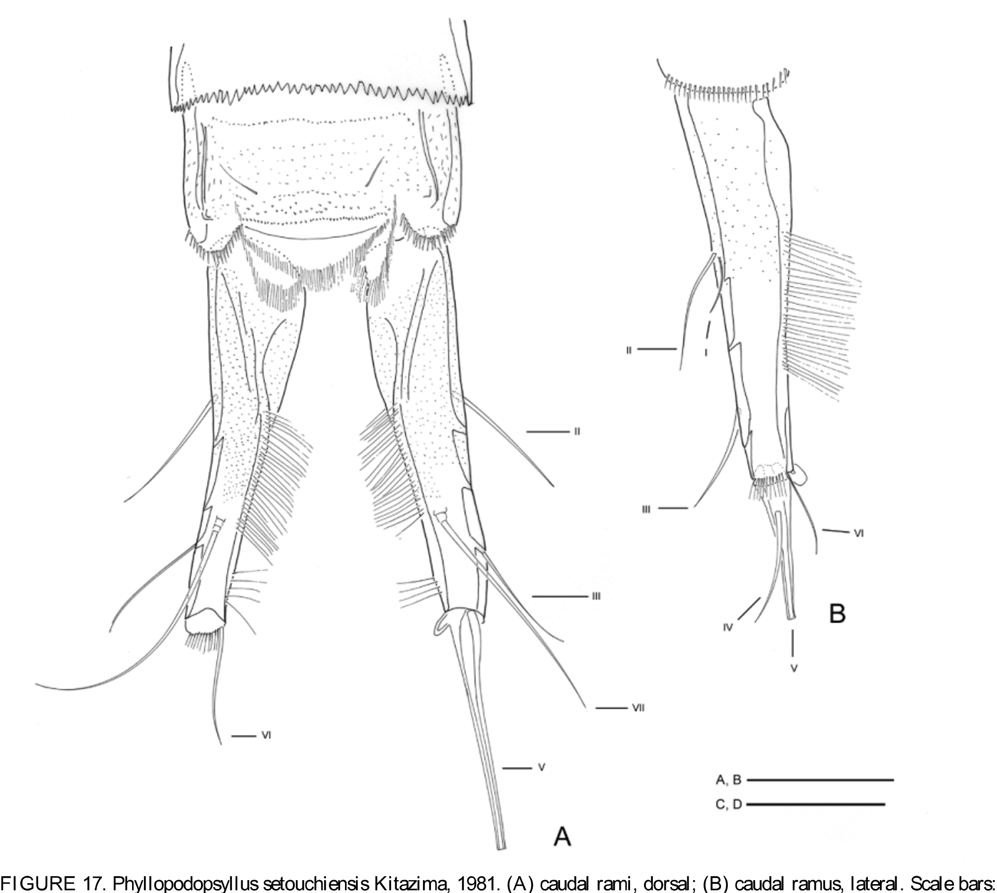
( Figs. 17 View FIGURE 17 & 18 View FIGURE 18 )
Material examined. São Sebastião, Guaecá Beach, Itaçucê ( 23º50’00”S 15º26’62”W): One female, 0 3 Mar. 2005; 3 females, 2 males, 1 copepodite and 5 nauplii, 18 May 2005; 2 females and 2 males, 0 8 Sep. 2005, in the intertidal zone; A. Pepato coll.
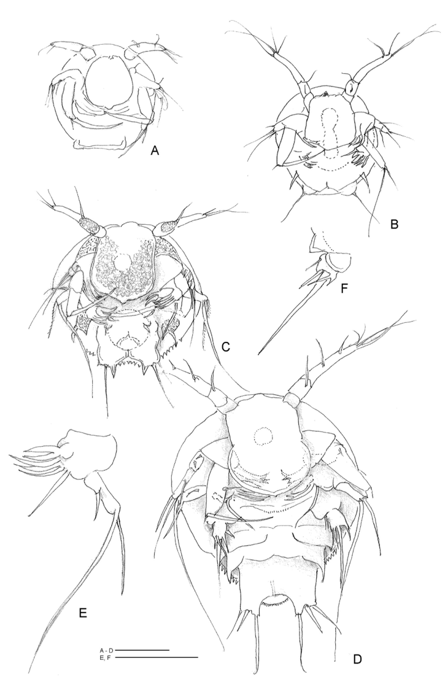
Description of naupliar stages. The nauplii of P. setouchiensis were briefly figured and described by Kitazima (1985). The Brazilian nauplii ( Figs 18 View FIGURE 18 A–F) are like the Japanese nauplii. We add a few details: NI ( Fig. 18 View FIGURE 18 A) prae-eclosion, 98 µm long, dorsal shield almost round, without ornaments. Labrum more or less round, extending from between antennule insertions to about middle area of ventral body wall. Antennular segmentation not very clear, with one seta proximally and three setae distally. Antenna with exopod 1-segmented bearing three setae; endopod 1-segmented with an inner marginal seta and two terminal setae, one hook-like, another, very small, inserted at the basis of the bigger one. Mandible, not clearly visible, with 1-segmented exopod, bearing two long terminal setae, inner one about twice the length of the other one.
NII ( Fig. 18 View FIGURE 18 B) body round, about the same size as NI, differs from NI as follows: last antennular segment thinner Antennal basis: bears a curved process, terminally bifid, turned towards the labrum, added to the other already present. Exopod 1- or 2-segmented with two setae on first and three setae on second segment. Mandible with endopod bearing two fat pointed setae and three thin setules on basis and a seta on coxa. Maxillule anlage present in the form of a strong pointed seta situated laterally on each side of the ventral body wall. On each side of the anal indentation, there is a small rounded process limiting the distal edge of the anal region. The rounded processes carry a terminal seta each.
NIII ( Fig. 18 View FIGURE 18 C): Body 132 µm long, covered by a dorsal shield finely serrate marginally. Body divided by ventral suture line into frontal and back ventral body wall. Antennules with 3 segments, not always clearly delimited. First two segments very short, third segment twice the length or longer than the two first summed together. First segment glabrous; second segment has 1–2 setae; third segment bears 2 terminal, and a lateral, preterminal seta. Antenna: exopod 2- or 3-segmented: first segment with two setae second with three. Mandible: as in NII. Maxillule: a lateral lobe below ventral suture line bearing a pointed process posteriorly and an outer curved pointed spine. Between the two maxillule lobes the ventral body wall protrudes from the suture line backwards with a square shape bearing anteriorly and laterally two little round processes, with a setule each, and posteriorly, medially, the anal area indentation. This area shows posteriorly, on each side, a minute pointed process, a small spine and a seta.
NVI ( Fig. 18 View FIGURE 18 D) differs from NIII as follows: body length 199 µm, last antennular segment with six setae, antennal coxa basis bears a well-developed masticatory process, a 3-segmented exopod, with two setae on first segment, one seta on second, and two setae on last segment (in Fig. 18 View FIGURE 18 D represented on left side of nauplius). Mandible ( Fig. 18 View FIGURE 18 E) basis with a thin blade-like process directed medially; exopod with a short and a very long bipinnate seta; endopod with 2 blade-like (scissor-shaped) processes and three setae. Maxillule ( Fig. 18 View FIGURE 18 F) 2- segmented, with 5 spines on distal segment. Anlagen or primordia of maxillae and maxillipeds represented by lateral pointed processes Posterior region of the nauplius divided into 2 almost square pointed terminal processes, bearing 3 setae each (the inner seta, the longest) and the dorsal shield or scutum with denticles along the posterior margin and the anal operculum with a fine setulose margin.
Remarks. Phyllopodopsyllus setouchiensis is a widespread and variable species. Kitazima (1981) called attention to its variability showing the drawings of the holotype female P4 compared to the same appendage of the paratype collected in the same region. The Brazilian specimens are characterized by thinner furcal rami ( Figs 17 View FIGURE 17 A– B) and seta V insertion on the caudal rami is slightly different from the Japanese specimens. Described from the Inland Sea of Japan (Kitazima 1981) it was also collected in Costa Rican waters, both in the Caribbean Sea and in the Pacific Ocean (Mielke 1989, 1992). According to this author “the populations of Japan, Hawai ( Kunz 1984 ) and Costa Rica belong to one and the same species”. Following Mielke’s (1992) reasonings, we also consider the Brazilian specimens as belonging to the same species in spite of these slight differences. Brazilian specimens were identified as P. setouchiensis because the leg setal formulae, the genital structures and the nauplii (represented by Kitazima 1981, 1985) were the same in Japanese and Brazilian species.
No known copyright restrictions apply. See Agosti, D., Egloff, W., 2009. Taxonomic information exchange and copyright: the Plazi approach. BMC Research Notes 2009, 2:53 for further explanation.
|
Kingdom |
|
|
Phylum |
|
|
Class |
|
|
Order |
|
|
Family |
|
|
Genus |