
Phyllopodopsyllus iuanamai, Björnberg, Tagea & Kihara, Terue C., 2013
|
publication ID |
https://doi.org/10.11646/zootaxa.3718.6.1 |
|
publication LSID |
lsid:zoobank.org:pub:4ED0B842-AFD9-486F-A01F-01B633972B4A |
|
DOI |
https://doi.org/10.5281/zenodo.6157988 |
|
persistent identifier |
https://treatment.plazi.org/id/03BA87BB-FFD4-FFBF-35C7-ACE1FD4DFE66 |
|
treatment provided by |
Plazi |
|
scientific name |
Phyllopodopsyllus iuanamai |
| status |
sp. nov. |
Phyllopodopsyllus iuanamai sp. nov.
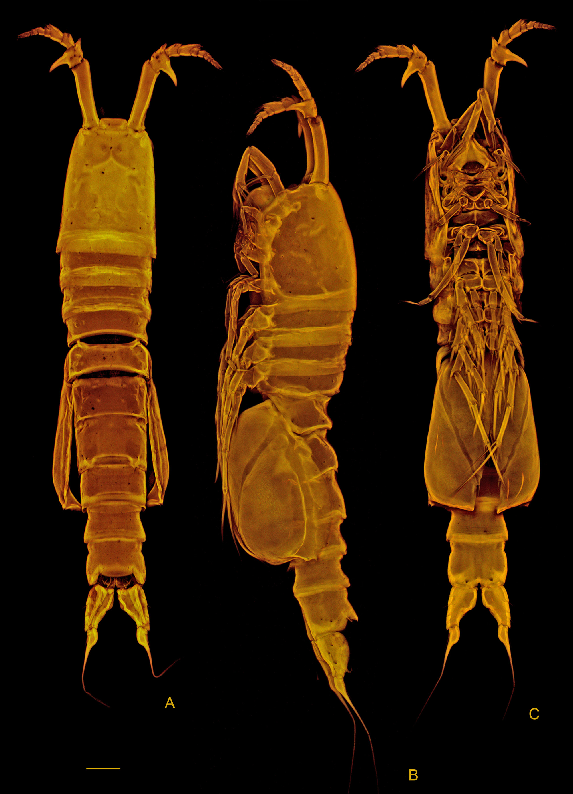
( Figs. 1–10 View FIGURE 1 View FIGURE 2 View FIGURE 3 View FIGURE 4 View FIGURE 5 View FIGURE 6 View FIGURE 7 View FIGURE 8 View FIGURE 9 View FIGURE 10 )
Type material. One female, holotype (MZUSP 28031) and 15 paratype females; 9 paratype males; 6 copepodites and 7 nauplii paratypes (MZUSP 28030). Collection sites: São Sebastião, Guaecá Beach, Itaçucê ( 23º50’00’’S 15º26’62’’W), Pepato, A. coll. São Sebastião Is., Parcel da Praia Grande ( 23º51’30’’S 45º25’00’’W), Oliveira, J. M. coll.
Type locality. Itaçucê, Guaecá Beach, São Sebastião ( 23º50’00’’S 45º26’62’’W).
Diagnosis. A Phyllopodopsyllus with paired dorsal spines on the urosomites; with hook-like processes on the second segment of the antennule; anal operculum margin fringed with a row of very fine setules, leg 4 endopod 2- segmented; female caudal ramus 1.4 times as long as anal somite and strongly dilated proximally; male caudal ramus 3.5 times as long as anal somite.
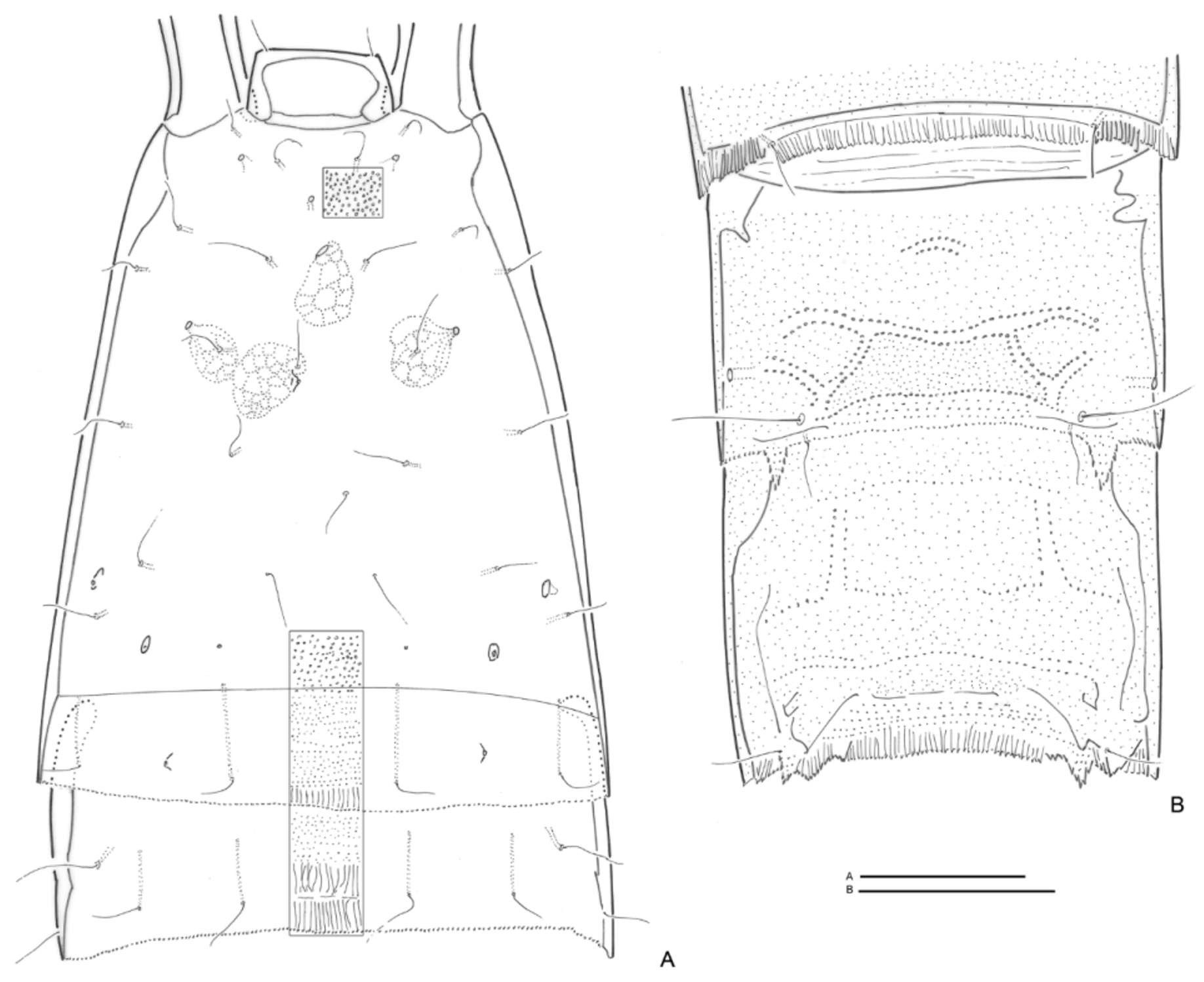
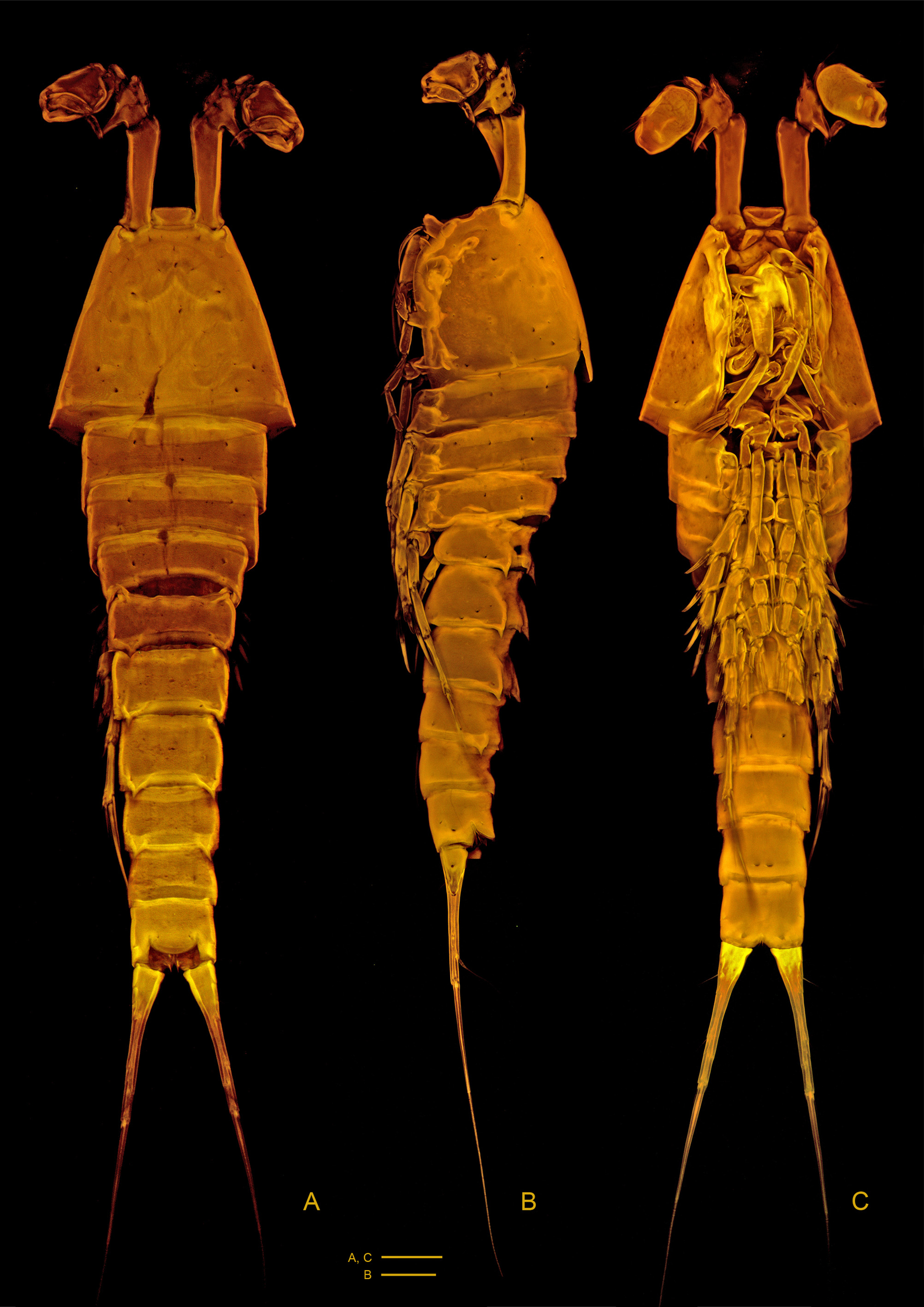
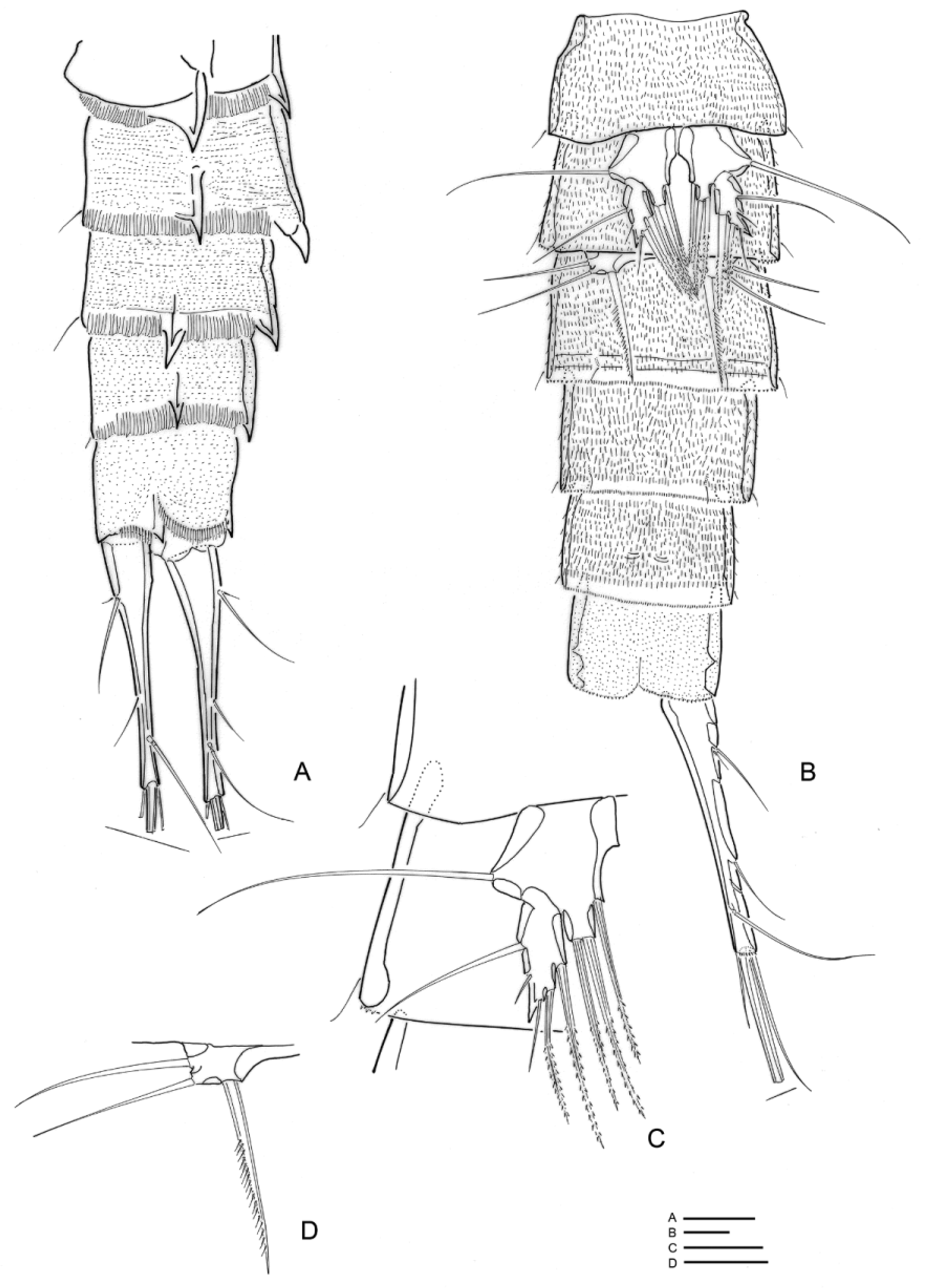
Description of adults. Female ( Figs 1 View FIGURE 1 A–C): Total body length from 770–950 µm (n = 6, X = 860 µm). Color when alive light brown. Dorsal red spot (“eye”) anteriorly. Body with 10 somites. Dorsal surface of cephalosome and first free somite as in Figure 2 View FIGURE 2 A. Pedigerous somites with 2 long sensilla dorsally, one on each lateral distal site. Numerous minute dots either following the striae or dispersed irregularly dorsally and laterally ( Fig. 2 View FIGURE 2 A). Posterior margin of somites ornamented with close packed transparent setules forming a hyaline fringe all around the segment. Epimeral lappets short, rounded. Somite bearing P5 and double genital somite with weak (somite bearing P5) or strong (double genital somite) pairs of dorsal spiniform processes ( Fig. 2 View FIGURE 2 B). Ventral margin below brood pouch (P5) with patch of setules. Genital double somite with suture visible dorsally, laterally and ventrally ( Figs 1 View FIGURE 1 A–B, 3C). Anal operculum ( Figs 2 View FIGURE 2 B, 3A) finely setulose and with a lateral small sensillum on each side.
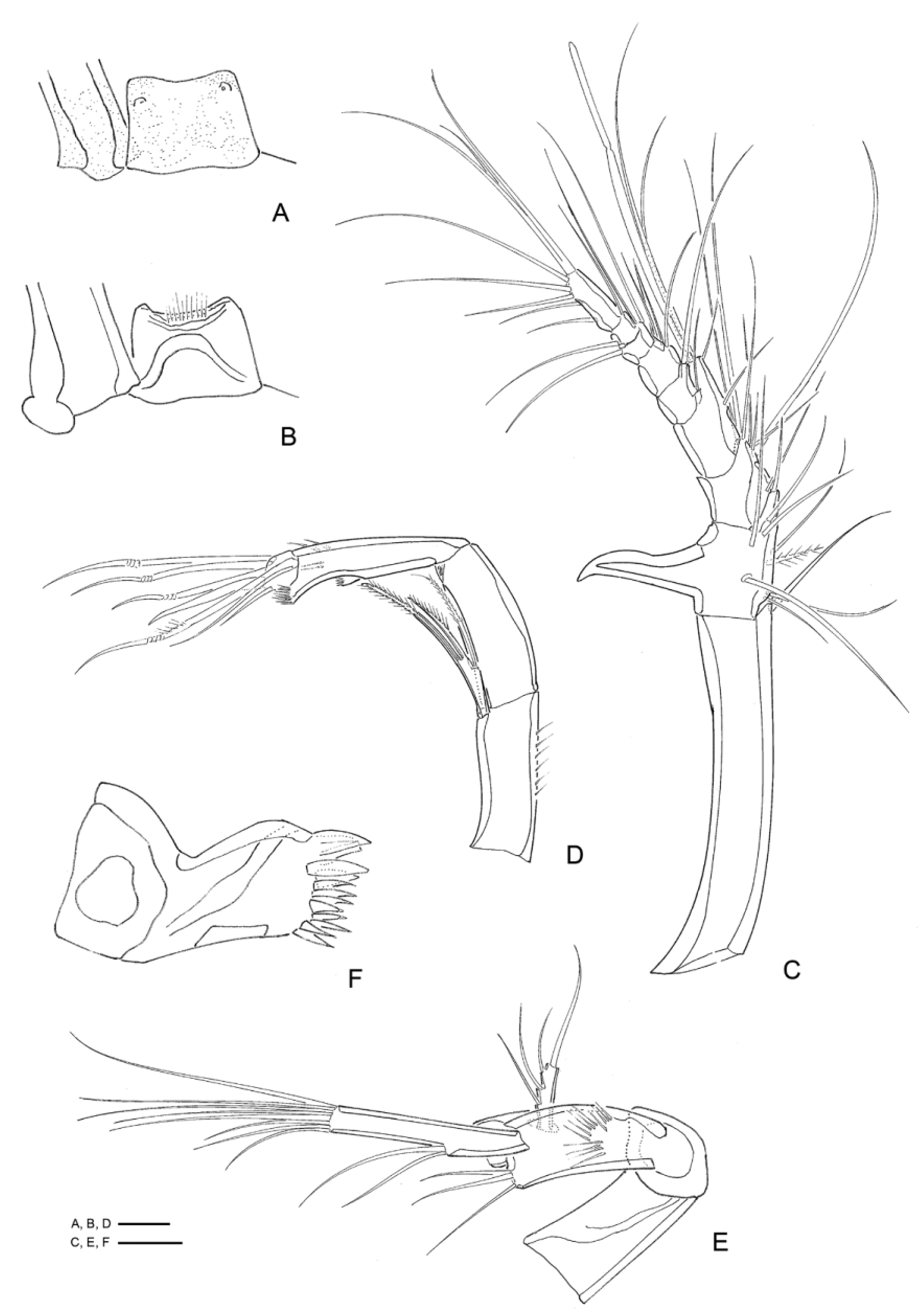
Caudal rami ( Figs 3 View FIGURE 3 A–C) pear shaped and with a dorsal ridge, inner margins developed into very thin lateral transparent expansions, with setulose ridge on dorsal and full of setulose striae on the inner side. Pores scattered along outer margin of the rami. Each ramus with 7 setae ( Figs 3 View FIGURE 3 A–C), seta V long and proximally globular.
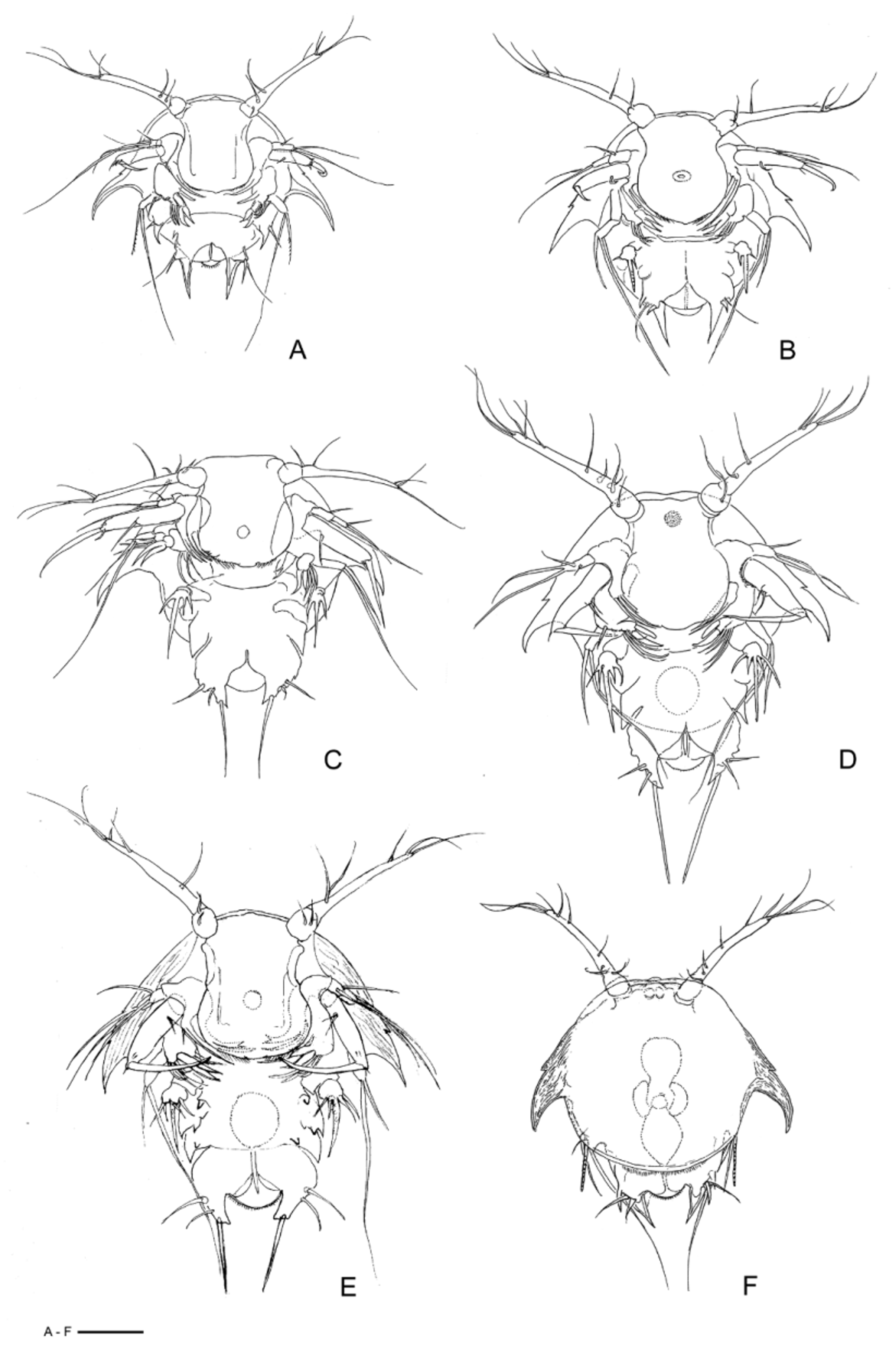
Rostrum ( Figs 2 View FIGURE 2 A, 4A–B): flat, defined at base, with two frontal sensilla and with setules along the ventral distal margin.
Antennule 9-segmented ( Fig. 4 View FIGURE 4 C). Segment-1 about as long as all following segments summed together. Segment-2 with prominent pointed outer hook about twice as long as supporting segment. Setal formula: 1, 9, 8, 4+1 ae, 2, 4, 2, 2, 7+1 ae.
Antenna ( Fig. 4 View FIGURE 4 D) biramous. Basis unarmed. Exopod 1-segmented with 3 elements: a bipinnate short, a bipinnate long seta and a unipinnate middle seta. Endopod 2-segmented, segment-1 unarmed, segment-2 with 7 setae (4 geniculated), 2 small setae laterally, pre-terminal spinules and 2 spinulose frills, one median and one preterminal.
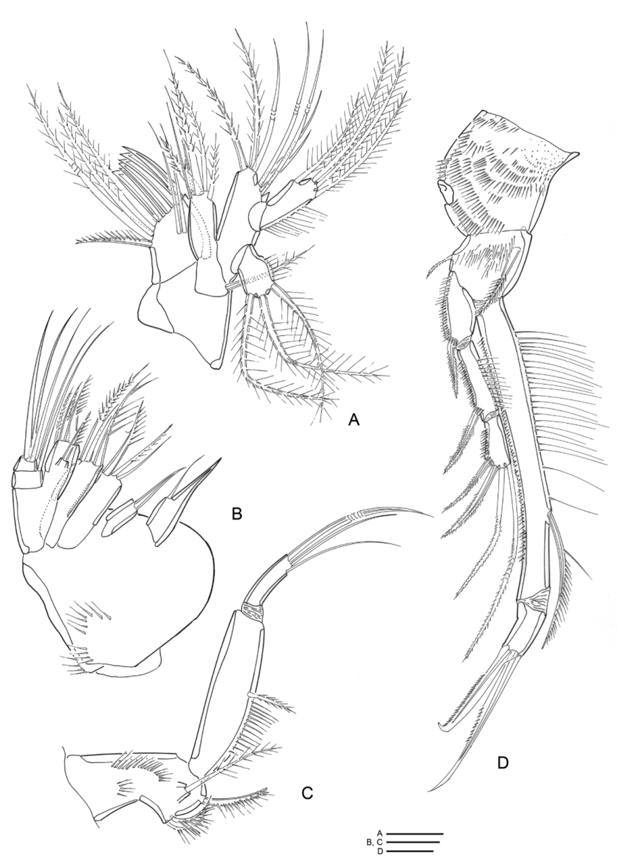
Mandible ( Figs 4 View FIGURE 4 E–F) with well-developed coxal gnathobase with 3 main tooth-like processes and a row of spines. Basis prominent, bearing 3 terminal setae and a tuft of setules on medial face. Exopod 1-segmented with 4 setae, 3 lateral and 1 apical. Endopod with 2 medial and 8 distal setae.
Maxillule ( Fig. 5 View FIGURE 5 A) with well-developed praecoxal arthrite bearing 10 distal elements, and 2 surface setae. Coxa with 6 setae on endite and with 1 long seta on exite. Basis with 7 setae. Endopod and exopod 1-segmented, with 4 and 3 setae respectively.
Maxilla ( Fig. 5 View FIGURE 5 B) with 4 syncoxal endites bearing 2, 1, 3, 3 setae on proximal to distal segment, respectively. Basis armed with 2 spines and 2 setae. Endopod 2-segmented, proximal segment with 2, distal segment with 3 setae.
Maxilliped ( Fig. 5 View FIGURE 5 C) prehensile. Syncoxa with 3 bipinnate setae. Basis with 1 seta, and a row of spinules along inner margin. Endopod 1-segmented with long geniculated claw and 2 slender setae.
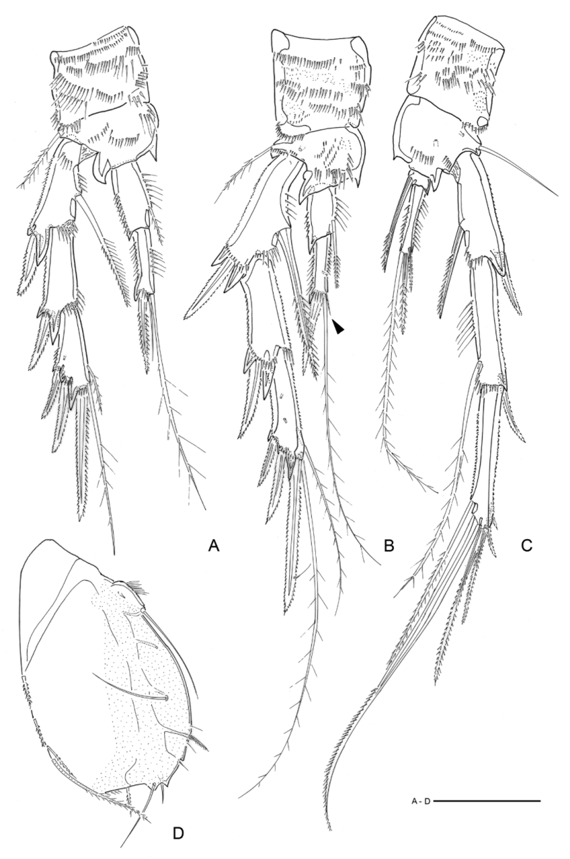
Leg 1 ( Fig. 5 View FIGURE 5 D) biramous. Coxa ornamented with rows of spinules. Basis with tufts of spinules, bearing 1 small seta on distal outer corner and 1 bipinnate spine on proximal inner margin. Exopodal segments with small spinules along outer margin and exp-2 with setules along inner margin. Enp-1 6.3 times longer than enp-2, with small spinules along outer margin and long setules along inner margin.
Legs 2–4 biramous ( Figs 6 View FIGURE 6 A–C). Coxa ornamented with rows of spinules. Basis with rows of spinules, bearing one seta on outer corner, and spiniform projection on distal rim between exopod and endopod and on distal inner corner. Exopod 3-segmented. Endopod 2-segmented. Inner and outer margins of exopods and endopods of P2–4 ornamented with setules and spinules, spiniform projections on distal outer corner of segments of P2–3 and on first segments of P4 exopod and endopod. Armature formula as follows:
Leg 5 ( Fig. 6 View FIGURE 6 D) large and foliaceous. Outer margin from proximal to distal with row of setules, 1 proximal slender seta (remnant of outer setophore), 1 seta inserted medially directed to inwards, 1 pinnate spine, 1 seta, 1 pointed process, 2 setae, 1 pointed projection and 1 bipinnate seta. Distal area with fine setules. Outer margin with 4 bipinnate setae.
Genital field as in Fig. 3 View FIGURE 3 C.
Leg 6 ( Fig. 3 View FIGURE 3 C) represented by 3 setae on each side of genital pore.
Male: Total body length from 600–722 µm long (n = 6, X = 678 µm). Body cylindrical, 10 somites ( Figs 7 View FIGURE 7 A– C). Prosome tergites with rounded epimeral lappets and hyaline margin. Urosomites with very prominent paired pointed processes ( Figs 7 View FIGURE 7 A–B, 8A) dorsally, except on anal somite. Sensilla covering the whole body laterally and dorsally, except on wide prosome hyaline margin ( Figs 8 View FIGURE 8 A–C).
Caudal rami fusiform, about 8 times as long as wide, 2.5 times as long as anal somite, with proximal third 3 times wider than distal third and armed with 7 setae. Seta V well developed and remaining setae as in figures 8A– B.
Rostrum, antenna, mandible, maxillule, maxilla and maxilliped similar to those of female.
Antennule sub-chirocer. 6-segmented ( Fig. 9 View FIGURE 9 A). Segment 1: 3 times as long as segment 2; segment 2 with spiniform process. Setal formula: 1, 12, 10, 11+1 ae, 1, 11+1 ae.
Leg 2 exopod as in female. Endopod ( Fig. 9 View FIGURE 9 C) first segment with naked seta and setules along inner margin; second segment slender, with setules along inner and outer margin and 3 apical elements: 2 naked setae and 1 outermost setae strong and basally fused to the segment.
Leg 3 ( Fig. 9 View FIGURE 9 D) exopod-1–2 as in female, exopod-3 with reduced setae. Endopod first segment with naked seta and setules along inner margin; second segment with setules along inner and outer margin and 2 apical elements.
Leg 4 ( Fig. 9 View FIGURE 9 E) exopod as in female. Endopod first segment with delicate unipinnate seta; second segment with 2 apical elements.
Leg 5 ( Fig. 8 View FIGURE 8 C), with distinct basoendopod carrying outer seta and 3 elements; exopod tapering distally and with 5 elements: 2 inner subdistal, 1 apical and 2 outer ones.
Leg 6 ( Fig. 8 View FIGURE 8 D) a process bearing 3 setae (innermost strong and unipinnate).
Description of naupliar stages. The naupliar stages obtained were NII–VI.
NII ( Fig. 10 View FIGURE 10 A): body 132 µm long, roundish, dorsal shield or scutum with two lateral, pointed acute processes and two posterior pointed acute processes limiting a deep round indentation area surrounding the anal region ventrally. The body is divided into cephalic or front area and hind body by a transversal suture. A small round area with setulose edge protrudes beyond the cephalic shield of the nauplius between the two acute posterior processes. A small lateral process next to each of the acute posterior processes carries two setae. Labrum (posteriorly rounded) is wide and reaches the middle of the body. Two minute pointed processes are present in the ventral body wall laterally below the transversal suture. Antennules uniramous, 3-segmented with first segment very short unarmed, second segment bears one ventral seta, third segment very long and thin, with 7 setae. Antenna: coxa (probably with masticatory process not seen), basis with two long spines turned medially, 1-segmented endopodite and 1-segmented exopodite. Endopodite carries a stout claw terminally extending medially and a little seta proximally. The insertion point of the claw is surrounded by a row of setules. The exopodite has a lateral outer seta and two long terminal setae. Mandible with coxa, basis, 1-segmented endopod and exopod. Basis carries a fingerlike process turned medially. Exopodite with 2 long setae terminally and one short lateral seta proximally. Endopodite 2-segmented with first segment carrying two small setae and second segment with a scissor-like structure composed of two foliaceous or spatulate pointed setae. Maxillule: Anlage present in the form of a pointed lateral process bearing a little lateral outer spine.
NIII ( Fig. 10 View FIGURE 10 B): Differs from NII as follows: 144 µm long. A1 with 4–5 instead of 7 setae (their number varies in the same specimen and from nauplius to nauplius), two additional small round lateral lobes on ventral body wall in place of the two minute posterior pointed processes below the transversal suture. Antennal exopod with two segments (one seta on first and two setae on second segment). Mandible endopod with additional seta on first segment. Maxillule anlage with changes, three setae appear instead of one. The middle seta is longer and stouter than the other two. Masticatory process not clearly seen.
NIV ( Fig. 10 View FIGURE 10 C): Differs from NIII as follows: 180 µm long from anterior cephalic region to end of posterior spines; cephalic shield much shorter; posterior pointed processes shorter and wider, bearing a longer inner caudal seta and two lateral small setae. Posterior edges or margins of hind body with deeper lateral indentations in ventral area forming a lateral lobe with pointed edge on each side; antennal exopod with one seta added terminally. Coxal maxilllular lobe extends transversally posterior to transversal suture. Maxillule divided into two segments, with addition of one more seta to distal segment.
NV ( Fig. 10 View FIGURE 10 D): Differs from NIV as follows: 250 µm long. A1 distal segment with 7–9 setae. A2 with one inner seta added medially on exopod, one seta added to endopod in A2. Terminal seta on posterior region longer
NVI ( Fig. 10 View FIGURE 10 E): Differs from NV as follows: 290 µm long. Terminal caudal seta shortened. Second segment of A1 with 2 setae. A2 with masticatory process clearly visible; number of setae on endopod diminishes to 3 or 4. Posterior edge or margin of the dorsal shield finely setulose. Marginal indentations on the ventral body wall separate the anlagen of the future maxilla, maxilliped, and legs.
Etymology. The species name is derived from the Tupi-Guarani (a South-American native language) iu (meaning spine, projection) and anama (meaning strong, stout).
Biological note. One of the most common and abundant species along São Sebastião coastal substrates. Reproduction most intense during spring time.


No known copyright restrictions apply. See Agosti, D., Egloff, W., 2009. Taxonomic information exchange and copyright: the Plazi approach. BMC Research Notes 2009, 2:53 for further explanation.
|
Kingdom |
|
|
Phylum |
|
|
Class |
|
|
Order |
|
|
Family |
|
|
Genus |