
Gromia, 2009
|
publication ID |
https://doi.org/ 10.1111/j.1096-3642.2009.00540.x |
|
DOI |
https://doi.org/10.5281/zenodo.5114962 |
|
persistent identifier |
https://treatment.plazi.org/id/03BA87C4-FF82-C176-FCE2-FEC670E4056C |
|
treatment provided by |
Carolina |
|
scientific name |
Gromia |
| status |
sp. nov. |
GROMIA MARMOREA View in CoL View at ENA SP. NOV. ( FIGS 2–4 View Figure 2 View Figure 3 View Figure 4 )
Diagnosis: species of Gromia with a rounded test, which is spherical, to droplet-shaped, to ovoid in shape; diameter 1.0– 3.4 mm, length: width ratio 0.6– 1.9. Overall colour in fresh specimens, greenish with silvery patches, giving marble-like mottling of wall; preserved specimens, brown. Single, prominent, mound-like oral capsule.
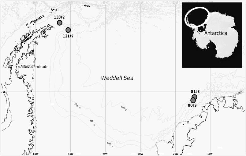
Type material and locality: The holotype and paratypes are from an EBS deployment at RV Polarstern station 133#2, 62°46.95 ′ S, 53°1.72 ′ W, 1584 m water depth, 16th March 2005 ( Table 1 View Table 1 ). They are deposited at the Research Institute and Natural History Museum Senckenberg, Frankfurt am Main. GoogleMaps The holotype is catalogued under reg. no. SMF XXVII 7398 . GoogleMaps The paratypes are catalogued under reg. no. SMF XXVII 7399 . The type specimens were extracted from> 300-Mm residue and are preserved in 4% formaldehyde solution buffered with borax GoogleMaps .
Additional material: Station 133#2: approximately 130 specimens.
Derivation of name: From the Latin marmoreus, meaning ‘like marble’, alluding to the marbled pattern of the test surface.
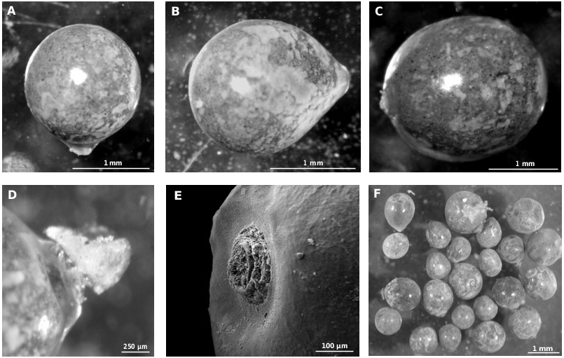
Overall appearance: The 130 specimens range in length from 1.0 to 3.4 mm (mean 1.9 ± 0.4 mm), and range from 0.8 to 3.7 mm in width (mean 1.7 ± 0.4 mm). The length: width ratio varies from 0.6 to 1.9 (mean 1.1 ± 1.0). They vary from nearly spherical, to droplet-shaped, to ovoid in lateral outline. Ovoid specimens are widest behind the midpoint, with a rounded posterior end, and a narrower anterior end terminating in the oral capsule. The degree to which the test narrows towards the aperture varies between specimens, giving rise to the different morphologies. Freshly collected, unfixed specimens were greenish-grey, sometimes with silvery, shiny patches giving the test a mottled appearance. After formalin fixation, specimens were predominantly brown in colour, although the mottling persisted ( Fig. 2 View Figure 2 ).
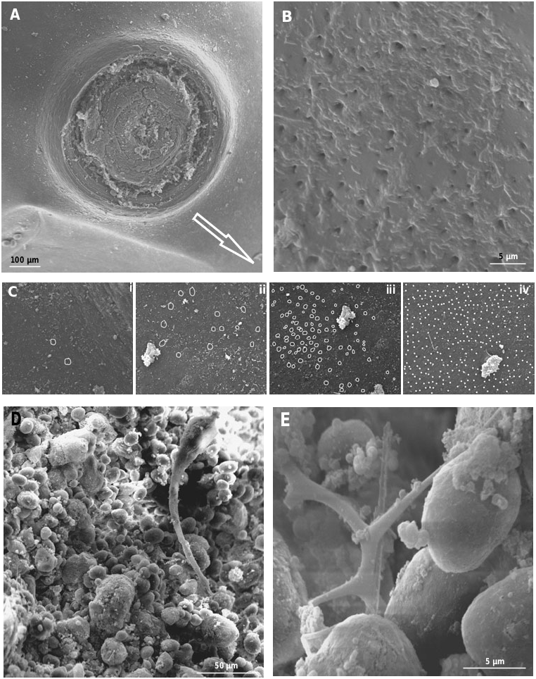
Oral capsule: The single oral capsule is a very distinct, golden brownish-coloured structure, and is roughly circular in plan view ( Figs 2E View Figure 2 , 4A View Figure 4 ). In lateral view it appears as a broad, mound-like structure, and is penetrated by a central canal ( Fig. 2D View Figure 2 ). In the ovoid- and droplet-shaped specimens, the oral capsule is located at the narrower end of the test ( Fig. 2B View Figure 2 ). The height of the oral capsule (i.e. the distance it protrudes from the test surface) ranges from 100 to 600 Mm (mean 140 ± 100 Mm, N = 129), and the diameter ranges from 200 to 800 Mm (mean 400 ± 100 Mm, N = 129). In 18 specimens, a flaccid extension (up to a maximum length of 200 Mm), composed of organic material, protrudes from the central canal through the aperture opening ( Fig. 2D View Figure 2 ).
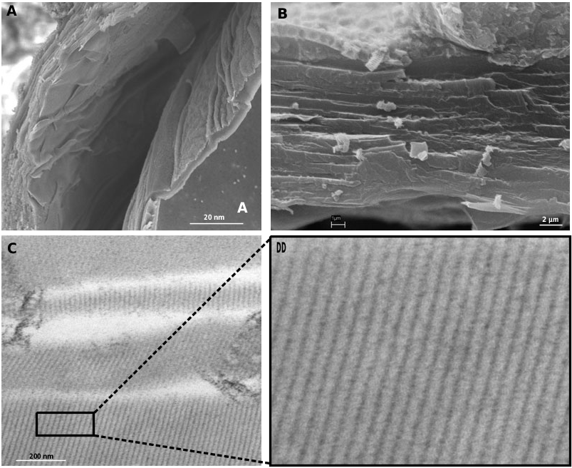
Test wall: The test is delicate and tears easily. The organic wall appears featureless and smooth when viewed under a binocular microscope. It is usually partially translucent, sometimes with a milky appearance, and displays a characteristic marble-like pattern ( Fig. 2A–C View Figure 2 ). Viewed using SEM, the wall is composed of multiple layers, and each layer is a fraction of a micron thick ( Fig. 3A, B View Figure 3 ); TEM revealed the existence of an inner layer of honeycomb membranes ( Fig. 3C, D View Figure 3 ). In cross section, the membranes appear as a series of very regular lines. The pores form openings on the test surface, ranging in diameter from ~0.3 to 3.1 Mm (N = 14; only clearly defined pores were measured; Fig. 4B View Figure 4 ). They occur across the entire surface, but are usually less frequent around the aperture. In the illustrated specimen, their density increases from about one pore per 10 Mm 2, within 40 Mm of the aperture, to about 12 pores per 10 Mm 2, at ~250 Mm from the aperture ( Fig. 4C, i View Figure 4 –iv). Concurrently, the pore diameter tends to increase from ~0.3 Mm close to the oral capsule to about 3.1 Mm at a distance of ~250 Mm from the capsule. However, the density of pores also varies between specimens. In one of the four individuals investigated by SEM, the pores were smaller and less frequent than in the other specimens, and did not display the decrease in size described above.
Test contents: The test contents are visible through the translucent test wall, and consist mainly of a densely packed mass of small, oval, and brownish stercomata ( Fig. 4D View Figure 4 ). These range from 6 to 22 Mm (N = 140) in length, and from 3.7 to 20 Mm in width (N = 140), and are characterized by a very smooth surface. Additional particles visible in SEM include possible mineral grains and sponge spicules ( Fig. 4E View Figure 4 ).
Distribution: Powell Basin, east of the tip of the Antarctic Peninsula, 1584-m depth ( Fig. 1 View Figure 1 ).
Remarks: Gromia marmorea sp. nov. is by far the most abundant gromiid in the ANDEEP-III material. The new species encompasses a range of shapes, from spherical, to ovoid, to droplet-shaped, which are also exhibited by G. oviformis ( Jepps, 1926) . However, it differs from the latter in the mottled appearance of the test wall and the dark-greenish, rather than lightbrownish, colour of fresh, live specimens. In addition, the organic test wall of G. marmorea sp. nov. is very delicate, and tears easily. Gromia marmorea sp. nov. ranges in length from 1.0 to 3.4 mm, and is therefore larger than G. pyriformis (<1-mm long; Gooday & Bowser, 2005) and smaller than G. schulzei (8–9-mm long; Schulze, 1875), as well as being smaller than G. sphaerica (maximum length 38 mm; Gooday et al., 2000). Like most other gromiids, this new species has a single, large oral capsule rather than many small capsules scattered across the test, as in G. sphaerica ( Gooday et al., 2000) . The capsule is a prominent, relatively low, broad, mound-like structure, and is larger (200–800 Mm in diameter) than in other Weddell Sea species. The test is perforated by numerous pores that have a minimum diameter of 0.3 Mm (300 nm), compared with 73 nm in G. pyriformis (Gooday & Bowser, 2005) .
| RV |
Collection of Leptospira Strains |
| SMF |
Forschungsinstitut und Natur-Museum Senckenberg |
No known copyright restrictions apply. See Agosti, D., Egloff, W., 2009. Taxonomic information exchange and copyright: the Plazi approach. BMC Research Notes 2009, 2:53 for further explanation.