
Fulvius attenboroughi Masłowski, Taszakowski & Wolski, 2023
|
publication ID |
https://doi.org/10.11646/zootaxa.5330.1.3 |
|
publication LSID |
lsid:zoobank.org:pub:D9311153-69E4-4688-8409-08511EFE09CA |
|
DOI |
https://doi.org/10.5281/zenodo.8249152 |
|
persistent identifier |
https://treatment.plazi.org/id/03BB87CD-126B-FFBB-FF6D-FF40FE4DC50C |
|
treatment provided by |
Plazi |
|
scientific name |
Fulvius attenboroughi Masłowski, Taszakowski & Wolski |
| status |
sp. nov. |
Fulvius attenboroughi Masłowski, Taszakowski & Wolski sp. nov.
( Figs 1 View FIGURE 1 , 2 View FIGURE 2 )
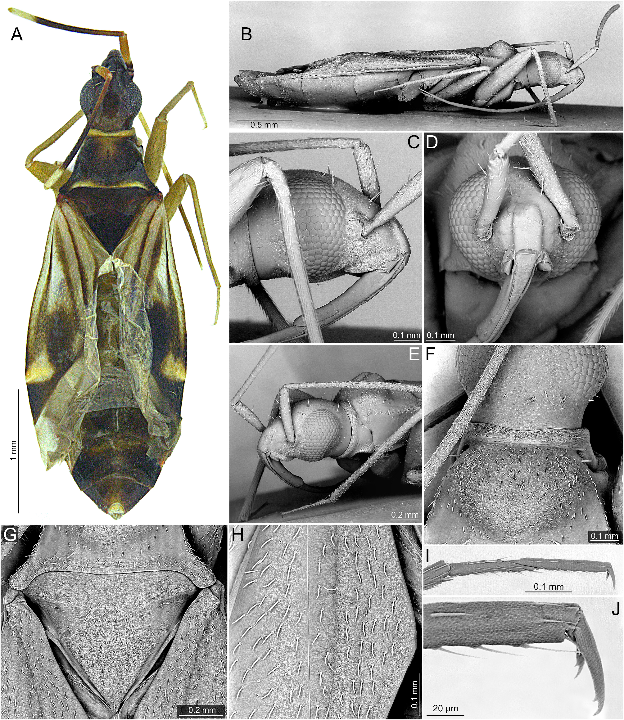
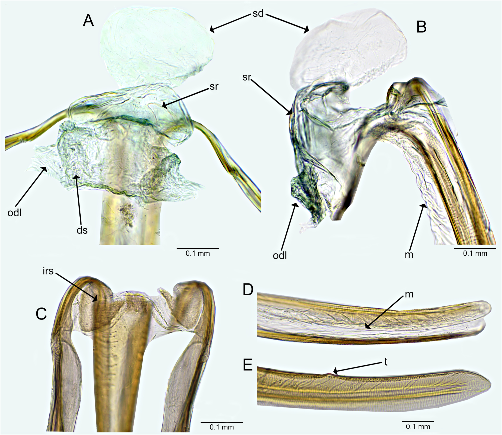
Diagnosis. The new species belongs to the anthocoroides -group (see discussion below) and can be distinguished by combination of the following set of characters: vertex convex medially, sloping onto posterior margin of vertex and frons ( Figs 1A, C, E View FIGURE 1 ); pronotum mostly black with contrastingly yellow collar and posterior margin ( Fig. 1A View FIGURE 1 ); anterior lobe of pronotum with calli distinctly upraised ( Figs 1B, E, F View FIGURE 1 ); scutellum entirely black ( Fig. 1A View FIGURE 1 ); hemelytron mostly yellow with large brown stripes and patches ( Fig. 1A View FIGURE 1 ); fore and middle legs entirely yellow ( Fig. 1A View FIGURE 1 ); sclerotized rings paired, subellipsoid, situated laterally on genital chamber ( Fig. 2A View FIGURE 2 ); dorsal sac of genital chamber membranous, situated laterally ( Fig. 2A View FIGURE 2 ); interramal sclerite of the posterior wall subtriangular ( Fig. 2C View FIGURE 2 ), apices of gonapophyses 8 and 9 obtuse ( Figs 2D, E View FIGURE 2 ).
Description. Female. Macropterous. COLORATION. Head. Mostly black, weakly paler, dark brown posteriorly ( Fig. 1A View FIGURE 1 ); antennomere I mostly pale gold, basal part narrowly black; antennomere II dark brown on basal one third, remainder of antennomere black except for apical, relatively broad, pale yellow annulation; antennomere III (glued to separate card attached to specimen) black; antennomere IV missing ( Fig. 1A View FIGURE 1 ); labium dark brown on basal two thirds, remainder of segment yellow; segment II entirely yellow; segments III and IV dark brown. Thorax. Pronotum. Mostly black; collar and posterior margin yellow; calli with dark yellowish patch anteriorly ( Fig. 1A View FIGURE 1 ). Mesoscutum and scutellum. Black ( Fig. 1A View FIGURE 1 ). Thoracic pleura. Dark brown ( Fig. 1A View FIGURE 1 ). Hemelytron. Basal one third yellow with brown, longitudinal stripes, two on clavus (one along medial part and other along inner margin), other two on corium (one along claval suture, other along inner margin of embolium) and one along outer margin of embolium; apical one third dark brown except for relatively large yellow patch on apex of embolium and exocorium; clavus dark brown ( Fig. 1A View FIGURE 1 ). Legs. Yellow ( Fig. 1A View FIGURE 1 ); forecoxae with narrow dark brown annulation basally. Abdomen. Mostly dark brown, weakly tinged with dark yellow. TEXTURE AND VESTITURE. Head, pronotum, thoracic pleura, mesoscutum and scutellum with net-like sculpturation based on microsetae ( Figs 1F, G View FIGURE 1 ); hemelytron shagreened ( Fig. 1H View FIGURE 1 ). Head. Vertex with transverse row of four long, erect, cylindrical apically serrate bristles, such bristles also present on anterior portions of vertex and frons; remainder of head with fine, short, semirecumbent setae ( Figs 1C–F View FIGURE 1 ); antennomeres I and II clothed with short, fine and recumbent setae, antennomere I with few long semirecumbent bristles, apical two thirds of antennomere II also with scale like, closely fitting short setae ( Figs 1C–F View FIGURE 1 ); antennomere III covered with long, dense, semirecumbent setae. Thorax. Pronotum. Collar with several fine, short, semirecumbent setae and with transverse row of four long, erect, cylindrical apically serrate bristles ( Figs 1E, F View FIGURE 1 ); remainder of pronotum with fine, short, semirecumbent, moderately dense setae, each anterior angle with single erect, stiff bristle ( Figs 1E, F View FIGURE 1 ). Mesoscutum and scutellum. Covered with sparse setae similar to those present on pronotum ( Fig. 1G View FIGURE 1 ). Thoracic pleura. Glabrous. Hemelytron. Shagreened, covered with relatively dense, uniformly distributed, moderately long, fine, scale-like setae ( Figs 1G, H View FIGURE 1 ). Legs. Coxae with several fine, short setae; remainder of leg covered with moderately dense, short, semirecumbent setae. STRUCTURE. Body elongate oval ( Fig. 1A View FIGURE 1 ).
Head. Eyes somewhat removed from pronotal collar ( Fig. 1A View FIGURE 1 ); vertex convex medially, sloping onto posterior margin of vertex and frons ( Figs 1C, E View FIGURE 1 ); antennomeres I and II nearly cylindrical, antennomere I weakly narrowed basally, antennomere II weakly broadened toward apex ( Figs 1A, C, E View FIGURE 1 ); labium reaching hind coxae ( Fig. 1B View FIGURE 1 ); segment I subdivided near medial part ( Figs 1B, C View FIGURE 1 ); segment II subdivided near apex ( Fig. 1B View FIGURE 1 ). Thorax. Anterior lobe of pronotum strongly convex, large, occupying two thirds of pronotum ( Figs 1A, B, E View FIGURE 1 ); humeral angle thin and relatively long; posterior margin strongly concave ( Figs 1A, G View FIGURE 1 ). Mesoscutum and scutellum. Mesoscutum weakly convex; scutellum flat ( Fig. 1G View FIGURE 1 ). Thoracic pleura. Scent gland evaporative area of metepisternum narrow, situated posteriorly. Legs. Tarsus two segmented ( Fig. 1I View FIGURE 1 ); pretarsal claw with subapical tooth ( Fig. 1J View FIGURE 1 ). Abdomen. Female genitalia. Genital chamber membranous ( Figs 2A, B View FIGURE 2 ); sclerotized rings paired, relatively large, subellipsoid, situated laterally ( Fig. 2A View FIGURE 2 ); dorsal sac of genital chamber membranous, relatively broad, situated laterally ( Fig. 2A View FIGURE 2 ); lateral oviducts removed from each other, conflating into dorsal sac ( Fig. 2A View FIGURE 2 ); ovipositor thin; gonapophyses 8 connected by distinct membranous structure along entire length ( Figs 2B, D View FIGURE 2 ); apices of gonapophyses 8 and 9 obtuse, without any teeth apically ( Figs 2D, E View FIGURE 2 ); gonapophysis 9 dorsal margin with single tooth medially ( Fig. 2E View FIGURE 2 ).
Measurements. Female (n=1)
Body. Length, clypeus-apex of abdomen: 3.85; width: 1.28.
Head. Length: 0.63; width, including compound eyes: 0.53; interocular width: 0.25; eye dorsal width: 0.16; height in lateral view: 0.42.
Antenna. Length of antennomere I: 0.38; II: 1.07; III: 0,55; IV:–.
Labium. Total length: 1.95; length of segment I: 0.50; II: 0.53; III: 0.55; IV: 0.38.
Pronotum. Mesal pronotal length: 0.47; lateral pronotal margin length: 0.58; posterior pronotal maximal width (straight): 0.88.
Scutellum. Anterior width: 0.46; length (mesoscutum excluded): 0.41, length (mesoscutum included): 0.63.
Hemelytron. Claval commissure: 0.84; corium length: 1.67; outer cuneal margin length (straight): 0.37; cuneus width: 0.42; cell length: 0.56; cell width: 0.18.
Material examined. Holotype ( ♀): ’NW MADAGASCAR, 23.-26.i.2015 \ Ankarafantsika N.P., 71 m, \ near Ampinjoroa village , \ circuit “Tour du lac”, \ S 16°18'13,0''; E 46°48'49.1'' \ M. Trýzna leg. ( MMBC) ( Fig. 8A View FIGURE 8 ). GoogleMaps
Biology. Unknown. Single known specimen was beaten from vegetation.
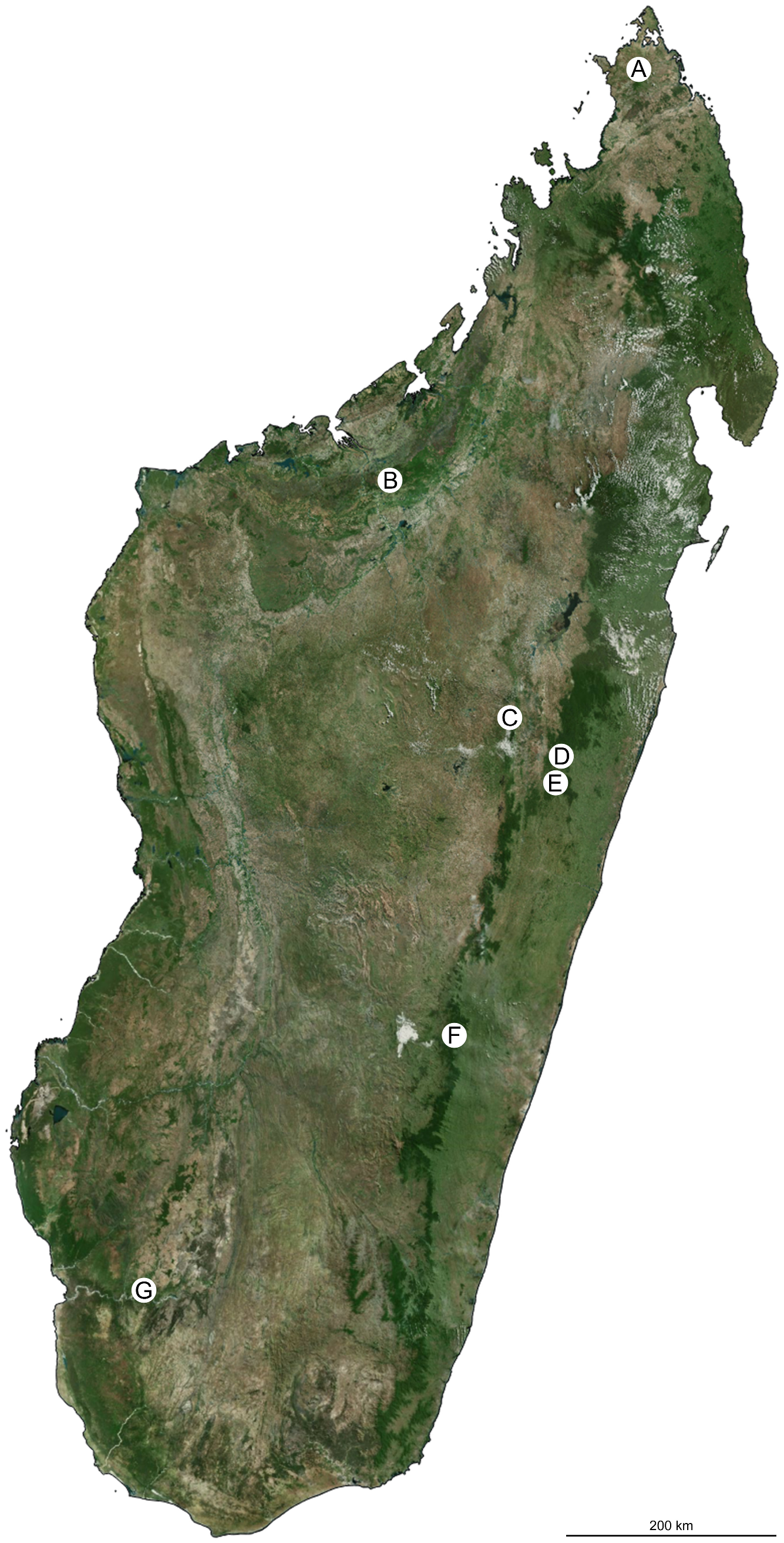
Distribution. North-west Madagascar ( Fig. 7B View FIGURE 7 ), Ankarafantsika National Park, seasonal forest.
Etymology. The new species is named in honor of Sir David Attenborough, whose outstanding documentaries have sparked a love for nature in many people.
Remarks. Fulvius attenboroughi sp. nov. is classified as a member of the anthocoroides -group as characterized by Gorczyca (2002a), Sadowska-Woda et al. (2008), and Wolski et al. (2018) based on the following characters: head, pronotum, thoracic pleura, mesoscutum and scutellum with net-like sculpturation based on microsetae ( Figs 1F, G View FIGURE 1 ) ( Wolski et al. 2018: figs 41–43; Gorczyca et al. 2020: figs 1A, C), second tarsomere not subdivided medially, subapical tooth present ( Fig. 1J View FIGURE 1 ) ( Wolski et al. 2018: figs 45–46; Gorczyca et al. 2020: figs 3A, B); membranous structure between the gonapophyses 8 present ( Figs 2B, D View FIGURE 2 ) ( Gorczyca et al. 2020; figs 5C, D). The new species is most similar to Fulvius carayoni Pluot-Sigwalt & Chérot, 2013 , F. kajae Gorczyca, 2000 , F. kerzhneri Gorczyca, 2000 , and F. webbi Gorczyca, 2000 in sharing the yellow stripes along hemelytra ( Fig. 1A View FIGURE 1 ; Pluot-Sigwalt & Chérot, 2013). It can, however, be easily distinguished by the convex vertex ( Figs 1A, C, E View FIGURE 1 ), eyes slightly removed from the pronotal collar ( Fig. 1A View FIGURE 1 ), the upraised pronotal calli ( Figs 1A, B, E View FIGURE 1 ), and the broader yellow stripes along hemelytra ( Fig. 1A View FIGURE 1 ). Within the abovementioned, possibly closely related group of species, F. attenboroughi sp. nov. is most similar to F. kerzhneri in sharing the pale yellow pronotal collar and posterior margin of pronotum and unicolored scutellum ( Figs 1A View FIGURE 1 , 3A View FIGURE 3 ).

| MMBC |
Moravske Muzeum [Moravian Museum] |
No known copyright restrictions apply. See Agosti, D., Egloff, W., 2009. Taxonomic information exchange and copyright: the Plazi approach. BMC Research Notes 2009, 2:53 for further explanation.