
Schmitzofulvius bigibber Gorczyca, 1998
|
publication ID |
https://doi.org/ 10.11646/zootaxa.5330.1.3 |
|
publication LSID |
lsid:zoobank.org:pub:D9311153-69E4-4688-8409-08511EFE09CA |
|
DOI |
https://doi.org/10.5281/zenodo.8249166 |
|
persistent identifier |
https://treatment.plazi.org/id/03BB87CD-126E-FFB7-FF6D-FC21FB72C3E1 |
|
treatment provided by |
Plazi |
|
scientific name |
Schmitzofulvius bigibber Gorczyca, 1998 |
| status |
|
Schmitzofulvius bigibber Gorczyca, 1998 View in CoL
( Figs 4 View FIGURE 4 , 5 View FIGURE 5 )
Schmitzofulvius bigibber Gorczyca (1998a) View in CoL : 10, figs. 1–6 (new species), 2000: 154, figs 2, 45 (redescription).
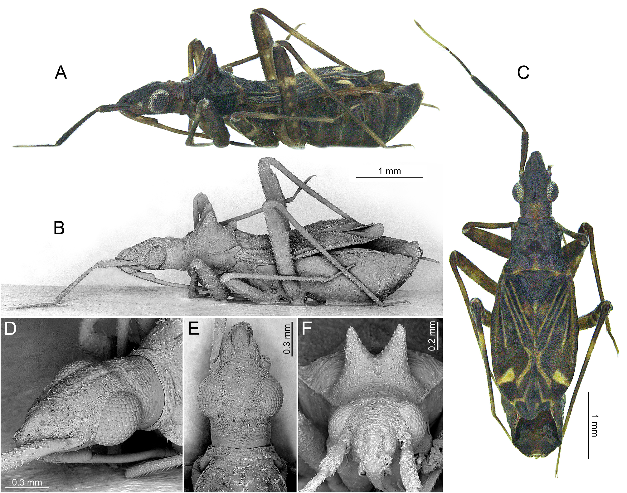
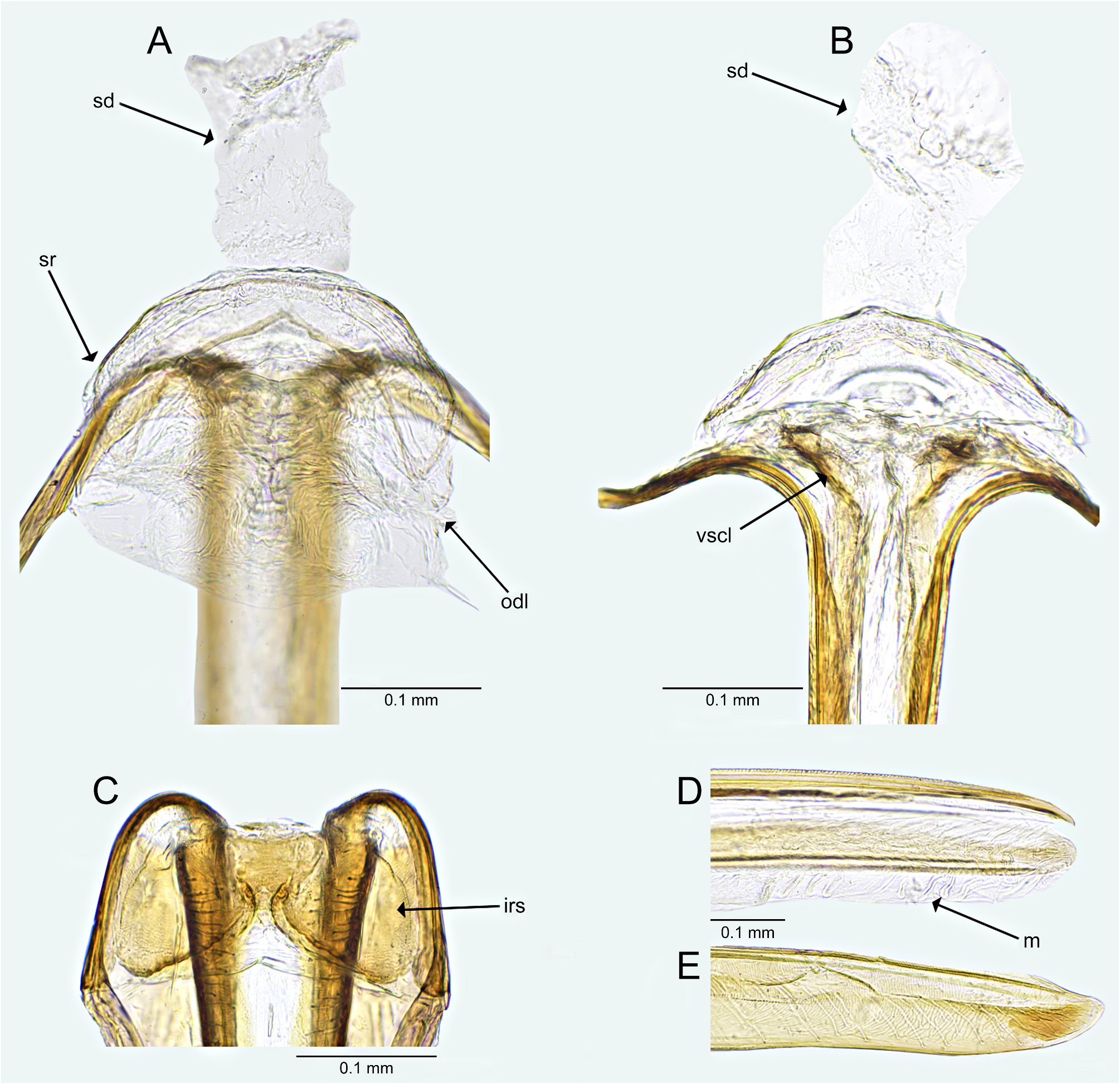
Diagnosis. Recognized by the following set of features: body brown to dark brown with large yellow areas ( Figs 4A, C View FIGURE 4 ); head with yellowish tinge behind eyes ( Figs. 4C View FIGURE 4 ); antennomere IV mostly yellow ( Figs 4C View FIGURE 4 ); posterior lobe of pronotum with yellowish tinges ( Figs 4C View FIGURE 4 ); pronotal calli projecting upward ( Figs 4A, B, F View FIGURE 4 ); sclerotized ring of dorsal labiate plate unpaired, relatively large, its lateral margin reaching margin of genital chamber; lateral oviducts situated close to each other, conflating into dorsal sac ( Fig. 5A View FIGURE 5 ).
Redescription. Female. COLORATION. Dorsum dark brown with large yellow areas ( Fig. 4C View FIGURE 4 ). Head. Dark brown, sometimes with relatively large, yellowish tinge on surface behind eyes ( Figs 4A, C View FIGURE 4 ); antennae mostly dark brown; antennomere IV yellow, narrowly dark brown basally and apically ( Figs 4A, C View FIGURE 4 ); labial segments I and II yellow to dirty yellow, segments III and IV fuscous ( Figs 4A View FIGURE 4 ). Thorax. Mostly dark brown, sometimes pronotal collar with indistinct, relatively small, yellowish tinge medially, posterior margin with indistinct, narrow, yellowish tinge, sometimes on surface between calli, reaching middle of pronotum ( Figs 4A, C View FIGURE 4 ). Mesoscutum and scutellum. Dark brown, sometimes with small, yellowish spot apically ( Figs 4C View FIGURE 4 ). Thoracic pleura. Dark brown ( Fig. 4A View FIGURE 4 ). Hemelytron. Dark brown with distinct yellow patches and stripes ( Fig. 4C View FIGURE 4 ); clavus with relatively large yellow patch medially, vein yellow along entire length, basal two thirds of claval vein yellow ( Fig. 4C View FIGURE 4 ); endocorium with yellow, relatively large patch medially, medial fracture yellow along entire length; exocorium with yellowish stripe originating basally and reaching medial part of hemelytron ( Fig. 4C View FIGURE 4 ); R+M vein slightly tinged with yellow medially ( Fig. 4C View FIGURE 4 ); apex of embolium and exocorium with relatively large, yellow patch bordering base of cuneus ( Fig. 4C View FIGURE 4 ); cuneus dark brown ( Fig. 4C View FIGURE 4 ); membrane fuscous with yellow veins, sometimes somewhat tinged with yellow ( Fig. 4C View FIGURE 4 ). Legs. Coxae dark yellow; femora dark brown with indistinct yellowish tinge ( Figs 4A, C View FIGURE 4 ); tibiae dark brown with indistinct, dark yellow, broad annulations: one situated near base and other apically ( Figs 4A, C View FIGURE 4 ); tarsi dark yellow ( Figs 4A, C View FIGURE 4 ). Abdomen. Dark brown with indistinct yellow tinge ( Fig. 4A View FIGURE 4 ). STRUCTURE. Female genitalia. Genital chamber membranous, suboval ( Figs 5A, B View FIGURE 5 ); sclerotized rings merged, relatively large, lateral margins reaching margin of genital chamber, weakly rimmed, nearly crescent-like, anterior margin semicircular, posterior margin distinctly concave medially, V-shaped ( Fig. 5A View FIGURE 5 ); lateral oviducts situated close to each other; vulva surrounded by two irregular, relatively long sclerotizations ( Fig. 5B View FIGURE 5 ); ovipositor thin; gonapophyses 8 connected by distinct membranous structure along entire length ( Fig. 5D View FIGURE 5 ); apices of gonapophyses 8 and 9 obtuse, without any teeth ( Figs 5D, E View FIGURE 5 ). Male genitalia. See Gorczyca (1998: figs 5, 6).
Measurements. Female (n=3)
Body. Length, clypeus-apex of membrane: 4.37–4.83; width: 1.32–1.41.
Head. Length: 0.99–1.03; width, including compound eyes: 0.66–0.72; vertex width: 0.32–0.36; eye dorsal width: 0.17–0.19; height in lateral view: 0.47–0.50.
Antenna. Length of antennomere I: 0.63–0.65; II: 1.11–1.22; III: 0.51–0.53; IV: 0.74.
Labium. Total length: 2.68–2.74; length of segment I: 0.59–0.67; II: 0.78–0.82; III: 0.77–0.82; IV: 0.63–0.56.
Pronotum. Mesal pronotal length: 0.75–0.77; lateral pronotal margin length: 0.87–0.90; posterior pronotal maximal width (straight): 1.06–1.20.
Scutellum. Anterior width: 0.50–0.56; length (mesoscutum excluded): 0.48–0.53, length (mesoscutum included): 0.61–0.68.
Hemelytron. Claval commissure: 0.65–0.68; hemelytral length: 2.29–2.44; corium length: 1.77–1.87; outer cuneal margin length (straight): 0.39–0.47; cell length: 0.44; cell width: 0.13.
Hind leg. Femur length: 1.81–1.84; tibia length: 2.59–2.63; tarsus length: 0.52–0.53; length of tarsal segment I: 0.23/0.24; II: 0.38/0.40.
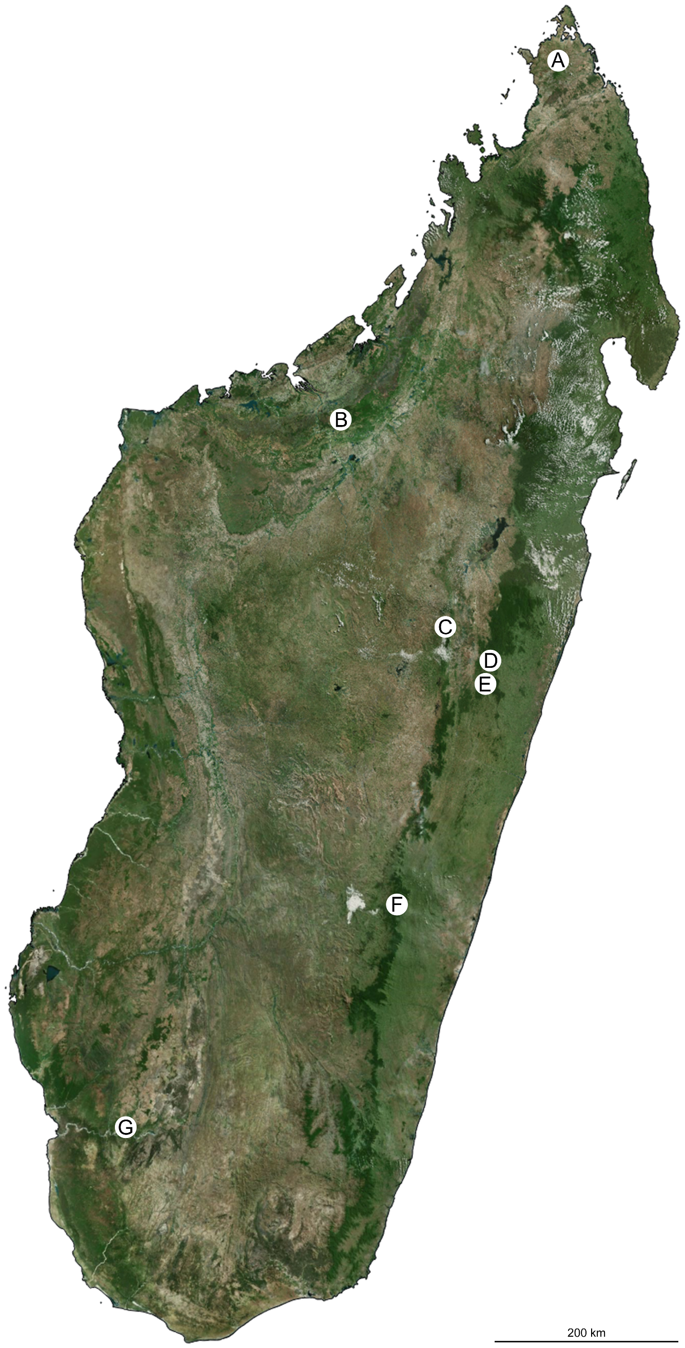
Material examined. ♀: ’ MADAGASCAR, 1000m, \ Ranomafana N.P., circuit \ Talatakely , 8.-12.i.2019 \ S21°15'47.2'' E47°25'20.7''; \ primary forest, M. Trýzna leg.’ ( MMBC) ( Fig. 8B View FIGURE 8 ) GoogleMaps ; ♀: ’ MADAGASCAR, 947m, \ Andasibe N.P., 22.-29.i.2019 \ Analamazaotra, Aventure circ. \ S18°56'40.6'' E48°25'26.7''; \ primary forest, M. Trýzna leg.’ ( MMBC) ( Fig. 8D View FIGURE 8 ) GoogleMaps ; 2♀♀: ’ MADAGASCAR, 1328m, \ Anjozorobe forest , circuit \ Bambou, 15.- 19.i.2019 \ S18°24'54.9'' E47°56'45.2'' \ primary forest, M. Trýzna leg.’ ( MMBC) ( Figs. 8E, F View FIGURE 8 ) GoogleMaps .
Biology. Unknown; specimens were collected in the rainforest during beating of shrubs and branches of trees ( Figs 8B, D–F View FIGURE 8 ).
Distribution. Central east Madagascar ( Fig. 7C, E, F View FIGURE 7 ), evergreen rainy forests.
Remarks.Based on the results of our examination of the available material including the holotype of Schmitzofulvius niger Gorczyca, 2000 (very damaged specimen), and the literature data, we herein present a short comparison of both Schmitzofulvius species. S. bigibber can be distinguished from S. niger primarily by the hemelytron with regular yellowish lines along the medial fracture and claval and R+M veins. Additionally, both species can be distinguished by the position of the pronotal calli (projecting upwards in S. bigibber and directed backward in S. niger ) and the different shape of the parameres ( Gorczyca 1998a: figs 5, 6; Gorczyca 1998c: figs 12, 13).

| MMBC |
Moravske Muzeum [Moravian Museum] |
No known copyright restrictions apply. See Agosti, D., Egloff, W., 2009. Taxonomic information exchange and copyright: the Plazi approach. BMC Research Notes 2009, 2:53 for further explanation.
|
Kingdom |
|
|
Phylum |
|
|
Class |
|
|
Order |
|
|
Family |
|
|
SubFamily |
Cylapinae |
|
Tribe |
Fulviini |
|
Genus |
Schmitzofulvius bigibber Gorczyca, 1998
| Masłowski, Adrian, Baňař, Petr, Carapezza, Attilio, Chérot, Frédéric, Jindra, Zdeněk, Taszakowski, Artur & Wolski, Andrzej 2023 |
Schmitzofulvius bigibber
| Gorczyca, J. 1998: 10 |