
Microlaimus validus, Gagarin, Vladimir G. & Tu, Nguyen Dinh, 2014
|
publication ID |
https://doi.org/ 10.11646/zootaxa.3856.3.4 |
|
publication LSID |
lsid:zoobank.org:pub:EED65F9D-B55B-4152-A9E1-36BD4E6F9E19 |
|
DOI |
https://doi.org/10.5281/zenodo.5678515 |
|
persistent identifier |
https://treatment.plazi.org/id/03BB87D1-FFC1-FFAF-FF4C-623EFF76FD6F |
|
treatment provided by |
Plazi |
|
scientific name |
Microlaimus validus |
| status |
sp. nov. |
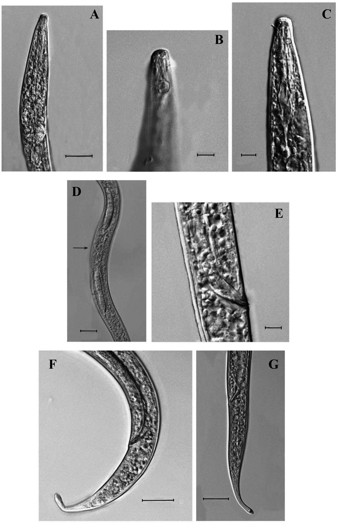
Microlaimus validus sp. n.
( Figs 3 View FIGURE 3 , 4 View FIGURE 4 ; Table 2)
Type material. Holotype male, slide reference 102/40, deposited in the RAS Helminthological Museum, Institute of Ecology and Evolution of Russian Academy of Sciences, Center for Parasitology (Moscow, Russia).
Paratypes. Three males and five females deposited in the collection of the department of Nematology of the Institute of Ecology and Biological Resource ( JEBR), Vietnam Academy of Science and Technology, 18 Hoang Viet Rd., Hanoi, Vietnam.
Measurements. Table 2.
ТABLE 2. Morphometric of Microlaimus validus sp. n.
Type habitat and locality. South China Sea, littoral zone of coastal Vietnam. Latitude: 21º25.712’– 21º27.382’ N; Longitude: 107º58.952’– 108º01.950’ E. Depth 1.5–3.0 m, sand, salinity 29.1–29.4 ‰. Collected on March 2010. Etymology. The specific epithet means “strong”, “valid”.
Description. Male. Body comparatively short and thin, brownish. Anterior body end strongly narrowed. Labial region 0.3 times as wide as body width at region of posterior pharynx end. Cuticle finely annulated, without dots and other punctuations 1.0–1.5 µm thick at mid-body. Cuticular pores and somatic setae not visible. Labial region high, well set off. Cephalic capsule not seen. Six inner labial sensillae papilliform. Six outer labial sensillae in the shape of short setae. Cephalic sensillae in the shape of thin and longer setae. Four cephalic setae about 25–33% of labial region width. Both rings of setae are arranged at a considerable distance from each other. Amphidial fovea round, relatively large (75–81% of corresponding body width) and located quite far from the anterior body end (1.6–2.0 labial region width). Cheilostoma with 12 longitudinal ribs; oesophastoma surrounded by pharyngeal tissue, its walls weakly sclerotized. Dorsal tooth small, badly developed; subventral teeth vague. Pharynx muscular, enlarged into almost spherical cardial bulb posteriorly. Lumen of terminal bulb dilated, its cuticle thickened and weakly sclerotized. Cardia small, triangular. Renette placed ventrally to the posterior part of cardial bulb. Excretory pore posterior to nerve ring.
Gonads diorchic; testes opposite and outstretched. Anterior testis situated to left of intestine, posterior testis to right of intestine. Two spicules equal in length, curved, 1.0–1.1 times as long as cloacal body diameter. Spicules proximally capitate and arcuate, distally acute. Gubernaculum lamellar, 8–10 µm long. Precloacal submedian supplements absent. Tail slender, elongate-conical, curved ventrally. Caudal glands incaudal, with terminal spinneret.
Female. Similar to male in general characteristics, but body thicker and tail comparatively longer. Structure of cuticle and anterior body end as in male. Labial region set off and high. Inner labial sensillae papilliform. Outer labial sensillae and cephalic sensillae in the shape of thin setae. Amphidial fovea round, located quite far from the anterior body end. Cheilostoma with longitudinal ribs. Oesophastoma weakly developed, its walls poorly sclerotized. Dorsal tooth small. Pharynx muscular, with spherical cardial bulb. Renette and excretory pore present.
Reproductive system didelphic, amphidelphic; ovaries homodromous, comparatively short. Anterior ovary situated to right side of intestine, posterior ovary to left side of intestine. Both uteri comparatively short, filled with numerous spermatozoa. Spermatheca not seen. Vagina short, extending to one quarter of the corresponding body diameter. Vulva a transverse slit, pre-equatorial. Vulva lips not sclerotized, not protruding outside the body contour. Tail elongate-conical, with caudal glands and terminal spinneret.
Diagnosis. Body short and thin (L = 562–683 µm, a = 24–34). Cuticle finely annulated. Labial region high, set off. Six inner labial papilliform sensillae; six outer labial sensillae and four cephalic sensillae in the shape of thin setae. Amphidial fovea relatively large, located 1.7–2.0 labial region width from anterior body end. Cheilostoma with 12 longitudinal ribs. Oesophastoma poorly developed, its wall weakly sclerotized. Dorsal tooth small; subventral teeth vague. Pharynx muscular, with sphaerical cardial bulb. Vulva a transverse slit, pre-equatorial. Ovaries paired outstretched. Vagina short. Spermatheca not observed. Testes paired, opposite. Precloacal supplements absent. Spicules equal in length, curved, 1.0–1.1 times as long as cloacal body diameter. Gubernaculum lamellar. Tail slender, elongate-conical.
Differential diagnosis. Microlaimus validus sp. n. belongs with the group of Microlaimus species having small body size (from 290 µm to 700 µm long) and finely annulated cuticle. The new species is morphologically similar to M. citrus Gerlach, 1959 and M. nanus Blome, 1982 . It differs from the former species in the more posterior location of the amphidial fovea from the anterior body end (at distance 0.7–2.0 labial region width vs 1.5 labial region width in M. citrus ) and absence of precloacal supplements in males ( Gerlach 1959). It differs from M. nanus in the shorter cephalic setae (1.5–2.0 µm long, 25–33% of labial region width vs 4 µm long, 50% of labial region width in M. nanus ) and shorter spicule length (15–16 µm long vs 18 µm long in males of M. nanus ) ( Blome 1982).

No known copyright restrictions apply. See Agosti, D., Egloff, W., 2009. Taxonomic information exchange and copyright: the Plazi approach. BMC Research Notes 2009, 2:53 for further explanation.