
Saccocalyx microhexactin, Gong, Lin, Li, Xinzheng & Qiu, Jian-Wen, 2015
|
publication ID |
https://doi.org/ 10.11646/zootaxa.4034.1.9 |
|
publication LSID |
lsid:zoobank.org:pub:7FDC215C-9722-41BB-B752-A9C38942CE82 |
|
DOI |
https://doi.org/10.5281/zenodo.5624233 |
|
persistent identifier |
https://treatment.plazi.org/id/03BB87FC-FFDC-5A2A-FF24-FE8E145D30DC |
|
treatment provided by |
Plazi |
|
scientific name |
Saccocalyx microhexactin |
| status |
sp. nov. |
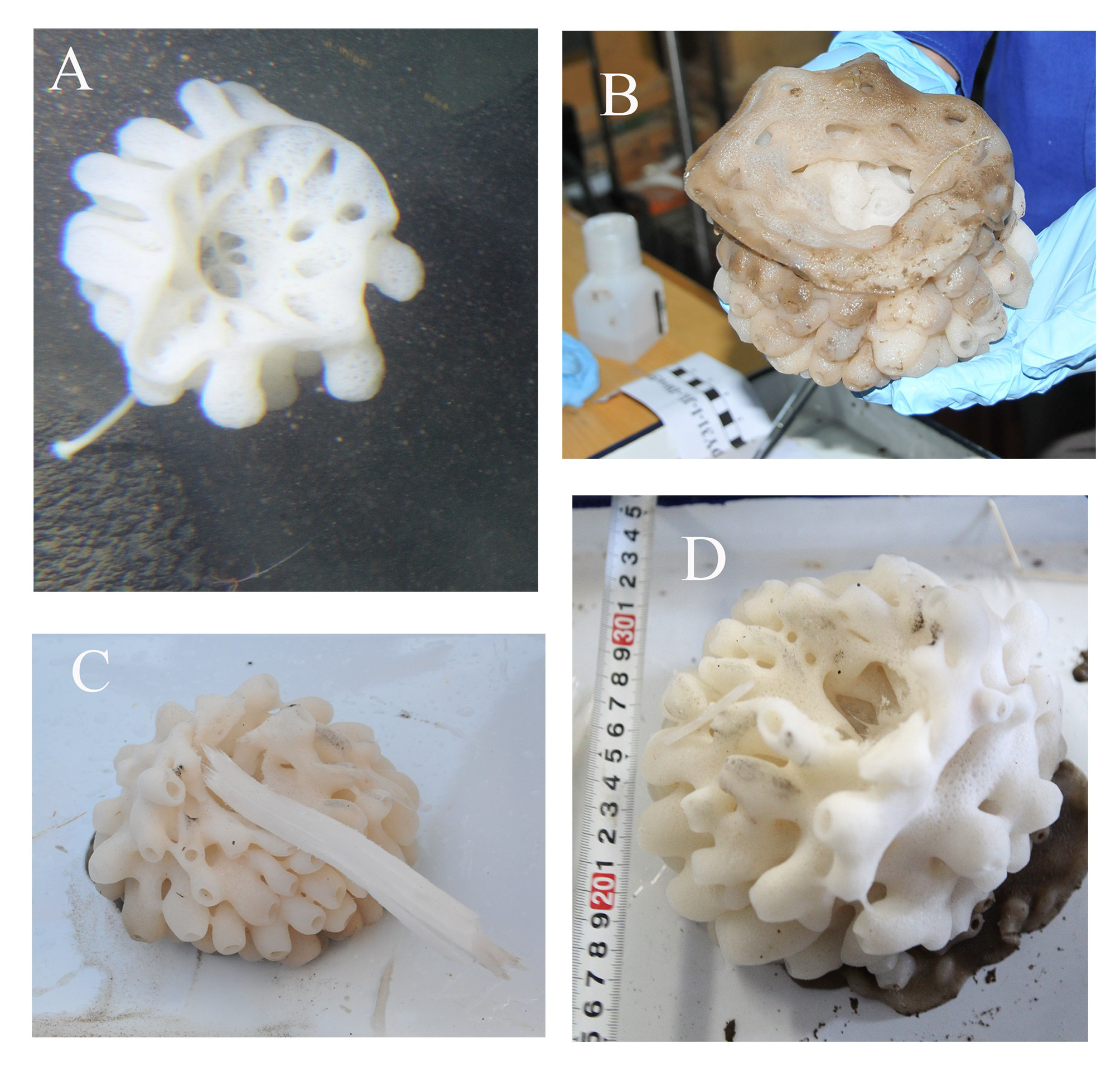
Saccocalyx microhexactin View in CoL sp. nov.
( Figures 3–4 View FIGURE 3 View FIGURE 4 )
Material examined. Holotype: MBM179994, South China Sea (17°33.95' N, 117°45.67' E), 5 July 2013, 3542 m depth, hard rock cliff above the mouth of an extinct volcano.
Description. Body is globular with several rows of radially arranged protrusions surrounding a central atrial cavity. The color is white. It looks like a beautiful flower with a long stalk growing on the cliff of a seamount ( Fig. 3 View FIGURE 3 A). Central atrial cavity is large, with a 100 mm diameter at the upper margin. Roughly 20 suboscula, 10–25 mm diameter open into the central atrial cavity. Radially arranged digitiform protrusions are up to 15 mm long and 10 mm in diameter. A lateral osculum with 5–10 mm diameter is present at the apex of each protrusion. In situ, the body is swollen, with protrusions pointing outward, like a blooming snow lotus herb. After being transported to deck, body with protrusions collapsed. Sponge attached to rock with a long tubular peduncle, 10 mm in diameter, 1 mm thick wall, and at least 250 mm in length.
Choanosomal skeleton solid ( Fig. 4 View FIGURE 4 O), with some spicules separated from skeleton only after digestion by concentrated nitric acid overnight. Spicules mainly contain hexactins and diactins. Pinular hexactins with rays having sparsely distributed short spines are present in dermalia and gastralia ( Fig. 4 View FIGURE 4 P), whereas diactins are present in the choanosome. Spirodiscohexasters are numerous and present in the whole body. Drepanocomes occur near dermal and gastral surfaces.
Spicules. Megascleres consist of diactins and hexactins. Diactins are smooth, 2120.3–3557.5/5.3–15.1 µm, usually with four tubercles in middle ( Fig. 4 View FIGURE 4 H). Dermalia and atrialia are similar in size and shape. Dermalia or atrialia ( Fig 4 View FIGURE 4 A) pinular rays ( Fig. 4 View FIGURE 4 I) are 245.8–450.6 µm long, tangential rays are 173.6–246.8 µm long, proximal rays are 188.2–585.8 µm long. Choanosomal hexactins (( Fig. 4 View FIGURE 4 O) are relatively less than diactins, they have short spines in terminal, rays are 126.5–355.8 µm. The skeleton of the peduncle ( Fig. 4 View FIGURE 4 N) is composed of diactines fused to each other by numerous synapticulae.
Microscleres consist of spirodiscohexasters, drepanocomes, plumicomes and microhexactins. Drepanocomes are in two types. Drepanocomes I ( Fig. 4 View FIGURE 4 B) 254.4–318.6 µm in diameter, with 6 clusters of 8 hook-like secondary rays ( Fig. 4 View FIGURE 4 J). Drepanocomes II ( Fig. 4 View FIGURE 4 C) 123.0–137.5 µm in diameter, with 6 clusters ( Fig. 4 View FIGURE 4 K) of 4–6 hook-like secondary rays. Structure of drepanocomes easy to destroy during digestion, and hard to isolate completed ones. Spirodiscohexasters ( Fig. 4 View FIGURE 4 D) 112.4–149.8 µm in diameter, formed by 6 spirally twisted clusters of roughly 8–12 terminal rays ending in discs ( Fig. 4 View FIGURE 4 L) with approximately 18 marginal teeth. Plumicomes ( Fig. 4 View FIGURE 4 E) have six shield-like primary termination, one on the top and one on the bottom, the other four are in the middle area. A single sheild ( Fig. 4 View FIGURE 4 M) have approximately 70 marginal sigma-like secondary rays. Each sigma-like secondary rays has prominent teeth on the inside curve. The diameter of Plumicomes is 35.8–58.5. Microhexactins ( Fig. 4 View FIGURE 4 F–G) with rays covered short and minute spines, rays are 21.4–70.3 µm long.
Etymology. “ mikros ”, Greek, small; “ hex ”, Greek, six; “ aktis ”, Greek, ray, light beam. Microhexactin is a kind of sponge microscleres. The specific name refers to the presence of microhexactins in the new species.
Remarks. Only two species of Saccocalyx are known in the world: Saccocalyx pedunculatus Schulze, 1896 and Saccocalyx careyi (Reiswig, 1999) . The main difference between the two species is that all pinular rays of S. careyi are clavate, while only part of the pinular rays of S. pedunculatus are clavate. However, since these spicules have a big variety in size and shape, and their ranges overlap, S. careyi was considered a doubtful species and could be a junior synonym of S. pedunculatus (Tabachnick, 2002) . Because there are no described microsclere structure details available for S. pedunculatus , we cannot tell what other differences between both species there may be. But our specimen has two distinctive characteristics which are different from the two known species: (a) presence of drepanocomes II, (b) presence of microhexactins. Both types of microscleres have not been found in the other two species of Saccocalyx . The new species has all pinular rays clavate as reported for S. careyi . However, there are other differences: S. microhexactin sp. nov. has less terminal discs in spirodiscohexasters, and more marginal sigma-like secondary rays in plumicomes than S. careyi .

No known copyright restrictions apply. See Agosti, D., Egloff, W., 2009. Taxonomic information exchange and copyright: the Plazi approach. BMC Research Notes 2009, 2:53 for further explanation.