
Izecksohniella puri Sperber et al. 2003
|
publication ID |
https://doi.org/ 10.5281/zenodo.191447 |
|
DOI |
https://doi.org/10.5281/zenodo.6218809 |
|
persistent identifier |
https://treatment.plazi.org/id/03BB87FE-1D31-FFFD-FF27-FF01C8BDF867 |
|
treatment provided by |
Plazi |
|
scientific name |
Izecksohniella puri Sperber et al. 2003 |
| status |
|
Izecksohniella puri Sperber et al. 2003
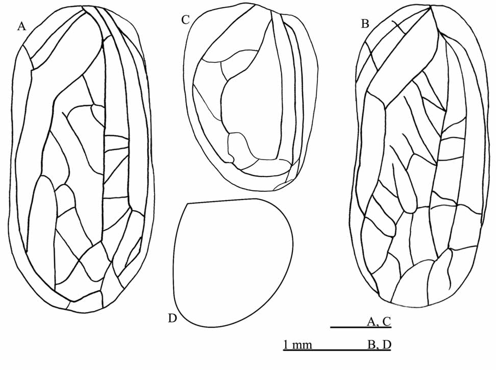
( Figs. 1 View FIGURE 1 C, 4 and 5)
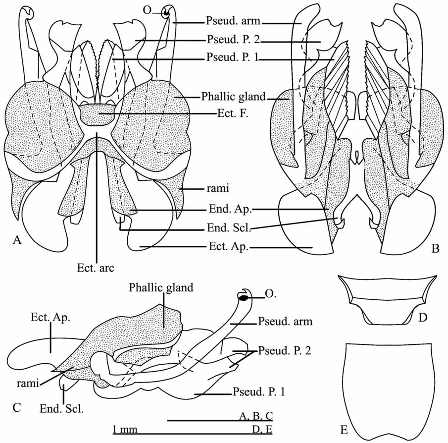
Diagnosis: Forewing as in figure 1C; (ii) head reddish, Male terminalia: pseudepiphallic arms short, with enlarged extremity slightly curved at apex ( Fig. 4 View FIGURE 4 A-B); pseudepiphallic paramere 2 formed by three dorsumapical lines that are parallel and teethed ( Fig. 4 View FIGURE 4 A), ventrally sclerotized with one small spine ( Fig. 4 View FIGURE 4 B); ectophallic fold ventral and anterior to the parameres, and dorsal and posterior to the endophallic sclerite ( Fig. 4 View FIGURE 4 B); ectophallic apodeme long and cylindrical ( Fig. 4 View FIGURE 4 A-C); endophallic sclerite heart-shaped ( Fig. 4 View FIGURE 4 B); female copulatory papilla funnel shaped; with apex membranous in dorsal and lateral side ( Figs. 5 View FIGURE 5 A-C); totally sclerotized in ventral side ( Figs. 5 View FIGURE 5 B).
Male. Terminalia: phallic gland present with secretor duct and small orifice present ( Figs. 4 View FIGURE 4 A-C). Pseudepiphallic arms short, with enlarged extremity slightly curved to apex ( Fig. 4 View FIGURE 4 A-B); pseudepiphallic paramere 2 formed by three dorsum-apical parallel lines teethed ( Fig. 4 View FIGURE 4 A); ventrally sclerotized with one small spine ( Fig. 4 View FIGURE 4 B). Pseudepiphallic paramere 1 large ( Fig. 4 View FIGURE 4 B). Ectophallic fold ventral and anterior to the parameres, and dorsal and posterior to the endophallic sclerite ( Fig. 4 View FIGURE 4 B); ectophallic apodeme long and cylindrical ( Fig. 4 View FIGURE 4 A-C); ectophallic arc as in figure 4A; endophallic sclerite heart-shaped ( Fig. 4 View FIGURE 4 B); rami short ( Fig. 4 View FIGURE 4 C). Supra-anal plate small ( Fig. 4 View FIGURE 4 D). Subgenital plate large, with rounded apex and not compressed laterally ( Fig. 4 View FIGURE 4 E).
Female. Terminalia: copulatory papilla funnel shaped, with apex membranous in dorsal and lateral side ( Figs. 5 View FIGURE 5 A and C); totally sclerotized in ventral side ( Fig. 5 View FIGURE 5 B). Median valve of ovipositor as in figure 5D. Spermatheca large ( Fig. 5 View FIGURE 5 E). Supra-anal plate rounded in the apex ( Fig. 5 View FIGURE 5 F). Subgenital plate quadrangular, laterally compressed ( Fig. 5 View FIGURE 5 G).
Material examined. Holotype; Five male and five female paratypes: Brazil, Minas Gerais, Viçosa. Mata da Biologia, 09.iv.2006. Sperber, CF leg.
Marliella Mews & Mól gen. nov. et sp. nov.
Type species: Marliella titai Mews & Mól sp. nov.
Etymology. The genus name is in honor to Marli Marta Mews.
Diagnosis. Head with three ocelli, forming an equilateral triangle. Male forewing reduced ( Fig. 1 View FIGURE 1 B); forewing without glandular hair, stridulatory vein and glandular border. Tympanum present in internal face of tibia I. Tibia III with four internal dorsal spurs (the first and fourth smaller than the second and third) and four external, (the first smaller than the others, that are similar). Three internal apical spurs, the first is half of the size of the others and three external apical spurs, first and third smaller than second. Metanotal gland absent. Male terminalia: with phallic glands; pseudepiphallic arms long, curved ventral-apically, fining gradually, with teeth on all extension ( Fig. 6 View FIGURE 6 A); pseudepiphallic paramere 2 cut ( Fig. 6 View FIGURE 6 A), teethed ventrally ( Fig. 6 View FIGURE 6 B); pseudepiphallic paramere 1 in “L” shaped, with small denticles on the internal side; female copulatory papilla sclerotized, conical shaped ( Fig. 7 View FIGURE 7 A-C).
Marliella titai Mews & Mól sp. nov. ( Figs.1 View FIGURE 1 B, 6 and 7)
Etymology. The specific name is in honor to the biologist Cândida Lahís Mews, nickname “Tita”, that collected this species for the first time.
Holotype: Brazil, Mato Grosso, Nova Xavantina, UNEMAT, Parque do Bacaba, Cerradão, Male holotype: 09.viii.2006. Mews, CL. leg. MZSP.
Diagnosis: Male forewing without glandular hair, stridulatory vein and glandular border ( Fig. 1 View FIGURE 1 B); Male terminalia: pseudepiphallic arms long, fining gradually, curved ventral-apically, with teeth on all extension ( Fig. 6 View FIGURE 6 A); pseudepiphallic paramere 2 cut ( Fig. 6 View FIGURE 6 A), teethed ventrally ( Fig. 6 View FIGURE 6 B); pseudepiphallic paramere 1 in “L” shaped, with small denticles on the internal side; female copulatory papilla sclerotized, conical shaped ( Fig. 7 View FIGURE 7 A-C); without specialized area.
Male. Head: evenly medium brown. Vertex dark brown with two light medium stripe. Fastigium dark brown; gena medium brown with dark brown patch below to the eyes. Clypeus, labrum medium brown, labial palpi light brown. Maxillary palpi: light brown of the first to the third segment, fourth segment medium brown, fifth segment dark brown, apex either narrow as the base, resembling a scythe. Antennae: medium brown, scape and pedicel medium brown; flagellum medium brown with white in some segments. Pronotum: medium brown, lateral lobes rectangular dark brown. Metanotum without glands, but with two broad humps on its anterior-lateral borders and two smaller callus-shaped ones between then. Forewings covering one abdominal tergite, medium brown, apical border pale yellow, with lateral field reduced; without glandular hairs, inflated border, stridulatory vein and specialized veins ( Fig. 1 View FIGURE 1 B). Abdominal tergites medium brown; abdominal sternites pale yellow. Legs with thick dark hair. Coxa and trochanter light brown with medium brawn marks. Femur I medium brown; femur II medium brown; femur III: proximal portion medium brown with transversal pale yellow stripe; distal portion dark brown. Tibia I medium brown; tibia II medium brown; tibia III medium brown, serrulated: 22 spines on inner and 24 on outer margin, among dorsal spurs. Tarsi I light brown with eight spines in outer dorsal margin and six spines in inner dorsal margin; Tarsi II light brown and III medium brown. Cerci light brown. Terminalia: Phallic gland, secretor duct and orifice present. ( Fig. 6 View FIGURE 6 A-C). Pseudepiphallic arms: the left crosses (raisin on) on the right. Pseudepiphallic arms long curved ventral-apically, fining gradually, with teeth on all extension ( Fig. 6 View FIGURE 6 A). Pseudepiphallic paramere 2 cut, membranous in dorsal side ( Fig. 6 View FIGURE 6 A); teethed ventrally ( Fig. 6 View FIGURE 6 B). Pseudepiphallic paramere 1 in “L” shaped, with small denticles on the internal side. Ectophallic fold reaching the base of parameres ( Fig. 6 View FIGURE 6 A, B). Ectophallic apodeme: base curved for the center of the genitalia, apex with two apical projections that reach the middle of the parameres, giving to the apodeme, with "H"-shaped. Endophallus small. Rami short ( Fig. 6 View FIGURE 6 B, C). Supra-anal plate medium brown on the base and dark brown apex, longer than wide ( Fig. 6 View FIGURE 6 D). Subgenital plate light brown on the base and medium brown on the apex, bilobed apically ( Fig. 6 View FIGURE 6 E).
Female. Coloration as adult male. Terminalia: copulatory papilla conical dorsum-ventrally ( Fig. 7 View FIGURE 7 A-C); base and apex membranous in dorsal side ( Fig. 7 View FIGURE 7 A), sclerotized in ventral side ( Fig. 7 View FIGURE 7 B), laterally cylindrical, continuing to distal extremity ( Fig. 7 View FIGURE 7 C). Median valve of ovipositor as in figure 7D. Supra-anal plate wider than long with rounded apex ( Fig. 7 View FIGURE 7 E). Subgenital plate narrow and convex, with pronounced medium depression ( Fig. 7 View FIGURE 7 F).
Measurements in mm. (Male (n=10) min. – max. values, followed by female (n=10) min. – max. values): body length: 5.40–5.65, 4.90–6.00; head width: 1.10–1.20, 1.15–1.30; intra-ocular distance: 0.60– 0.70, 0.60–1.20; pronotum length: 0.70–0.85, 0.90–1.30; pronotum width: 1.20–1.40, 1.40–1.60; femur III length: 4.50–4.85, 4.00–5.20; tibia III length: 4.80–5.20, 4.00–5.00; male wing length: 0.90–1.05; male wing width: 0.90–1.00; female ovipositor length: 0.70–2.10.
Material Examined. Holotype; five male and five female paratypes Brasil, Mato Grosso, Nova Xavantina. UNEMAT. Parque do Bacaba, Cerradão, 02-30.i.2008 Mews, CL leg; six male and five female paratypes: some local and collector, 02-20.iii.2008.


| MZSP |
Sao Paulo, Museu de Zoologia da Universidade de Sao Paulo |
No known copyright restrictions apply. See Agosti, D., Egloff, W., 2009. Taxonomic information exchange and copyright: the Plazi approach. BMC Research Notes 2009, 2:53 for further explanation.