
Postbursoplana donoraticensis, Meini, Gianluca, 2015
|
publication ID |
https://doi.org/ 10.11646/zootaxa.3947.3.9 |
|
publication LSID |
lsid:zoobank.org:pub:28501A93-1BBD-4133-A7A3-D6A740121A6E |
|
DOI |
https://doi.org/10.5281/zenodo.5624449 |
|
persistent identifier |
https://treatment.plazi.org/id/03BC87B5-FFDD-FFFB-FF23-FBC6FD3C628A |
|
treatment provided by |
Plazi |
|
scientific name |
Postbursoplana donoraticensis |
| status |
sp. nov. |
Postbursoplana donoraticensis sp. nov.
( Figs 11–19 View FIGURE 11 View FIGURES 12 – 15 View FIGURES 16 – 17 View FIGURES 18 – 19 )
Holotype. A sagittally-sectioned specimen (EMLC-UP 161) deposited in the Electron Microscopy Laboratory Collection of the Dipartimento di Biologia, Unità di Etologia (Università di Pisa).
Type locality. Italy, Tuscany, Ligurian Sea: loc. Marina di Donoratico (Livorno), (43°09’14’’ N, 10°32’22’’ E). Otoplanen-Zone characterized by medium-fine sand. Coll. April 2007.
Additional material. At least 35 specimens were studied in vivo, including drawings and photographs, all from the type locality. Paratypes (EMLC-UP 161-A,B,C,D,E): three specimens from the type locality, sagittally sectioned. Two whole mounts (EMLC-UP 161-F,G) from the type locality.
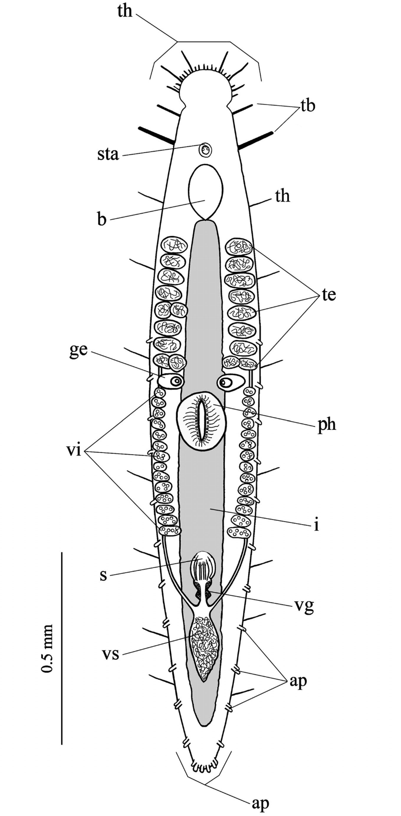
Description. The sexually mature animals are 1.6–1.8 mm in body length ( Figs 11 View FIGURE 11 , 12 View FIGURES 12 – 15 ). Anterior tip characterized by a cephalic elliptic swelling, provided with numerous tactile hairs (th) and two lateral couples of prominent tactile bristles (tb), retractable into the respective wide pockets ( Figs 11 View FIGURE 11 , 12, 13 View FIGURES 12 – 15 ). The anterior tactile bristles are relatively conspicuous, while the posterior ones are longer and more robust. Tactile hairs are present on the lateral body sides.
Behind the cephalic swelling are present the statocyst (sta) and the ovoidal brain (b).
The testes (te), located anterior to the pharynx, consist of two series of follicles stretched along the longitudinal axis. There are 8–10 per side, generally single, rarely coupled and of medium-large size ( Figs 11 View FIGURE 11 , 12, 13 View FIGURES 12 – 15 ). Two germaries (ge) of medium-large dimensions, at some distance from each other, are present in front of the pharynx ( Fig. 11 View FIGURE 11 ), posterior to the last testis follicles. They are globoid and contain numerous egg cells. Behind the ovaries two rows of medium–small vitellaries (vi), in single line and laterally placed, and adjacent to the penis papilla opening ( Figs 11 View FIGURE 11 , 12 View FIGURES 12 – 15 ).
The pharynx (ph), perpendicularly situated in the body center, shows the so-called collar-shaped organization (‘kragenförmig’, Ax, 1956), typical of the genus. The sacciform intestine (i) is a caecum at both extremities ( Figs 11 View FIGURE 11 , 12 View FIGURES 12 – 15 ). The caudal end is characterized by a narrow and blunt plate provided with a few finger-shaped adhesive papillae (ap) of conspicuous size. These bi-glandular structures are also present in the ventro-lateral epidermis along the body length ( Figs 11 View FIGURE 11 , 12, 14, 15 View FIGURES 12 – 15 ).
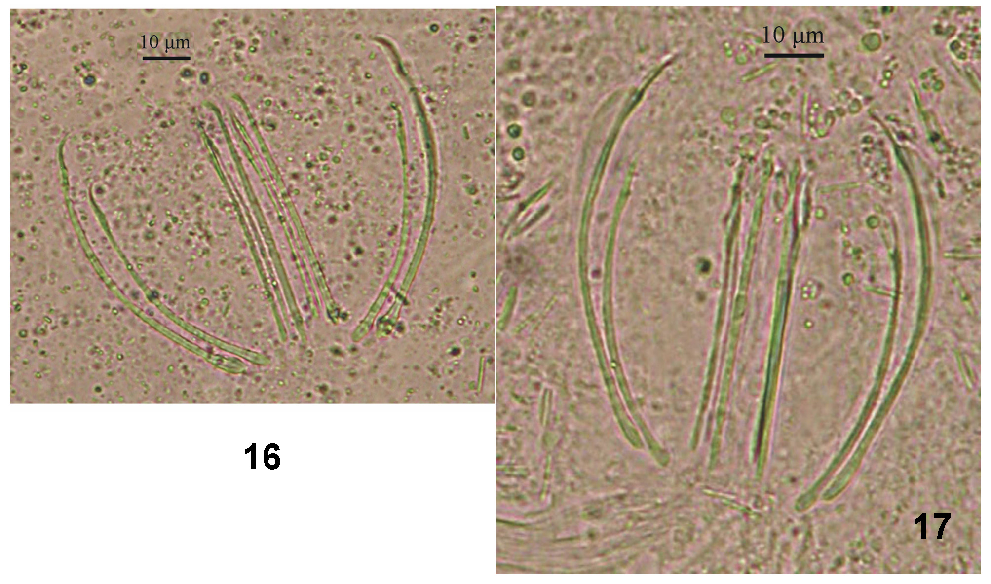
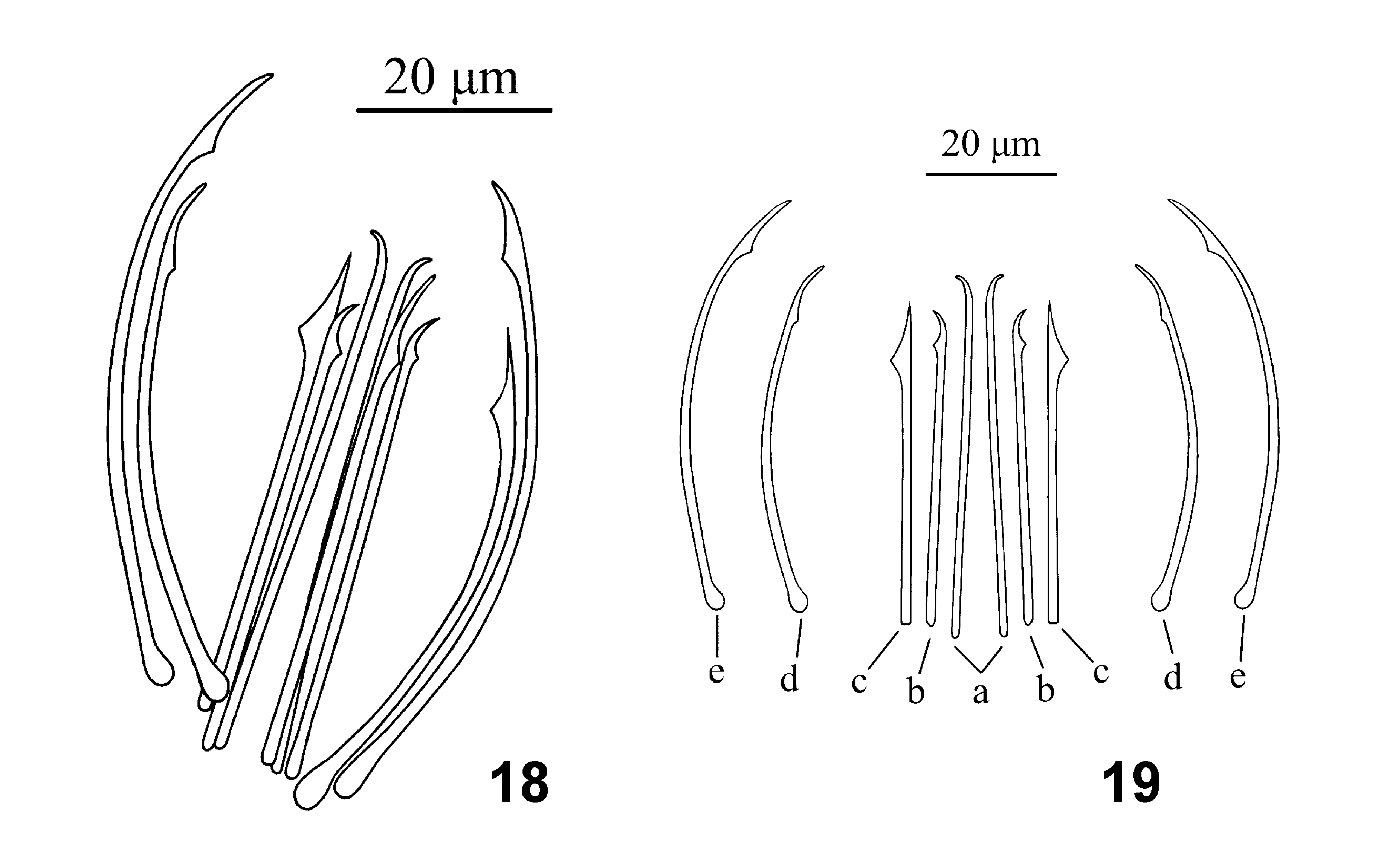
The male copulatory organ presents a sclerotic apparatus (s) with 10 spines of variable shape and length ( Figs 15 View FIGURES 12 – 15 , 16, 17 View FIGURES 16 – 17 , 18, 19 View FIGURES 18 – 19 ):
• 1 pair (a), 55 Μm long, placed in the center of the sclerotic complex with a straight body and pointed tip bent outwards;
• 1 pair (b), 46–48 Μm long, arranged laterally with respect to the previous one, with a small cuneiform subterminal prominence and an outwardly bent pointed tip;
• 1 pair (c), 46 Μm long, external to the previous pair (b), with a straight pointed tip. These spines are characterized by the presence of a large cuneiform sub-terminal prominence on the outward side;
• 1 pair (d), 52–55 Μm long, situated externally with respect to the central spine complex, internally curved with pointed and thin distal end. These spines present a small sub-terminal cuneiform prominence on the concave side;
• 1 pair (e), 68–70 Μm long, the outermost one placed on both sides of the previous pair (d), with pointed distal tip and sub-terminal cuneiform prominence on the concave side. Similarly to the former couple, these spines have a proximal extremity bulb-shaped.
Remarks. The new species shows a body length similar but slightly larger than P. tyrrhenica (1.5-1.6 mm), while all the remaining species of the genus have a smaller length (between 0.4 and 1.4 mm). The organization of the cephalic bulge is similar to that observed in P. propontica , while it is much less similar to that found in P. fibulata , P. minima and P. pontica . The distribution of the testes is typical of the genus, but their path and number are similar to those described in P. fibulata . Moreover, the dimensions of the testicular follicles in the new species are similar to those present in P. fibulata , P. pontica , P. tyrrhenica and P. parafibulata .
The paired germaries, located frontally to the pharynx, show a localization similar to those observed in P. fibulata and P. parafibulata , while their sizes are comparable to those found in P. pontica , P. propontica and P. parafibulata .
The position of the vitellaria, arranged in a single longitudinal row on both body sides, is typical of the genus; however, their arrangement is similar to that observed in P. fibulata , P. propontica and P. tyrrhenica . In P. donoraticensis , the medium-small size of vitellaries, which increase towards the penis papilla, are similar to those found in P. pontica and P. propontica . The pharynx displays the typical characteristics observed in all the genera of the subfamily, although its location in the organism is more central, similarly to that described in P. fibulata and in P. parafibulata . The intestine is similar to that of all the genus species, sacciform and without a cephalic intestine. The locations of vesicula seminalis, vesicula granulorum and penis papilla appear to coincide with those of the other species.
The spines of the male copulatory organ of the new species display a different organization from that of all the other species. The overall number of spines is 10, as in P. fibulata , P. macromystax and P. parafibulata , while all the other Postbursoplana species have only eight spines. In P. donoraticensis , the central pair (a) shows a pointed distal end with a short curvature, similar to that present in P. fibulata , P. macromystax , P. pontica and P. propontica . These two spines are larger (55 Μm) than those found in previously described species, where their length varies from 29 to 48 Μm, with the exception of P. parafibulata in which these spines are longer (61–62 Μm). The pair of spines (b), at both sides of this central longer pair, is very similar to the equivalent spines of P. fibulata . The length of these spines in P. donoraticensis (46–48 Μm) exceeds that of P. minima (24 Μm) and that of P. tyrrhenica (26–27 Μm), and is lower than the equivalent spines present in P. parafibulata (52–53 Μm).
The third spine pair (c) presents a shape similar to those observed in P. fibulata , P. minima , P. pontica and P. propontica , in which are obvious slight differences in the distal part with a sub-terminal cuneiform prominence less wide. In the new species, the length (46 Μm) of these spines is greater than that measured in P. fibulata (43 Μm), P. minima (35 Μm), P. pontica (26–27 Μm) and P. propontica (38 Μm), with the exception of P. parafibulata that instead shows a greater length (52–53 Μm) of these spines. The pointed tip of these spines reminds that observed in P. propontica , while the cuneiform sub-terminal prominence is similar to those described in P. macromystax , P. propontica , P. t y r rh e ni c a and P. parafibulata . In P. donoraticensis , the spines of the pair d (52–55 Μm long) are similar to the homologous spines present in P. fibulata (56 Μm) and P. parafibulata (52–53 Μm). In all other species the equivalent spines have a shorter length (between 30–32 Μm and 44–48 Μm). The length (68–70 Μm) of the most external pair of spines (e), exceeds that observed in the other species (40–60 Μm), with the exception of P. parafibulata (70 –73 Μm).
On the base of the data presented, P. donoraticensis differs from the previously described species in body dimensions and, above all, the characteristics of the sclerotic apparatus. The new species has a close relationship with P. fibulata , but represents a new species.
Etymology: The name donoraticensis refers to the type locality where this species has been collected. The site of sampling is very close to the village of Marina di Donoratico (Tuscany, Ligurian Sea, Italy), and donoraticensis means “coming from Donoratico”.
Habitat. Surf zone.
Distribution. Known only from type locality.

No known copyright restrictions apply. See Agosti, D., Egloff, W., 2009. Taxonomic information exchange and copyright: the Plazi approach. BMC Research Notes 2009, 2:53 for further explanation.