
Tupala, Stroiński, Adam & Szwedo, Jacek, 2015
|
publication ID |
https://doi.org/ 10.11646/zootaxa.4033.3.3 |
|
publication LSID |
lsid:zoobank.org:pub:5A0985CE-3B17-4BD0-866F-7763F1E2120B |
|
DOI |
https://doi.org/10.5281/zenodo.6097611 |
|
persistent identifier |
https://treatment.plazi.org/id/03BC87C1-3828-0217-E9E4-F885FC616A42 |
|
treatment provided by |
Plazi |
|
scientific name |
Tupala |
| status |
gen. nov. |
Tupala View in CoL gen. nov.
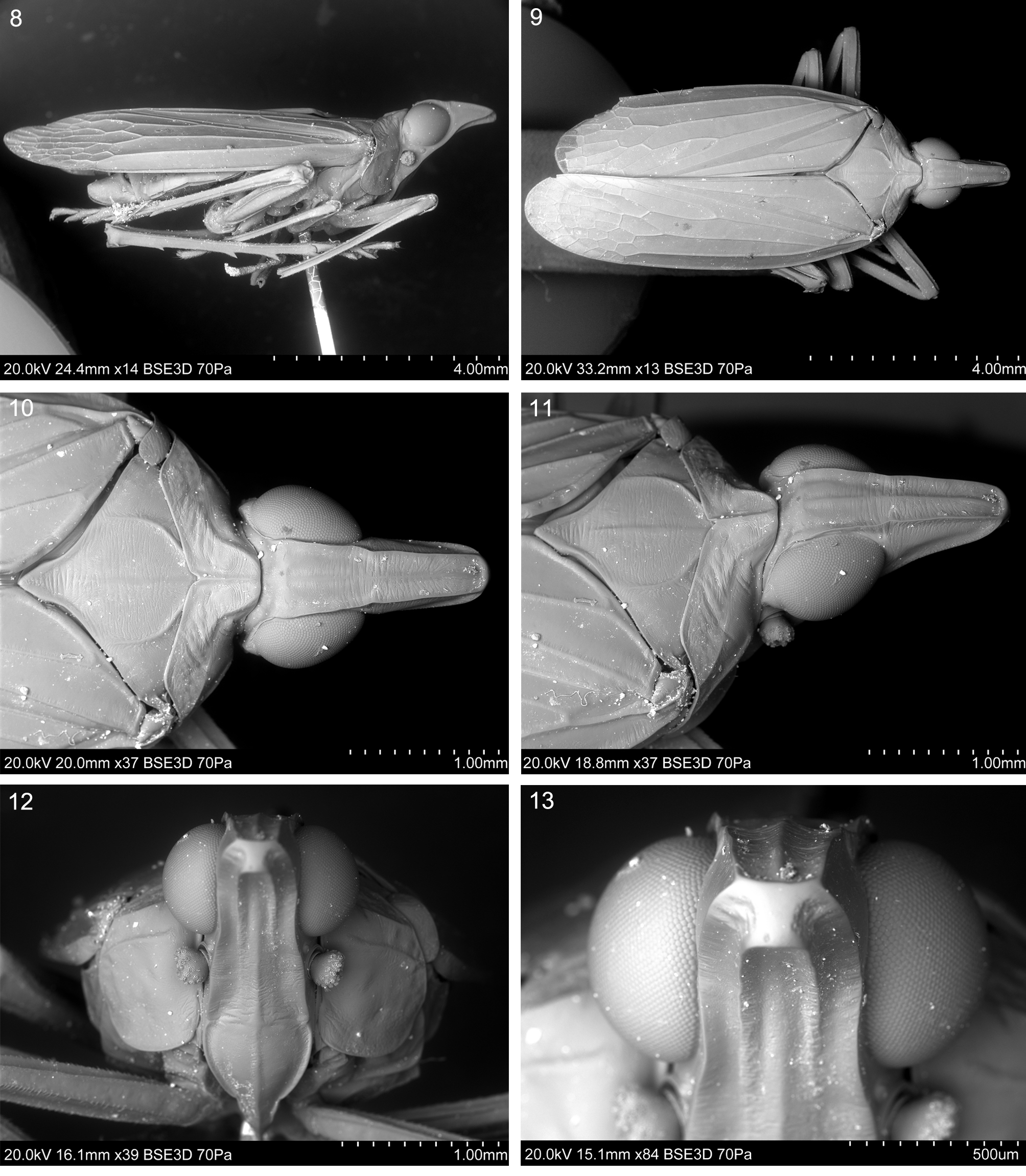
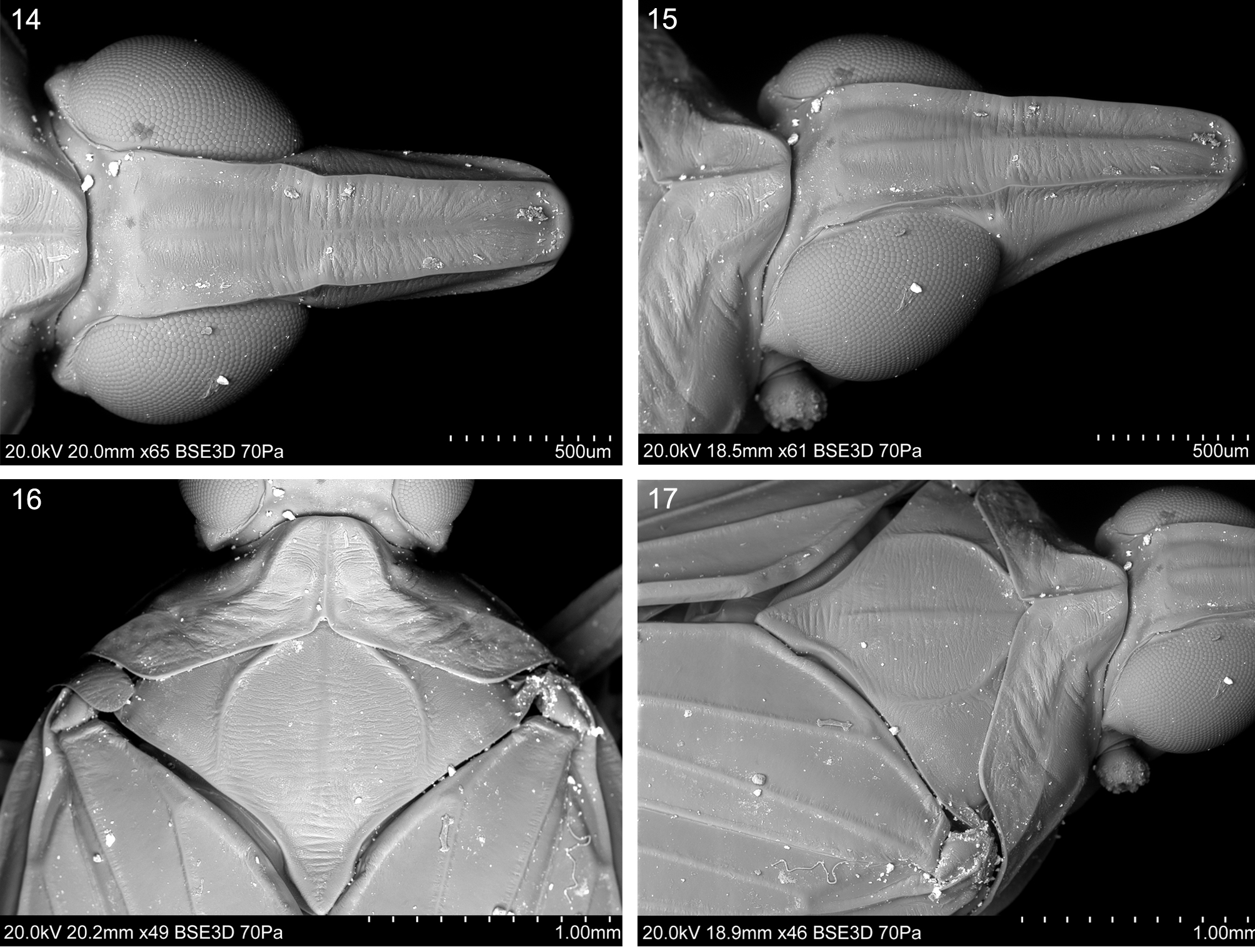
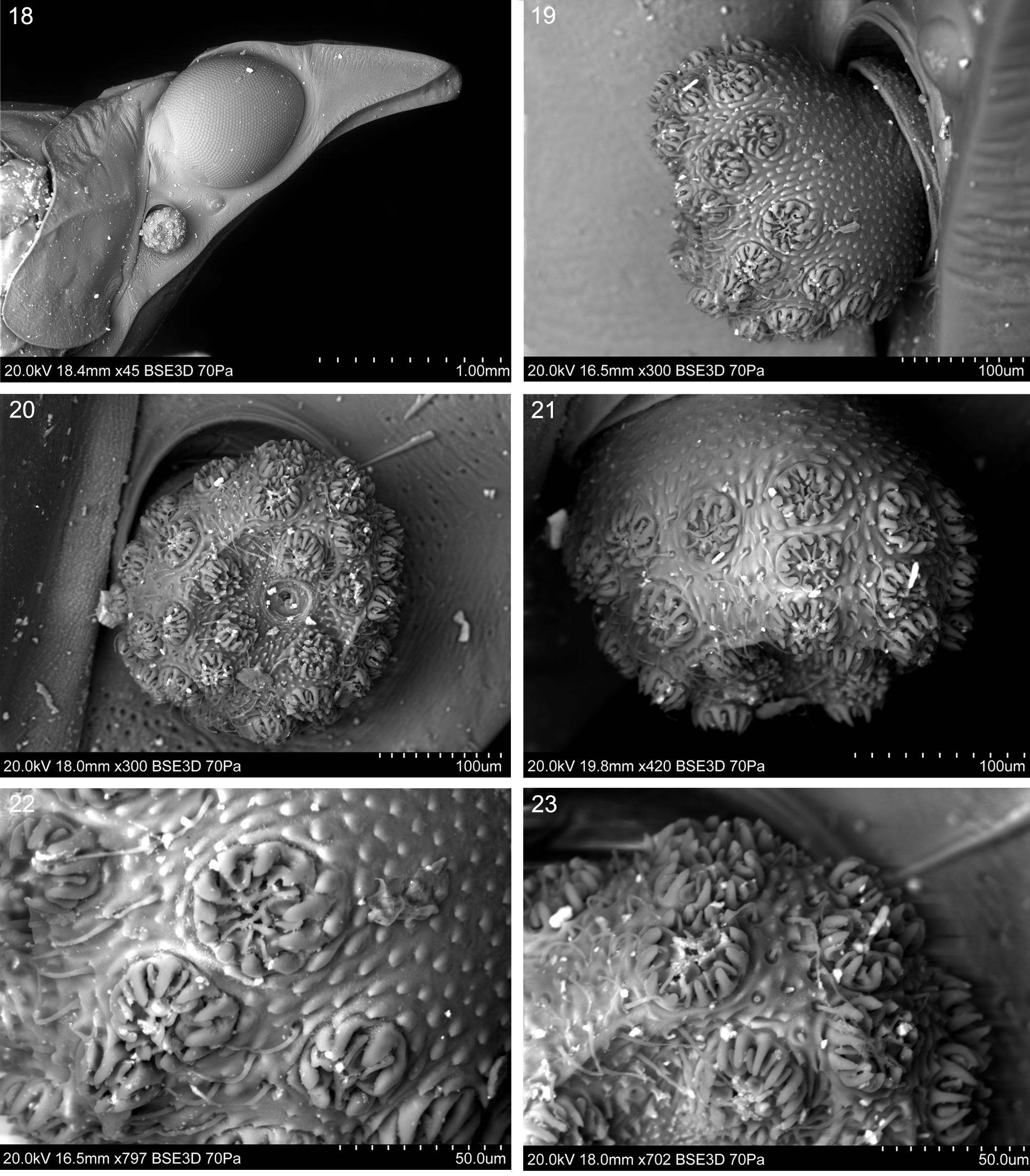
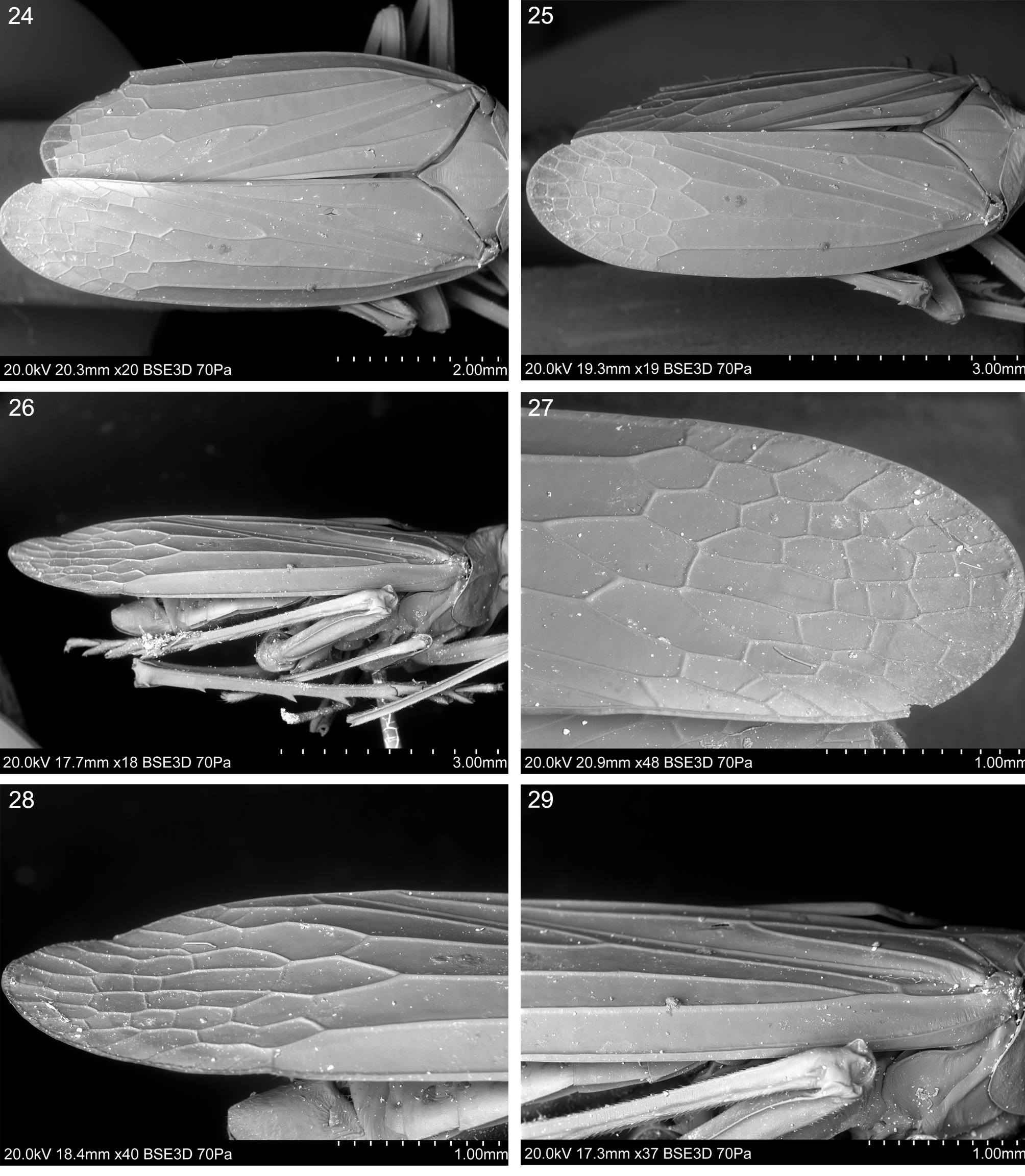
( Figs 1–50 View FIGURES 1 – 7 View FIGURES 8 – 13 View FIGURES 14 – 17 View FIGURES 18 – 23 View FIGURES 24 – 29 View FIGURES 30 – 33 View FIGURES 40 – 43 View FIGURES 44 – 50 , 55)
Type species. Tupala occulta sp. nov., here designated.
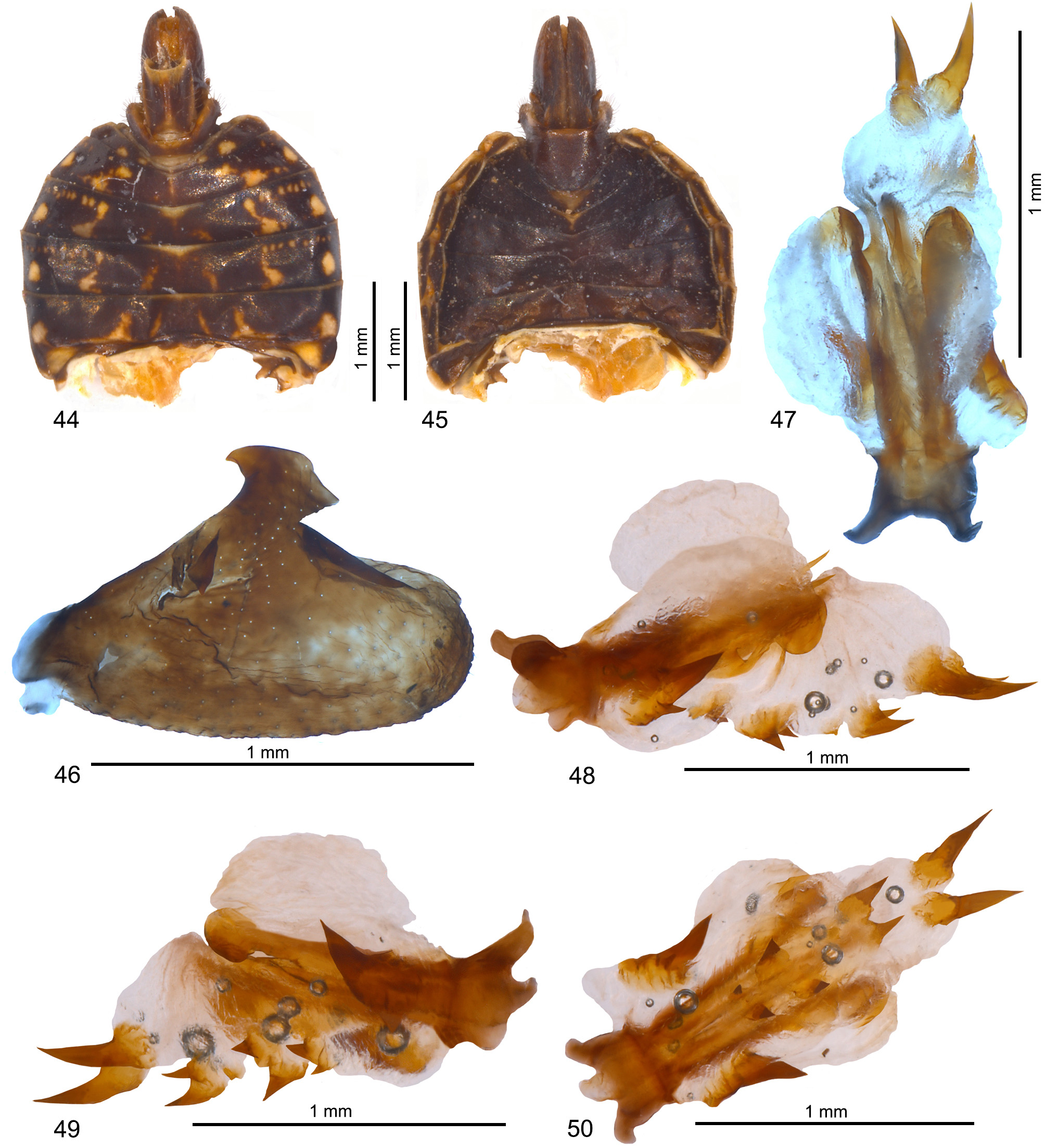
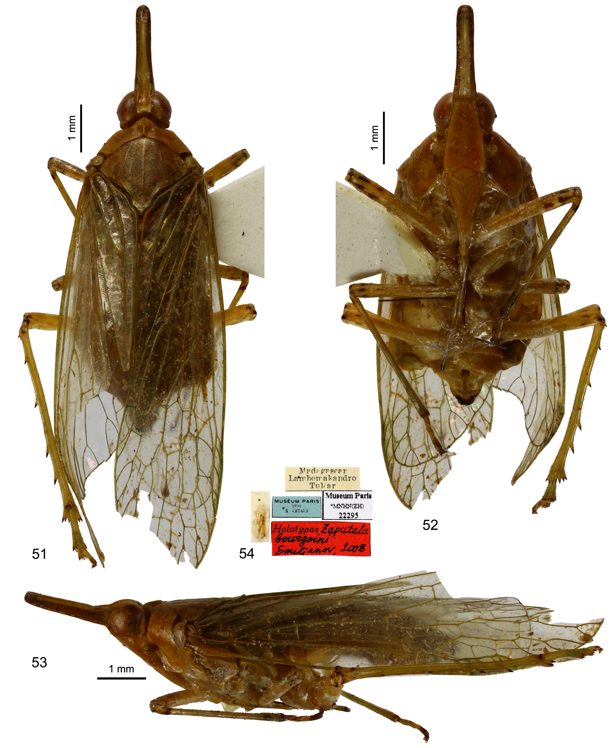
Etymology. Generic name is an anagram of dictyopharid generic name Putala . Gender: feminine. Diagnosis. Differs from Zaputala ( Figs 51–54 View FIGURES 51 – 54 ) by: presence of longitudinal carina of vertex (absent in Zaputala ); tricarinate pronotum (single carina in Zaputala ); frons with median carina (absent in Zaputala ); different organization of head capsule extension. Lateral portions of pronotum with obsolete granulation (lateral portions of pronotum smooth in Zaputala ).
Description. Head with compound eyes distinctly narrower than pronotum. Vertex longer than wide at base, tapering apicad; anterior margin arcuate, lateral margins with shallow incision at about half of their length, posterior margin shallowly convex, indistinct; lateral margins carinate, anterior margin thickened, callose; disc of vertex flat, with faint longitudinal carina. Posterior margin of vertex at posterior ⅓ of compound eyes length.
Frons longer than wide at frontoclypeal suture, with lateral margins carinate, merely sigmoid, diverging towards frontoclypeal suture, with breaking point above the level of antennae base, then more distinctly diverging, lateral carinae reaching to half of frons length, in upper portion, forming obtuse extension, convex in upper portion towards margin with vertex, concave in lower portion towards disc of frons, median carina reaching frontoclypeal suture; disc of frons concave laterad, with median convexity at lower ⅓ of its length. Frontoclypeal suture straight, not distinct. Postclypeus with distinct lateral carinae and median carina, disc of clypeus convex, anteclypeus distinguishable, convex, with lateral and median carinae. Compound eyes with small posteroventral callus. Lateral ocelli present. Antennal fovea deep, emarginated; scapus short, pedicel bulbiform, with sensory plates in upper portion distributed in apical half, and sensory plates of lower hemisphere distributed in basal and apical parts; trichoid sensilla type I present. Rostrum with apical segment almost as long as subapical one, apex of rostrum reaching to metacoxae.
Pronotum heptagonal, with disc slightly elevated, delimited by straight anterior margin, incomplete anterolateral carinae, strongly diverging posteriad, not reaching to posterior margin, with distinct median carina, posterior margin of pronotum widely triangularly incised, with small but distinct triangular incision at terminus of median carina; lateral portions of pronotum descending lateroposteriad, with oblique postocular carina, reaching posterior margin, and lateral carina reaching posterior margin.
Tegulae large, without carinae.
Mesonotum wider than long in mid line, with distinct median carina, reaching to level of scutellum and lateral carinae reaching posterior margin, delimiting flattened disc, lateral margins descending.
Tegmen membranous, macropterous, with venation distinct; apex of clavus exceeding ⅔ of tegmen length. Costal margin arcuate, veins of costal complex fused, anteroapical angle wide, apex rounded, apical point at level of middle of tegmen width, posteroapical angle wide, tornus long. Basal cell narrow, elongate, stems ScP+R and M leaving basal cell with a short common stalk, about half as long as basal cell; stem ScP+R long, slightly curved, subparallel to costal margin, forked posteriad of stems M and CuA forkings, merely basad of level of claval apex, branch ScP+RA reaching margin with three terminals, terminal ScP+RA1 reaching margin apicad of claval apex; branch RP forked apicad of RA3 terminus at margin, reaching margin with two terminals. Stem M merely sigmoid, forked slightly basad of claval apex, basad of stem ScP+R forking, branch M1+2 forked slightly apicad of branch M3+4 forking, then M1 forked again on membrane, forking of branch M3+4 slightly apicad of claval apex, then branches M3 and M4 forked again on membrane, then stem M reaching margin with 7 terminals. Stem CuA straight, long, forked apicad of claval veins junction, branch CuA1 forked subapically on membrane, branch CuA2 single. Claval veins Pcu and A1 fused before half of claval suture (CuP) length. Veinlet pccacp-scra delimiting anterior margin of ‘stigmal area’ placed slightly apicad of transverse veinlet 1r-m, apicad of claval apex, callose at costal margin. Nodal veinlet 1r-m at level claval apex; nodal veinlet 1m-cu slightly oblique, basad of 1r-m; veinlet icu oblique, reaching margin of tornus apicad of claval apex. Costal cell without transverse veinlets. Membrane with net of transverse veinlets delimiting polygonal cells. ‘Stigmal area’ wide, dissected by ScP+RA1 and RA2 terminals. Cell C1 pentagonal, slightly longer than wide; cell C3 elongately pentagonal, longer than wide, about twice as long as C1; cell C5 lanceolate, tapering posteriad, more than twice as long as cell C3.
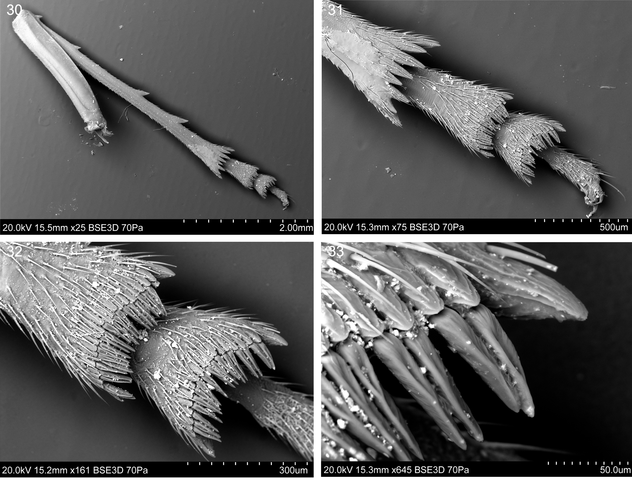
Legs long, slender, tibiae not flattened. Profemur longer than mesofemur. Metatibia longer than metafemur, with four lateral spines and 8 apical teeth arrange in row. Metabasitarsomere about as long as cumulative length of mid and apical metatarsomeres; Basimetatarsomere with row of apical teeth; subconical lateral ones and row of 22 dorsoventrally flattened and elongated teeth with subapical needle-shaped platellae, laterally compressed, with dorsal fuller in median portion. Midmetatarsomere with row of apical teethe, subconical lateral ones, and row of 18 dorsoventrally flattened and elongated teeth with subapical needle-shaped platellae, laterally compressed, with dorsal fuller in median portion. Apical metatarsomere about as long as preceding one; tarsal claws distinct, arolium wide.
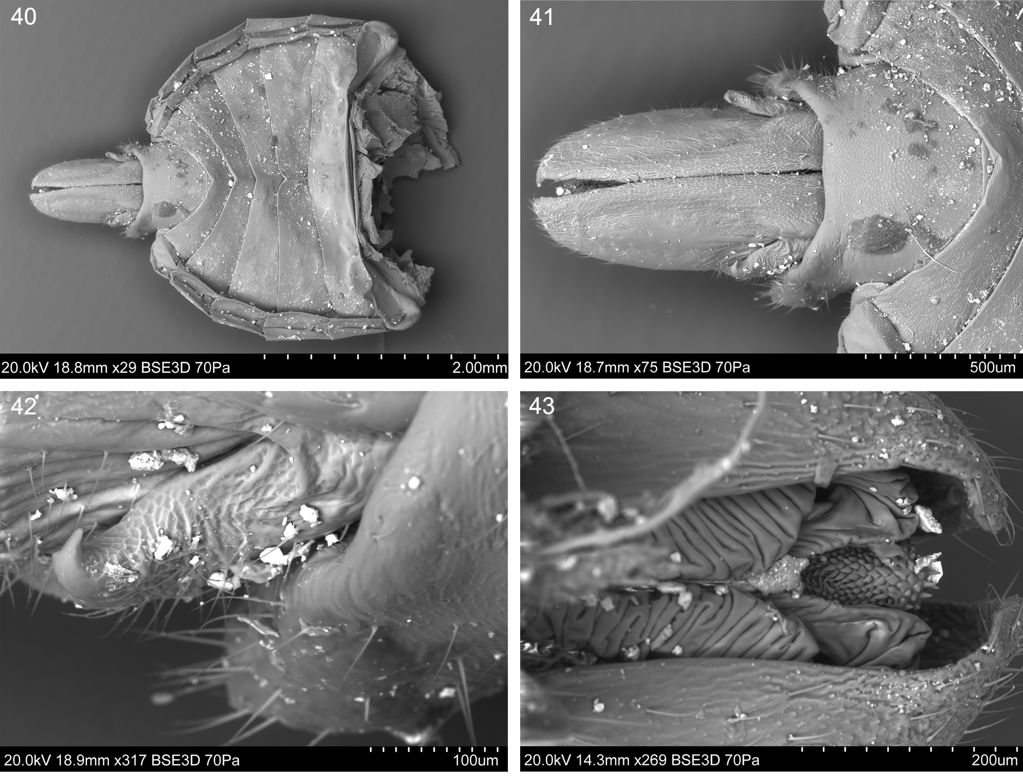
Male terminalia: pygofer laterally compressed, distinctly higher than long in lateral view, posterior margin smooth, with posterodorsal portion declining posteriad. Anal tube distinctly shorter than gonostyles, tubular, with short, triangular, posteroventral projections; anus placed apically. Gonostyles symmetrical, not fused; base narrow, expanded towards middle, than mildly tapering towards apex; dorsobasal margin straight, dorsoapical margin straight, apical portion bluntly rounded, ventral margin widely arcuate; upper margin with dorsal process in the middle, acutely tapering basad, hook-like process near sub-middle in lateral view, closer to dorsobasal margin.
Phallic complex almost symmetrical, with periandrium composed of two dorsolateral membranous sacs, with ventroapical sclerotized hemicircular lobes and sclerotized, double spine like processes present in laterobasal portion, dorsal spine distinctly bigger than ventral one; apical part of dorsal periandrium with two elongate, tapered, sclerotized processes. Aedeagus membranous sac-like, distinctly longer than periandrium, with double row of longitudinal pine-like processes, increasing in size from aedeagus base and posterodorsal portion covered with spiniferous microsculpture.
Female unknown.
Distribution. Madagascar, Antsiranana Province, Sava Region ( Fig. 55).

No known copyright restrictions apply. See Agosti, D., Egloff, W., 2009. Taxonomic information exchange and copyright: the Plazi approach. BMC Research Notes 2009, 2:53 for further explanation.