
Cordagalma rugosum, P. R. Pugh, 2016
|
publication ID |
https://doi.org/ 10.11646/zootaxa.4095.1.1 |
|
publication LSID |
lsid:zoobank.org:pub:690FFEBE-F71B-4EFD-865A-944D81A12897 |
|
DOI |
https://doi.org/10.5281/zenodo.6087817 |
|
persistent identifier |
https://treatment.plazi.org/id/03BC87D0-FF8E-FFC3-FF6E-F9A5F9CED087 |
|
treatment provided by |
Plazi |
|
scientific name |
Cordagalma rugosum |
| status |
sp. nov. |
Cordagalma rugosum sp. nov.
Diagnosis. Small heart-shaped nectophores, usually with relatively long, narrow basal extension. Extent of the axial wings very variable, from almost non-existence to having a deep indentation between them. Distinct lateral ridges extending from outer corners of axial wings down ostio-lateral sides of the nectophore and ending approximately on a level with the ostium. Two weaker ridges divide from them and curve inwards and downwards to end above ostial level. Up to six patches of cells can be present on each side of the velum. Relatively large bracts somewhat rounded with distinct, but small distal facet divided by a ridge, with a pair of patches of ectodermal cells. The edges of the distal facet frilled, and small nematocysts are present along them. Bracteal canal to c. half the length of bract, ending in a small swelling inflected into the mesogloea. Gastrozooids with pinkish pigmentation. Palpons possibly attached by their bases or slightly to one side of it. Structure of tentillum not known.
Material examined. A total of five specimens have been collected by the ROV Tiburon; all in the region of Monterey Bay, California, U.S. A:
Tiburon Dive 986 35°38.01'N 122°44.46' W 15-May-2006 depth 1228m GoogleMaps Tiburon Dive 1040 34°17.23'N 124°03.10'W 01-Oct-2006 depth 1201m GoogleMaps Tiburon Dive 1109 35°50.00'N 122°40.00'W 31-Jul-2007 depth 1120m GoogleMaps Tiburon Dive 1154 35°49.97'N 122°39.99'W 28-Nov-2007 depth 1040m GoogleMaps Ricketts Dive 421 35°33.74’N 123°44.97’W 20-Dep-2012 Depth 1318m GoogleMaps
The specimen collected during the Tiburon Dive 1109 was frozen for molecular studies.
Holotype: The specimen collected during Tiburon Dive 1154 has been designated the holotype, and will be deposited at the United States National Museum (Smithsonian Institution), Washington, DC.
The remaining specimens will be placed in the collections of Dr Casey Dunn, at Brown University, Providence, Rhode Island, USA.
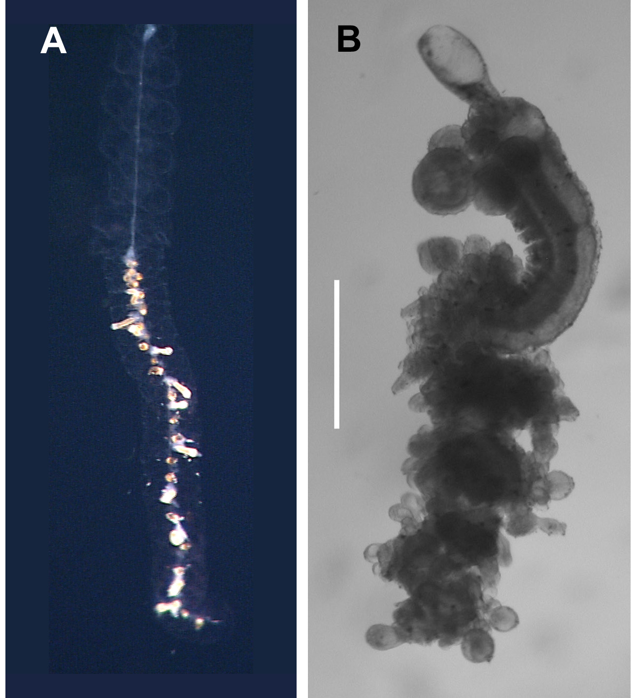
Description. Specimens of Cordagalma rugosum sp. nov. were small and fragile and they fell apart in the collecting devices and, as with Cordagalma abyssorum sp. nov., it was inevitable that some pieces were missed despite a thorough search. An in situ frame grab of the type specimen is shown in Figure 22A View FIGURE 22. A . It was difficult to gauge the length of the colony, but it was certainly less than the diameter (16.5 cm) of the collector. The frame grabs indicated that the stomach region of the gastrozooids was lightly pigmented (see below), while between each gastrozooid there were one or two groups of brownish coloured appendages. It can only be presumed that one of these represented a palpon, possibly with some gonophores, and that the other represents a gonodendron.
Pneumatophore: A pneumatophore and denuded stem ( Figure 22 View FIGURE 22. A B) was present with the preserved holotype and Tiburon Dive 986 specimens. It was featureless and colourless, and measured c. 5 mm in length and 3.5 mm in diameter.
Nectosome: It was clear from the denuded stems that the nectophores were developed on the ventral side of the nectosome ( Figure 22 View FIGURE 22. A B). Several buds of nectophores were present at the apical end of the nectosome.
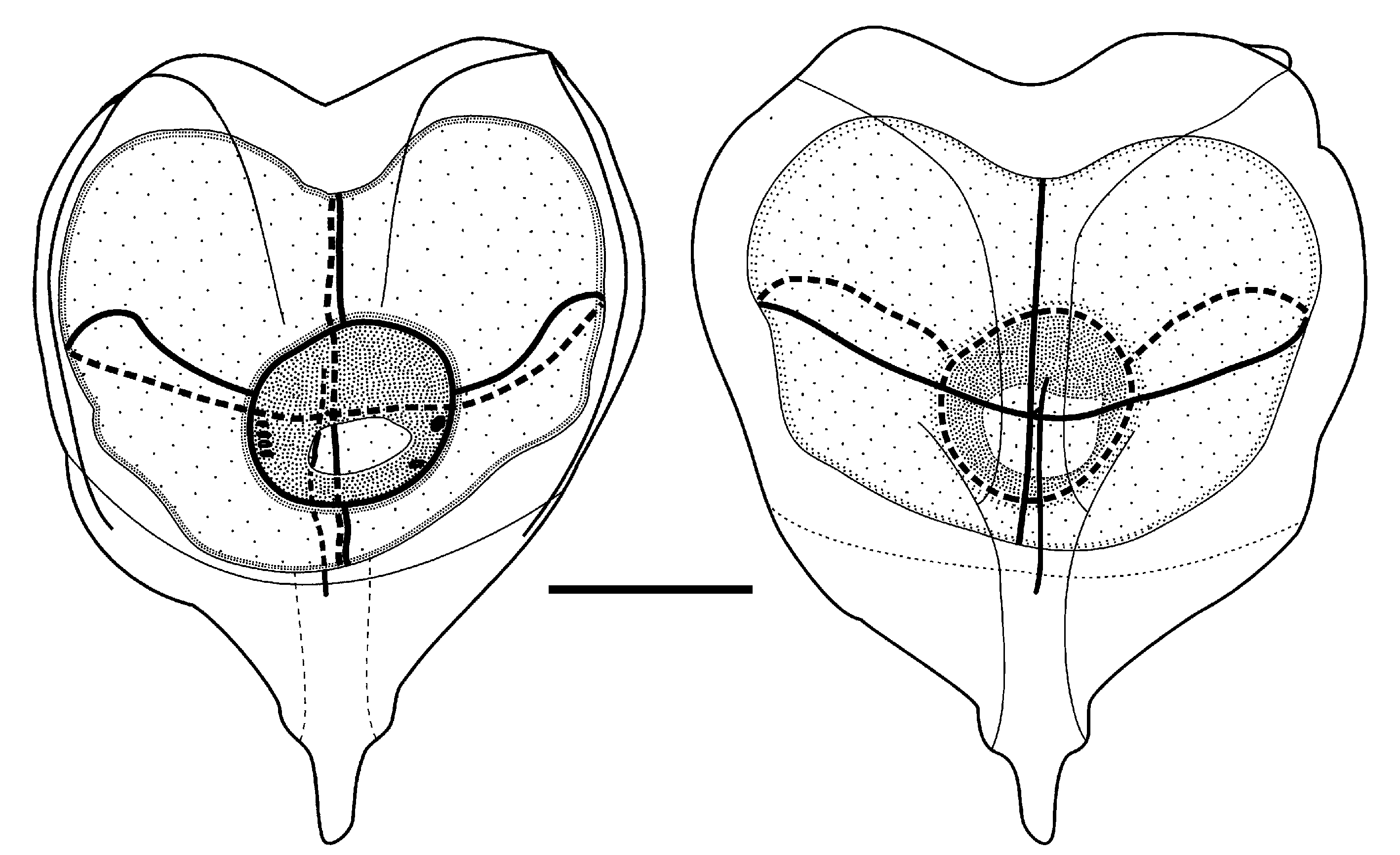
Nectophore: Only three nectophores were found with the holotype specimen, each of which had a characteristic form. The largest one ( Figure 23 View FIGURE 23 ) measured 5.1 mm in length and 4 mm in width. The axial wings of this nectophore were very small, with only a shallow central indentation separating them. From their lateral apices a pair of obvious ridges ran down close to the lateral margins of the nectophore, but abruptly disappeared at about the level of the base of the nectosac. Close to their origin, another pair of less distinct ridges divided from them and ran inwards and then downwards, defining the edges of a shallow furrow, to end just above the nectosac. In the region immediately below the nectosac, the nectophore narrowed considerably, while its depth, on the ostial side, decreased rapidly to c. 2/3rd its deepest. The distal process was thus quite narrow, like an inverted isosceles triangle, except that its basal tip was turned to one side. On the axial side of the nectophore there was a very shallow median furrow that narrowed and deepened considerably in the region of the pedicular canal. It then shoaled to form a broad shallow gutter, and continued down the basal process.
The nectosac, in ostial view, was almost circular and had no lateral extensions. The upper and lower radial canals were straight, while the laterals were only slightly looped. The descending mantle canal was much longer than the ascending one. The pedicular canal immediately gave rise to the four radial canals on reaching the nectosac. There were no obvious patches of ectodermal cells in the region of the junction of the lateral radial canals with the ostial ring canal. However, there were distinct patches of cells on the velum itself. A row of six such patches were found on the left-hand side of the velum, below the insertion point of the lateral radial canal, but only one was found on the right.
Outwardly, the middle-sized nectophore ( Figure 24 View FIGURE 24 ), which measured 4 mm in height and 3.3 mm in width, differed in many respects from the largest one. The nectophore had pronounced ostial wings, while the basal process was broadly triangular with a rounded base, from which hung a small appendage. The apparent asymmetry in the basal process is a preservation artefact. However, in most other respects the characteristic features of the nectophore were similar to the largest nectophore. There were distinct lateral ridges running down the nectophore on its ostial side. A weaker pair of ridges arose from them and ran inwards and downwards to peter out above the ostium. The ostial side of the nectophore rapidly lost depth just below the ostium. The margins of the hydroecial gutter, on the axial side, were weak and its depth was very shallow in the basal half of the nectophore, but increased greatly in the upper half.
The presence of axial wings was mirrored by the development of latero-apical wings to the nectosac, which resulted in the lateral radial canals being more arched than on the largest nectophore. The ascending mantle canal remained much shorter than the descending one, and the origin of the pedicular canal was similar to that in the largest nectophore. On reaching the nectosac it immediately gave rise to all four radial canals. Patches of cells were again present on the velum of the nectosac, with one large and two smaller ones on the left-hand side and six small ones on the right.
The smallest nectophore measured 4 mm in height and 2.85 mm in width ( Figure 25 View FIGURE 25 ). It was quite similar to the largest nectophore in basic shape, but the axial wings were relatively larger and the shape of the basal process was somewhat different. Otherwise it shared the same basic characters as the other two nectophores, although the lateral ridges were relatively weak and, despite the apico-lateral extensions of the nectosac, the lateral radial canals were only slightly arched. The velum of the nectosac had four patches of cells on the left-hand side and one large and one small one on the right.
For the other specimens, nine nectophores were found with the Tiburon Dive 986 specimen, which measured up to 6 mm in length and 4.7 mm in width. All were in poor condition, with the nectosac having become completely detached in six, and partially so in another two. On the remaining one, the velum of the nectosac bore 3 spots on the left-hand side and four on the right. All these nectophores had small axial wings with a broad shallow depression between them. The form of the basal process varied between the two extremes found with the largest and smallest nectophores of the holotype. Thus in some it formed a narrow isosceles triangle, while in others the sides were almost parallel, although their width in this region was quite variable, before tapering down to a rounded base.
The eight nectophores found with the Tiburon Dive 1040 specimen were smaller, with maximum dimensions of 4.7 mm in length and 3.4 mm in width. They were all in reasonable condition, with their nectosacs largely intact, and were all of the form shown by the intermediated sized nectophore of the holotype, with more or less pronounced and pointed axial wings. In ostial view the inner margins of the wings came to overlap each other, leaving a narrow but deep incision between them, just above the median apex of the nectosac. They then diverged slightly and petered out above the ostium. The basal process was broad and basally rounded, with the basal protrusion much less pronounced than for the holotype nectophore; with six nectophores only showing a small pimple; one with no sign of it at all; and one with a small but distinct protrusion. The number of patches on the velum of the nectosac was quite variable, with up to six on each side.
Siphosome: The contracted siphosomal stem is shown in Figure 22 View FIGURE 22. A . The buds of palpons, bracts and gonophores were all that remained attached to it.
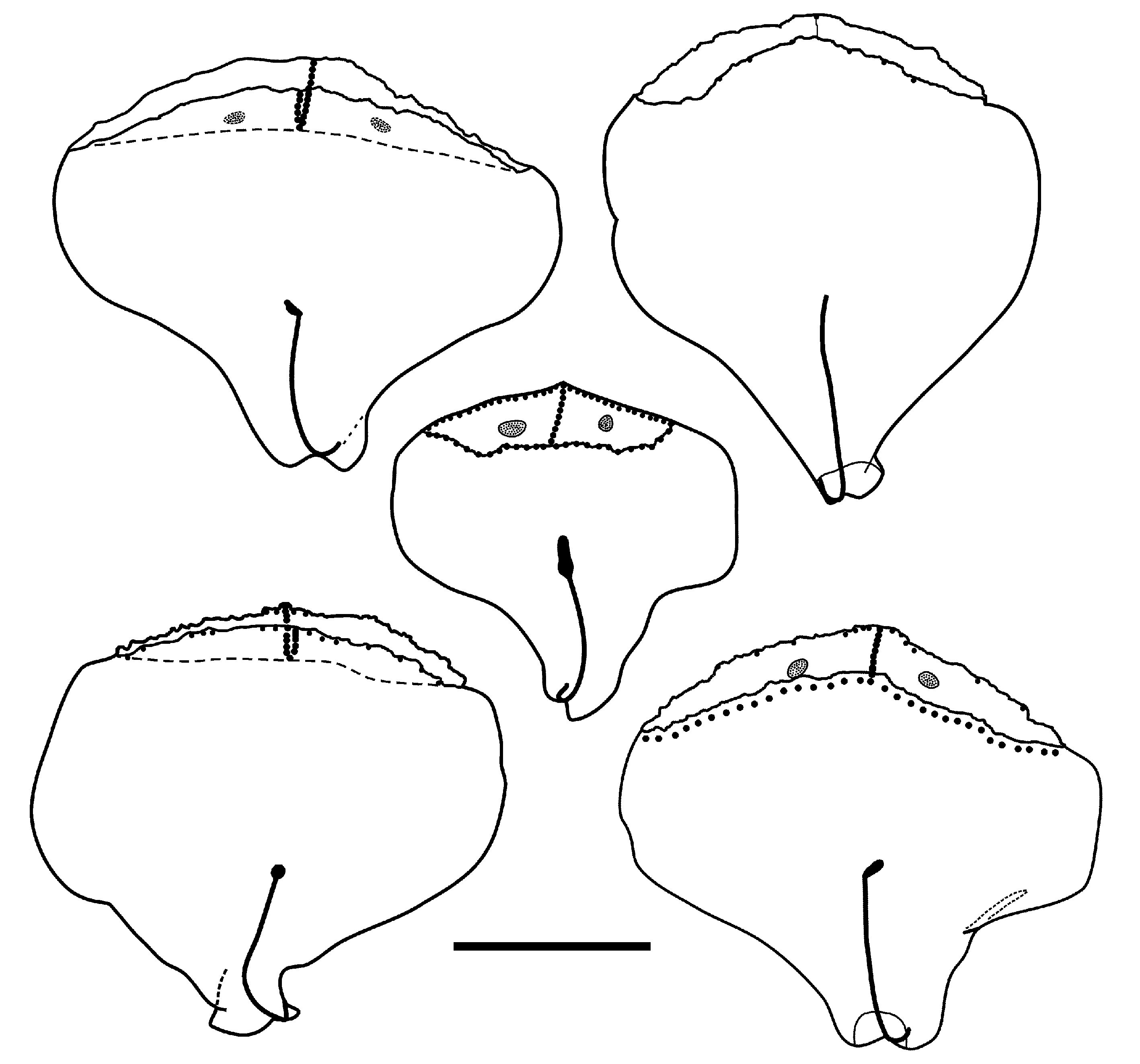
Bract: ( Figure 26 View FIGURE 26 ). The bracts measured up to 2.5 mm in length and 2.6 mm in width, and in general were broader than they were long. The broadest part was in the distal half to two-thirds of the bract, and proximal to this the bract narrowed rapidly. The proximal tip of the bract was turned up and formed a small process on the upper side of the bract. A narrow furrow ran along it, at the base of which was the proximal end of the bracteal canal. Occasionally in the proximal region the bracts were asymmetrical, narrowing more rapidly on their inner side, and only the outer side was turned up. The bracteal canal ran to just under half the length of the bract and usually its distal tip was swollen and inflected, often vertically, into the mesogloea for up to half the thickness of the bract itself. This was a very characteristic feature, when present, but in larger bracts it was often very difficult to discern, as was the narrow part of the canal that was in contact with the lower side of the bract.
The lower side of the bract was slightly concave, while the upper side was level for most of its length, tapering down slightly toward the proximal end. A distal facet was demarcated by a transverse ridge that arched across the upper surface just short of the distal end of the bract. The edges of both this ridge and the more rounded ridge forming the lower border of the distal facet often had a frilly appearance, but this probably was a preservation artefact. A very indistinct median region also divided the distal facet into two. The edges of the distal facet and median ridge were overlain by small, spherical nematocysts, but frequently these had been lost. Usually on each half of the distal facet there was an opaque patch comprised of a few ectodermal cells, but like the nematocysts these were often eroded in the larger bracts, although where they had been positioned could still be seen.
Gastrozooid and tentacle: Two small gastrozooids were found with the holotype specimen, and three with the Tiburon Dive 968 one. They were all narrow and tubular, with the largest measuring 2 mm in length, but were only 0.2 mm in diameter in the stomach region. From the in situ frame grabs, this region was a very pale orange in colour, while the proximal and distal ends were white. The proboscis region was distinct, with the mouth usually turned out and back on itself. The basigaster was about twice as wide as the stomach region, but usually quite short. All bore the proximal remnants of a tentacle.
Tentilla: None of the proximal remnants of the tentacle bore any mature tentilla, but numerous small, bag-like initial buds of them were present.
Palpon: The holotype specimen, and that from Tiburon Dive 968, included about four small tubular structures, some with an inflated middle section, that probably were palpons. They measured up to 0.5 mm in length and 0.3 mm in diameter, but otherwise showed no characteristic features. The preserved and contracted stem of the holotype showed a number of small buds that probably represent developing palpons. Most of these buds were slightly asymmetrical in shape, but appeared to be attached at their proximal ends. However, for one or two of the buds this attachment appeared to be slightly displaced to one side, as found for the preserved palpons of Cordagalma ordinatum . More material will be required before the structure and arrangement of the palpons can be ascertained with certainty.
Gonophores: Only a single mature male gonophore was found with the Tiburon Dive 968 specimen. It was typically ovoid in shape, milky white in colour and measured 5.75 mm in length by 3.5 mm in diameter. There were no distinguishing characters. The holotype specimen possessed buds of both male and female gonophores, but no mature ones were found.
Distribution. Only five specimens of Cordagalma rugosum sp. nov. have been collected; one of which was frozen for molecular analyses. All five specimens came from the vicinity of Monterey Bay, California, and were found within the relatively narrow depth range of 1040 to 1318 m.
Etymology. The specific name rugosum is derived from the Latin ruga, rugosus meaning wrinkled, and refers to the wrinkled appearance of the edges to the basal facet on the bracts, although this may indeed be a preservation artefact.


No known copyright restrictions apply. See Agosti, D., Egloff, W., 2009. Taxonomic information exchange and copyright: the Plazi approach. BMC Research Notes 2009, 2:53 for further explanation.
|
Kingdom |
|
|
Phylum |
|
|
Class |
|
|
Order |
|
|
Family |
|
|
Genus |