
Cellaria, Ellis & Solander, 1786
|
publication ID |
https://doi.org/ 10.11646/zootaxa.4801.2.1 |
|
publication LSID |
lsid:zoobank.org:pub:D69B752F-09F6-42ED-AADF-93E57421F3C7 |
|
DOI |
https://doi.org/10.5281/zenodo.5586883 |
|
persistent identifier |
https://treatment.plazi.org/id/03BCB65E-6D55-4F60-FF07-39E7FC38FD67 |
|
treatment provided by |
Plazi |
|
scientific name |
Cellaria |
| status |
|
Cellaria View in CoL sp. 2
( Fig. 10 View FIGURE 10 )
Cellaria sp. 2: Achilleos et al. 2019: [4–8].
Material examined. NIWA 133018 View Materials , cruise TAN0306, Stn 7, 51.0723º S, 164.6062º E, southwest of Auckland Island , 1030–1065 m, collected 14 April 2003 GoogleMaps .
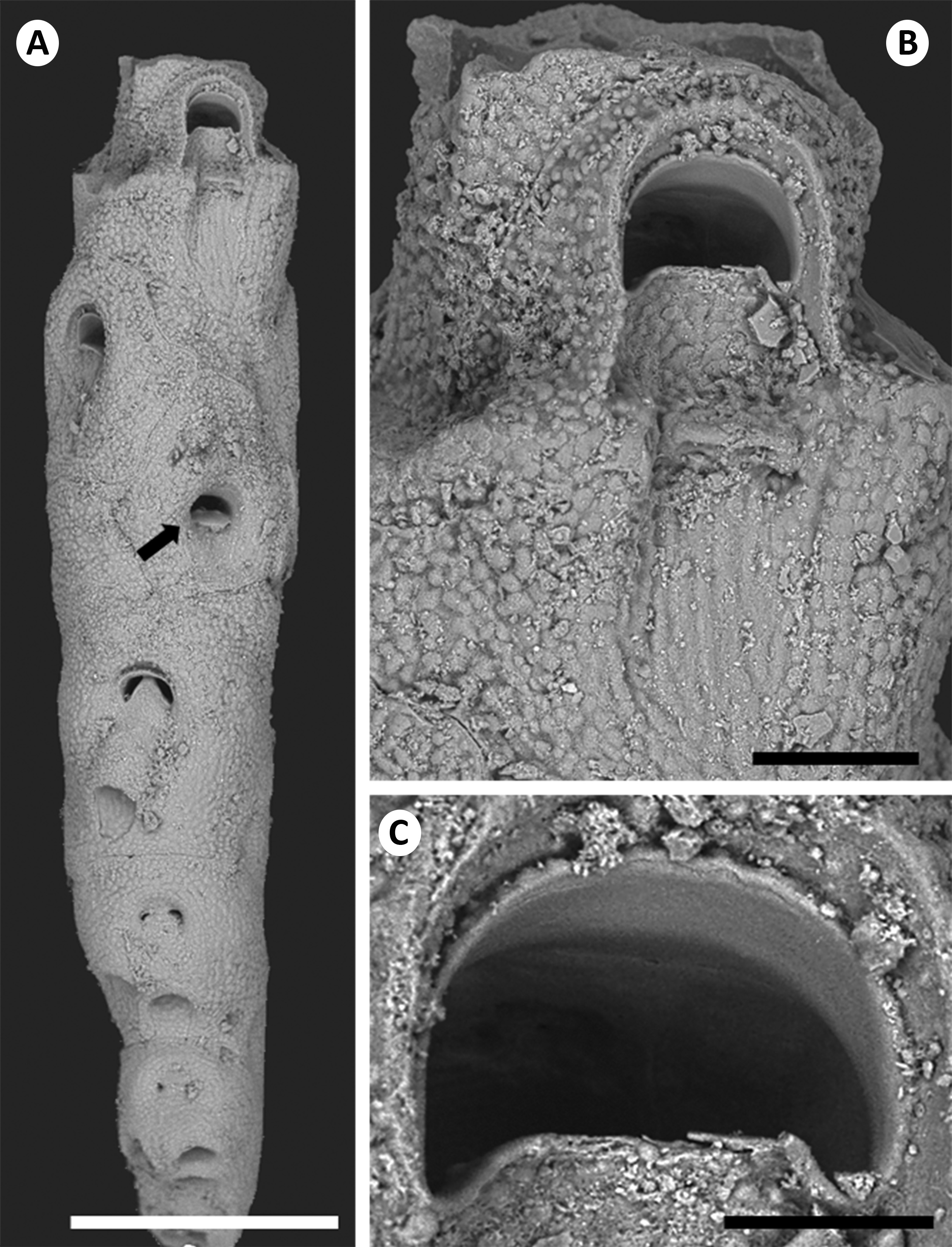
Description. Colony erect, comprising a single proximal stem fragment not> 2 mm in length, previously fractured distally but partially regenerated; stem cylindrical, tapered proximally (W, 275–477 μm). Zooids arranged back to back in alternating pairs, hence longitudinally quadriserial.
Autozooidal cystid with hexagonal outline, longer than wide (ZL, 415–583 μm; ZW, 347–387 μm; ratio 1.3). Cryptocyst densely and evenly granular-tubercular over entire surface; cryptocystal ridges continuous, rounded distally, converging but not meeting proximally, but precise surface configuration modified by secondary calcification; sunken area of cryptocyst lowest proximal to opesia. Moderate-sized area distal to opesia. Opesia wider than long (OpL, 65–75 μm; OpW, 86–96 μm; ratio 0.72); rim slightly elevated, the proximal median convexity upturned, straight; opesiular indentations with small condyles.
Avicularia and ovicells not seen.
Ancestrula not precisely determined; proximal-most zooids truncated and highly modified by secondary calcification, with 1–2 large rootlet pores in proximal half. Zooids immediately distal to ancestrular region with opesia occluded by distal extension of median convexity; frontal part of sunken cryptocyst modified by secondary calcification into rootlet tube.
Remarks. The specimen is too small and fragmentary to say much about its identity other than that it appears to be a different species from the others described herein. Distinguishing features include the wedge-shaped median convexity (not as developed as in Cellaria sp. 3), the closure of the opesia (from proximal rim, not distal rim) and the configuration of the modified proximal anchoring zooids.
Distribution. Southwest of Auckland Island, 1030–1065 m depth.
No known copyright restrictions apply. See Agosti, D., Egloff, W., 2009. Taxonomic information exchange and copyright: the Plazi approach. BMC Research Notes 2009, 2:53 for further explanation.