
Echiniscus ganczareki, Michalczyk, Łukasz & Kaczmarek, Łukasz, 2007
|
publication ID |
https://doi.org/ 10.5281/zenodo.176679 |
|
DOI |
https://doi.org/10.5281/zenodo.5612646 |
|
persistent identifier |
https://treatment.plazi.org/id/03BCDE48-FFDB-6B40-3ED4-1B29C9E2149E |
|
treatment provided by |
Plazi |
|
scientific name |
Echiniscus ganczareki |
| status |
sp. nov. |
Echiniscus ganczareki sp. nov.
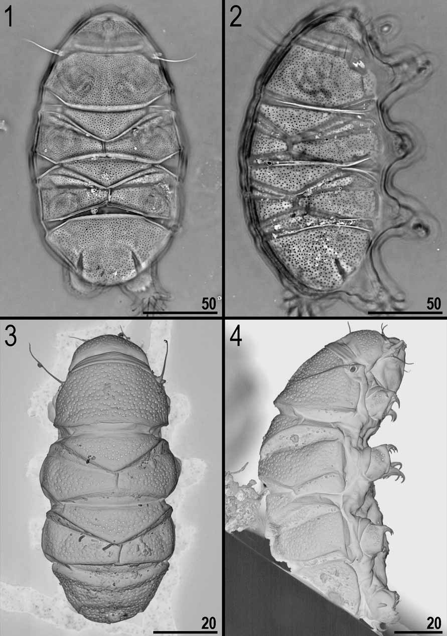
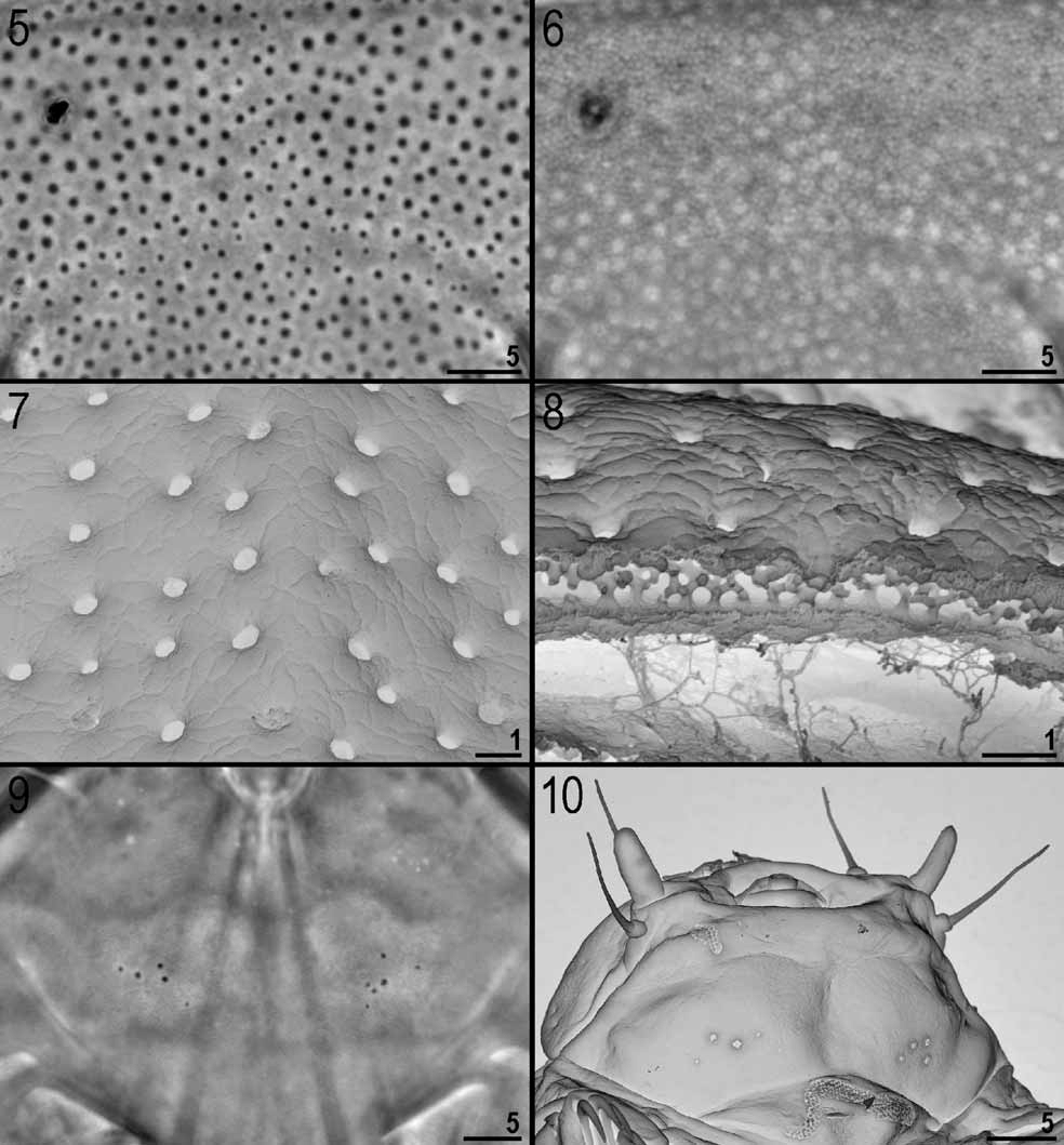
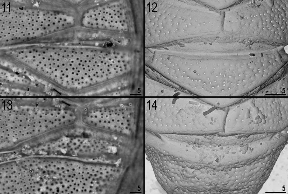
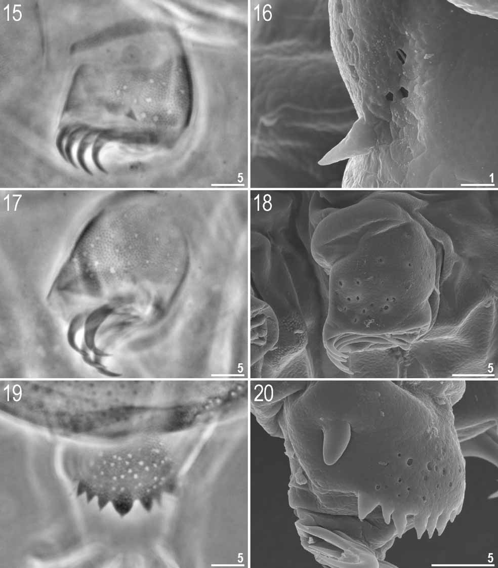
( Figs. 1–22 View FIGURES 1 – 4 View FIGURES 5 – 10 View FIGURES 11 – 14 View FIGURES 15 – 20 View FIGURES 21 – 22 , 25 View FIGURES 23 – 26 )
Material examined: Holotype (female) and 24 adults (18 females + 6 males) from the type locality in Costa Rica. The holotype and 14 paratypes (10 females + 4 males) were mounted in Hoyer’s medium and 10 paratypes (8 females + 2 males) were prepared for SEM.
Description: Adult female (measurements of the holotype): Body length 185.0 ( Figs. 1–4 View FIGURES 1 – 4 ). Body red. Red eyes visible in living specimens only. Internal cirrus 11.6 long, external cirrus 15.2 long; internal/external cirrus ratio = 0.76. Cephalic papilla 7.3 long ( Fig. 10 View FIGURES 5 – 10 ). Apart from head cirri and cirrus A no other appendices present. Cirrus A 31.6 long, ending in a point. Cirrus A /body ratio = 0.17. Clava 5.6 long.
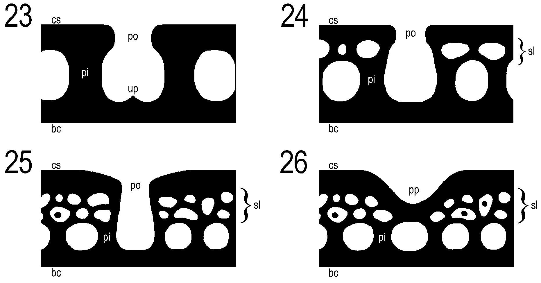
Dorsal (plate) cuticle with true pores and with true, but very fine granulation ( Figs. 5–20 View FIGURES 5 – 10 View FIGURES 11 – 14 View FIGURES 15 – 20 ). Granules (true granulation, on cuticle surface) present between scapular and first paired plates, on stripes in anterior portions of paired plates and in anterior part of second median plate ( Figs. 11–12 View FIGURES 11 – 14 ). Granules very small (0.2–0.3 in diameter). Cuticle appearance in SEM: Pores small (0.3–0.6 in diameter) ( Figs. 7 View FIGURES 5 – 10 , 12, 14 View FIGURES 11 – 14 ). ‘Sponge layer’ very well developed, small pillars present ( Figs. 8 View FIGURES 5 – 10 , 25 View FIGURES 23 – 26 ). Cuticle visible in PCM: Pores visible as bright/white dots; when focusing down through cuticle, cuticular pillars appear as faint dark dots ( Figs. 5–6 View FIGURES 5 – 10 ). Pores and cuticular pillars focusable. Ventral cuticle with very fine, dense and regular granulation. This granulation is caused by dense cuticular pillars. Several pores organised in 2 small irregular patches (left and right) present on ventral cuticle on head level (pores slightly smaller and shallower than those on dorsal plates) ( Figs. 9–10 View FIGURES 5 – 10 ).
Dorsal plates well developed. Head and scapular plates not faceted. In LM lateral portions of the scapular plate seem to be detached from the dorsal plate and form small shoulder plates (one on each side of the body) divided from the scapular plate by a thin bright stripe. This false division is caused by a bend of the plate where the cuticle is thinner; in SEM this pseudo division is not visible ( Figs. 2, 4 View FIGURES 1 – 4 ). Third median plate present, but relatively narrow, developed anteriorly ( Figs. 13–14 View FIGURES 11 – 14 ). Terminal plate faceted. Notches 18.2 long.
Spine on legs I in shape of narrow triangle, 2.2 long ( Figs. 15–16 View FIGURES 15 – 20 ). Papilla on legs IV finger-like, 4.0 long ( Fig. 20 View FIGURES 15 – 20 ). Outer cuticle of all legs with sculpture same as on dorsal plates, but with slightly smaller and shallower pores (0.1–0.6 in diameter) ( Figs. 15–20 View FIGURES 15 – 20 , see also Fig. 4 View FIGURES 1 – 4 ). Pores on leg often distributed irregularly. Dentate collar with 8 sharp, triangular teeth ( Figs. 19–20 View FIGURES 15 – 20 ).
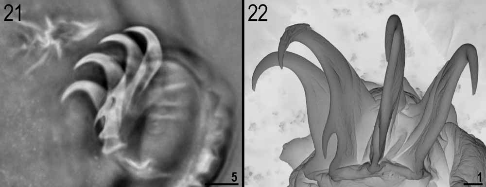
Claws of legs IV 12.6 long. External claws of all legs without spurs, internal claws of all legs with spur, directed downwards, growing out of claw at ca. 45o ( Figs. 21–22 View FIGURES 21 – 22 ). Spur on claws of legs IV relatively large (3.2 long; spur/claw ratio = 0.25). Claws of legs I–III slightly shorter.
Remarks: Extreme care is needed when determining the appearance of the anterior portion of the second median plate. When an animal is even slightly retracted, the porous posterior portions of the first paired plates ( Fig. 12 View FIGURES 11 – 14 ) may cover the anterior portion of the first median plate and may lead to an incorrect conclusion that the median plate is porous in its anterior part. The scapular plate can sometimes be slightly faceted in bigger animals. Because pores on legs and especially pores on the ventral head cuticle are less visible than those on the dorsal plates, they can be overlooked if an animal is not examined carefully enough.
Results of simple statistical analysis of measurements of selected morphological structures for 15 specimens (including the holotype) are given in Table 1 View TABLE 1 . No juveniles, larvae or eggs were found.
Even though the number of measured males is low (4 specimens), and the statistics with such a low number are not conclusive, it is worth noting that males seem to be smaller than females (p=0.003, t = 3.606, df = 13), have a shorter cirrus internus (p=0.016, t = 2.767, df = 13), externus (p<0.001, t = 4.936, df = 11.8) and cirrus A (p=0.048, t = 2.203, df = 12), also shorter claws (p=0.019, t = 3.038, df = 7) and spurs (p=0.035, t = 2.706, df = 6). The remaining characters do not differ significantly.
Type locality: Central America, Costa Rica, Cartago Province, Volcán Irazú National Park, liverwort from tree trunk, near the top of volcano, ca. 3400 m asl, 9o59' N, 83o51' W; 17 December 2002; leg. Ł. Kaczmarek.
Geographic distribution: Known only from the type locality.
Type depositories: Holotype and 7 paratypes are preserved at the Natural Sciences Collection, Faculty of Biology, A. Mickiewicz University, Umultowska 89, 61-614 Poznań, Poland; 7 paratypes are preserved in the collection of Ł. Michalczyk.
Etymology: The new species is dedicated to the first author’s very dear friend Joanna ‘ mzn ’ Ganczarek.
No known copyright restrictions apply. See Agosti, D., Egloff, W., 2009. Taxonomic information exchange and copyright: the Plazi approach. BMC Research Notes 2009, 2:53 for further explanation.