
Pealius satakshiae Dubey, 2019
|
publication ID |
https://doi.org/10.1080/00222933.2019.1691749 |
|
publication LSID |
lsid:zoobank.org:pub:9712A865-4DD6-457B-84B0-1C7873316DE4 |
|
DOI |
https://doi.org/10.5281/zenodo.3664761 |
|
persistent identifier |
https://treatment.plazi.org/id/03BDAE76-FFD9-FFBC-FE02-3FA0FBBE3500 |
|
treatment provided by |
Valdenar |
|
scientific name |
Pealius satakshiae Dubey |
| status |
sp. nov. |
Pealius satakshiae Dubey sp. nov.
( Figures 1 View Figure 1 (a) – 5(f))
Description
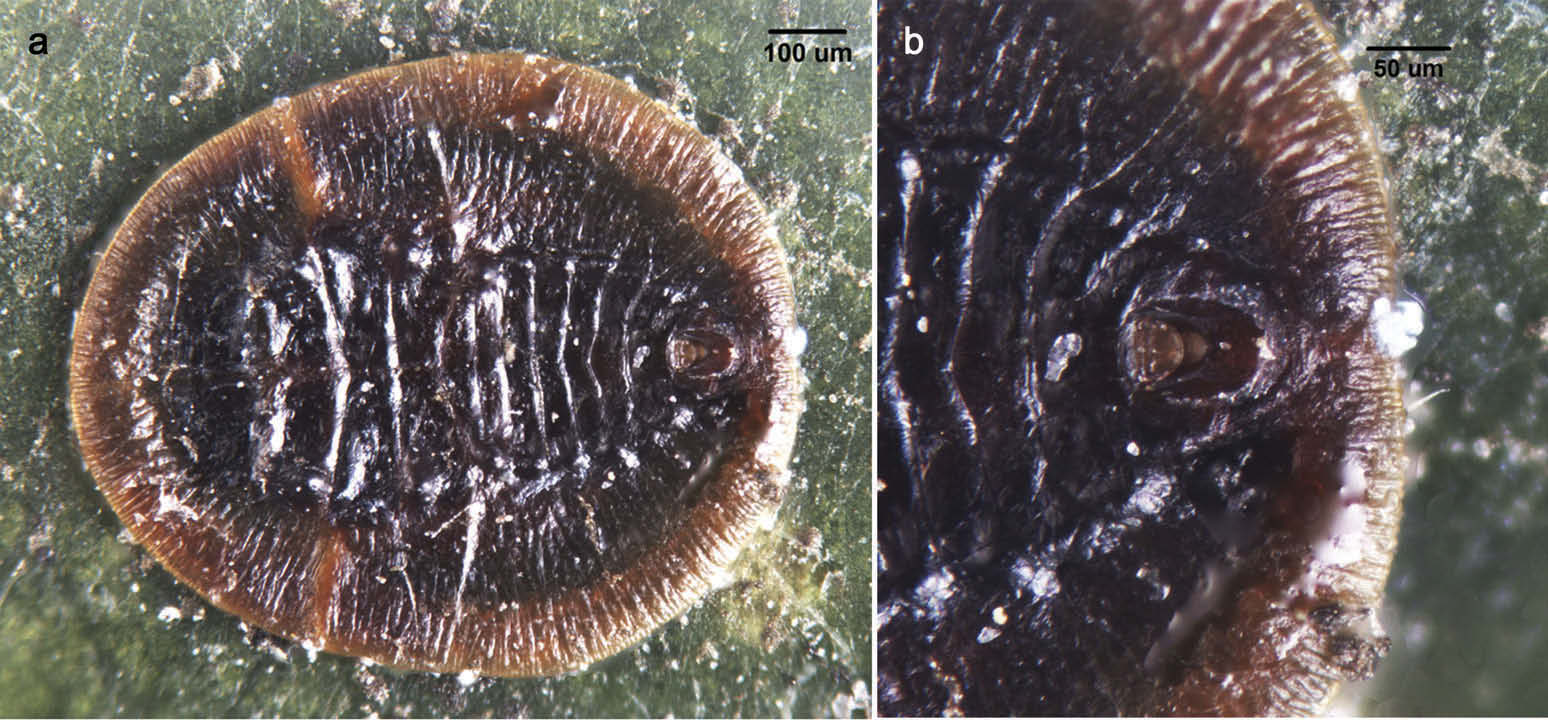
Puparium. Broadly oval; median and submedian area dark brown, submargin slightly raised, light brown ( Figure 1 View Figure 1 (a)); without secretion of wax over dorsum and periphery of margin, but small amount of white wax may be present in caudal and thoracic tracheal pore openings; vasiform orifice in a pit ( Figure 1 View Figure 1 (b)), but not so prominent in slide mounts; found singly on lower surface of leaves, one puparium per leaf; dimorphic, female 1175 – 1225 µm long, 1062 – 1100 µm wide; male 975 µm long, 825 µm wide.
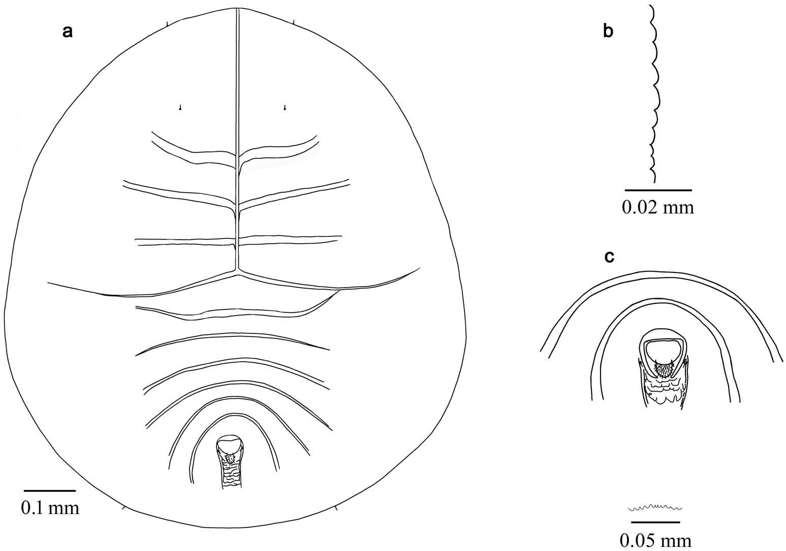
Margin. Regularly crenulate ( Figure 2 View Figure 2 (b)), 7 – 10 crenulations in 0.1 mm. Caudal and thoracic tracheal combs indicated in the margin ( Figure 3 View Figure 3 (c,d)) with a tuberclulate fold of dorsal cuticle. Thoracic tracheal furrows absent. Caudal tracheal furrow marked with transverse plates for some distance, posterior to vasiform orifice.
Dorsum ( Figures 4 View Figure 4 (a–h)ı 5d). Submargin broad, faintly demarcated from the dorsal disc by a faint ridge ( Figure 4 View Figure 4 (a)). Longitudinal moulting suture reaching margin and transverse moulting suture extending to and merging with submarginal lines. A pair of tubercles present on submedian area of pro- and metathorax ( Figure 4 View Figure 4 (e)). Cephalothoracic and abdominal segment sutures prominent, not reaching subdorsal area, abdominal segments rhachisform on submedian area ( Figure 4 View Figure 4 (f)). Intersegmental sutures prominently spaced. Submarginal lines prominent ( Figure 4 View Figure 4 (g)). Submedian depressions present on anterior part of each cephalothoracic and abdominal segment. Median length of cephalothorax and abdomen as measured equal, or the abdomen ( 489 – 610 µm long) is slightly longer than the cephalothorax ( 479 – 591 µm long). Abdominal segment VII much reduced medially as the pockets of segment VIII reaching anteriorly and overlapping intersegmental suture of VI/ VII. Median length of pro-, meso- and metathorax: 102 – 112, 67 – 80 and 72 – 75 µm, respectively, in female; 92, 62 and 60 µm, respectively, in male. Median length of abdominal segments I – VIII (A1 – A8): A1: 50 – 63, A2: 35 – 45, A3: 25 – 55, A4: 37 – 53, A5: 42 – 47, A6: 63 – 68, A7: 0, A8: 63 – 68 µm in the female, and A1: 48, A2: 35, A3: 43, A4: 40, A5: 38, A6: 48, A7: 0, A8: 55 µm in the male. Abdominal segment VIII forms a triangular figure. Distance between posterior end of vasiform orifice and caudal opening measured 150 – 157 µm long in the female ( 110 µm long in the male), furrow 40 – 48 µm wide posterior to vasiform orifice. Geminate pores large, each occupying an area 5 µm in length.
Vasiform orifice. Located in an elongate oval pit, posteriorly; elongate subcordate, posteriorly provided with transverse plates for some distance ( Figure 4 View Figure 4 (h)), 55 – 60 µm long, 55 – 57 µm wide (55 × 50 µm in the male); operculum subcordate, 30 – 33 µm long, 37 – 45 µm wide (27 × 37 µm in the male); lingula exposed, D-shaped, slightly constricted at base, not extending beyond posterior margin of the orifice.
Venter ( Figure 5 View Figure 5 (a–cıeıf)). Ventral submargin demarcated by a faint ridge ( Figure 5 View Figure 5 (a)). Thoracic tracheal folds smooth, without stipples ( Figure 5 View Figure 5 (b)). Caudal tracheal folds stipulated from spiracles to half length of the fold ( Figure 5 View Figure 5 (c)). A pair of ventral abdominal setae present, 7 – 15 µm long, 48 – 68 µm apart. Antennae 70 – 88 µm long (including keel of 5 µm long). Median and submedian area beneath intersegmental sutures with rows of fine stippling ( Figure 5 View Figure 5 (f)). Microsetae present at base of pro-, meso- and metathorax, approximately 3 µm long. Adhesive sacs and spiracles visible.
Chaetotaxy. Anterior marginal setae 11 – 15 µm long in female, 7 µm long in male. Cephalic setae small, pointed, 13 µm long. First abdominal setae absent. Eighth abdominal setae lateral to vasiform orifice, located almost equal to mid-width of operculum, 12 – 15 µm long. Caudal setae 20 – 25 µm long.
Host plant
Quercus leucotrichophora A. Camus (Fagaceae) .
Distribution
India: Himachal Pradesh.
Etymology
This species is named after Ms Satakshi, a high school student, who first indicated the occurrence of this species in the field.
Material examined
INDIA. Holotype ‘ puparium ’; Himachal Pradesh, Chail Wildlife Sanctuary, Indian Military Academy Campus Road ; 30.969°N and 77.197°E; 7375 ft above sea level etc.; 28 December 2014; A.K. Dubey & Satakshi ‘ leg. ’; on Quercus leucotrichophora , hand collection in paper covers ( NFIC-FRI) GoogleMaps . Paratypes: 18 puparia on 18 slides ( NFIC-FRI), 2 puparia on 2 slides with A.K. Dubey, data same as for the holotype .
Remarks
Puparia of the new species differ from all the described Pealius species in being larger in size, having the cephalus differentiated from the prothorax by a wide suture-like marking, prominent caudal and thoracic tracheal folds, and the medial length of seventh abdominal segment completely reduced, and by the absence of the first abdominal setae. The cephalus is differentiated in P. maculatus Takahashi (1942) and P. schimae Takahashi (1950) , but both differ in size and shape, and the former has a row of submarginal setae. The new species is placed in Pealius due to the shape of the lingula and because of the presence of a pyriform pit around the vasiform orifice. Pealius satakshiae sp. nov. also differs from P. schimae in its broader shape, and in the space from the hind end of the vasiform orifice to the caudal tracheal opening (i.e. equal to the length of the vasiform orifice and pit together), in the presence of a ventral fold, and by the absence of a median black patch on the abdomen.


No known copyright restrictions apply. See Agosti, D., Egloff, W., 2009. Taxonomic information exchange and copyright: the Plazi approach. BMC Research Notes 2009, 2:53 for further explanation.
|
Kingdom |
|
|
Phylum |
|
|
Class |
|
|
Order |
|
|
Family |
|
|
Genus |