
Hygrobates nigromaculatus Lebert, 1879
|
publication ID |
https://doi.org/ 10.11646/zootaxa.4277.1.2 |
|
publication LSID |
lsid:zoobank.org:pub:60FF6986-6FD7-473C-932E-9DF7AB1580FC |
|
DOI |
https://doi.org/10.5281/zenodo.6022536 |
|
persistent identifier |
https://treatment.plazi.org/id/03BE8793-2104-D654-FF7C-FC1152FCFC36 |
|
treatment provided by |
Plazi |
|
scientific name |
Hygrobates nigromaculatus Lebert, 1879 |
| status |
|
Hygrobates nigromaculatus Lebert, 1879
( Figs 1–26 View FIGURES 1 – 2 View FIGURES 3 – 10 View FIGURES 11 – 16 View FIGURES 17 – 23 View FIGURES 24 – 26 )
Material examined. 13 females, 14 males, 17 deutonymphs and 26 larvae: Russia, Yaroslavl Province, Nekouz District, Rybinsk reservoir, near village Pogorelka , June–August 2000–2002 ; 1 male, 1 deutonymph: Yaroslavl Province, Pleshcheevo Lake , 3 August 1975 ; 4 females, 6 males: Samara Province, Stavropol District, National natural Park “ Samarskaya Luka ”, Kuibyshev reservoir near village Koltsovo , June 1992. Larvae were reared from six females, the duration of the embryonic period was 10–15 days; 15 deutonymphs were reared from larvae in laboratory.
Diagnosis. Larva. Dorsal plate broad (L/W ratio 1.08–1.25) with convex lateral margins, coxal groups fused to each other medially but suture line between them present; tmas on coxae III absent; setae Pi and Pe equal in length; number of thickened distal setae from trochanter to tarsus on legs segments: I: 1-2-2-1-0, II: 0-2-3-4-0, III: 0-2-4-5-0.
Description. Dorsal plate broad (L/W ratio 1.08–1.25), covering large part of dorsum, acute anteriorly, with convex lateral margins ( Fig. 1 View FIGURES 1 – 2 ); simple setae long and thick, but setae Fch slightly longer than Vi, trichobothria Fp, Oi thin, short and equal in length. Eight pairs of setae (Oe, Hi, He, Sci, Sce, Li, Le, Si) situated in soft membrane; Oe, Hi and Sce subequal in length and longer than other setae situated in the membrane. Surface of dorsal plate with reticulation.
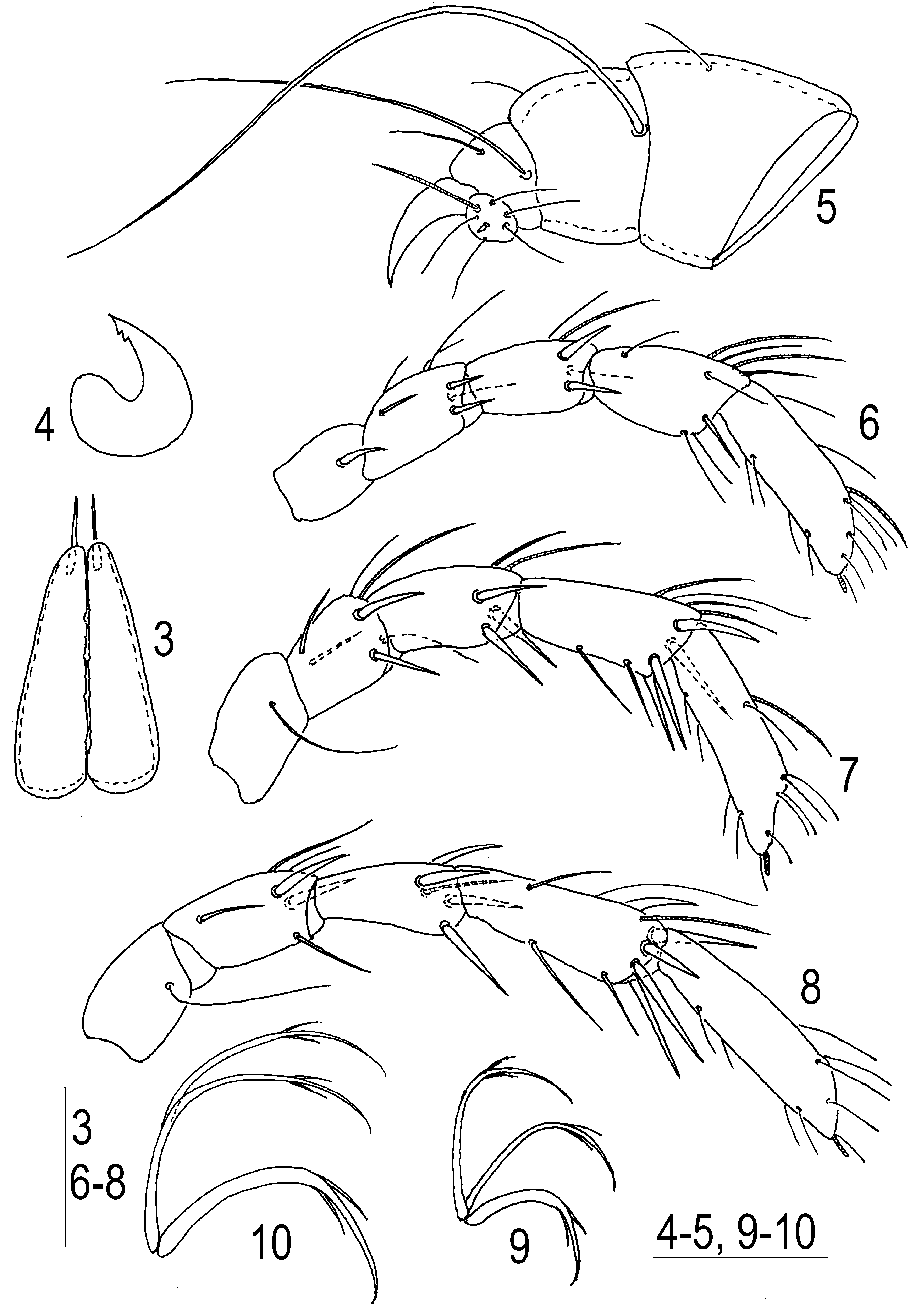
Both coxal groups fused to each other medially but suture line between them present ( Fig. 2 View FIGURES 1 – 2 ). Coxal setae C1 and C2 relatively short, thin and subequal in length; C3 located on coxae III anteriolaterally, slightly shorter than C4 but longer than anterior coxal setae. Transverse muscle attachment scar on coxae III absent. Setae Pi and Pe short, thin and equal in length. Urstigmae relatively large and oval shaped. Setae Ci ( Fig. 3 View FIGURES 3 – 10 ) very long and well thickened, inserted on moderately long projections. Excretory pore plate wider than long (L/W ratio 0.45–0.50), slightly convex anteriorly, “W”– shaped posteriorly and with muscle attachment scars anteriorly; excretory pore located near the center of plate between bases of setae Ae; bases of setae Ai located close to anterior margin; distance between bases of setae Ae–Ae two times larger than distance between Ai–Ai.
Basal segments of chelicerae fused to each other medially ( Fig. 3 View FIGURES 3 – 10 ), with slightly convex lateral margins, anterior edges more or less convex. Chela relatively small, crescent–shaped, with two small subapical teeth ( Fig. 4 View FIGURES 3 – 10 ).
Pedipalps stout ( Fig. 5 View FIGURES 3 – 10 ): P–1 very short, without setae; P–2 relatively large, with single dorsal seta near the middle of segment; P–3 with a very long, thick lateroproximal seta and a short, fine dorsoproximal seta; P–4 with two unequal setae and a massive dorsodistal claw; P–5 small, with one rather long solenidion and seven unequal simple setae, one of them very short.
Legs five–segmented. Shape and arrangement of setae, excluding eupathidia, as shown in Figs 6–8 View FIGURES 3 – 10 . Number of thickened setae from trochanter to tarsus: I–III-Leg: 1-2-2-1-0, II: 0-2-3-4-0, III: 0-2-4-5-0. Solenidion on II-Leg-5 located near the middle of segment ( Fig. 7 View FIGURES 3 – 10 ), and solenidion or solenidia on other segments of legs situated distally. Claws of legs III ( Fig. 10 View FIGURES 3 – 10 ) larger than claws of legs I and II ( Fig. 9 View FIGURES 3 – 10 ). Central claw and lateral claws in all legs nearly subequal in size and with two hair-like projections each.
Measurements (n=10). Dorsal plate L 195–240, W 180–220; setae Fch, Hi, Sci L 51–61,setae Fp and Oi L 9– 12, setae Vi L 48–55, setae Oe L 57–68; setae He and Sce L 41–48; setae Li, Le, Si and Se L 32–45, setae Ci L 145– 160, setae Pi and Pe L 19–23, setae C1 and C2 L 35–42, setae C3 L 41–55, setae C4 L 57–62; medial margins of coxae I–III L 1 53–175; gnathosomal bay L 44–48; urstigma L 19–22; excretory pore plate L 42–45, W 89–96; capitulum L 70–80; basal segments of chelicerae L 67–74, W 38–40, chela L 19–21; pedipalpal segments (P–1–5) L: 3–5, 30–32, 17–19, 6–7, 5–6; legs segments L: I–Leg-1–5: 25–32, 32–36, 32–36, 35–48, 41–55; II–Leg-1–5: 28–38, 35–39, 35–48, 44–52, 51–61; III–Leg-1–5: 38–42, 38–42, 41–48, 57–61, 57–71.
Deutonymph. Idiosoma oval in shape and somewhat flattened dorsoventrally. Integument soft and finely striated. Trichobothria Fp, Oi and setae Pi not associated with glandularia, other idiosomal setae associated with glandularia. Setae Fch ( Fig. 11 View FIGURES 11 – 16 ) much thicker than other idiosomal setae. Anterior coxal plates with short apodemes, posteromedial margin convexity rounded ( Fig. 12 View FIGURES 11 – 16 ). Coxal plate IV subtriangular in shape with rounded medial margin. Genital field with two plates, bearing two unequal acetabula (ac-2 slightly larger than ac-1) and three short, thin setae each; genital sclerite small and located between the plates.
Pedipalp moderately long ( Fig. 13 View FIGURES 11 – 16 ): P-1 without setae, P-2 with one dorsoproximal seta and two dorsodistal setae, ventral margin straight or slightly concave distally forming a right angle, a few denticles covering distal 3/5 of ventral margin of P-2; P-3 with two dorsodistal thick subequal setae, ventral margin convex with a few denticles covering distal half of segment; P-4 ventral setae on the same level near middle of segment.
Legs six–segmented without swimming setae. I-Leg-4 with two subequal relatively long distoventral setae, I- Leg-5 with two short unequal distoventral setae ( Fig. 14 View FIGURES 11 – 16 ); IV-Leg-4 with one long and three relatively short and thick distal setae, IV-Leg-5 with one short and two long, thick setae ( Fig. 15 View FIGURES 11 – 16 ). Claws of all legs with long external and short internal clawlets, lamella with concave ventral margin ( Fig. 16 View FIGURES 11 – 16 ).
Measurements (n=10). Idiosoma L 240–510; seta Fch L35–38; coxal plates I + capitulum mL 125–155; genital plates L 42–54, W 30–36, genital acetabula (ac-1–2) L/W 18–24/24–30, 24–30/30–33; cheliceral segments L: base 100–105, chela 42–50; pedipalp segments (P–1–5) L: 17–19, 48–54, 42–45, 65–72, 21–27; legs segments L: I-Leg-1–6: 30–42, 42–48, 42–55, 60–72, 65–78, 72–80; II-Leg-1–6: 30–42, 42–50, 54–60, 75–80, 85–105, 90–97; III-Leg-1–6: 30–45, 42–50, 60–66, 84–96, 95–100; IV-Leg-1–6: 60–66, 54–60, 72–90, 115–140, 125–145, 115–135.
Female. Dorsum similar to deutonymph. Setae Fch ( Fig. 17 View FIGURES 17 – 23 ) thicker than others idiosomal setae. Anterior coxal plates with short apodemes, posteromedial margin usually broadly rounded ( Figs 18 View FIGURES 17 – 23 ). Coxal plates IV subtriangular. Genital plates as long as or slightly shorter than gonopore, with 15–22 setae each; acetabula in a triangular position, medial margin slightly indented near the centre ( Figs 19–21 View FIGURES 17 – 23 ).
P-1 with a single dorsodistal seta; P-2 usually with six (occasionally five or seven) thick dorsal setae, ventral margin straight or slightly concave distally forming a right angle, denticles covering distal 2/3 or 3/5 of ventral margin of segment; P-3 with two dorsoproximal, two dorsodistal thick subequal setae and single thin distal seta, ventral margin straight, denticles covering distal 2/3 of segment; P-4 ventral setae on the same level near middle of segment ( Fig. 22 View FIGURES 17 – 23 ).
Legs slender, I-Leg-4 with two unequal, rather long, distoventral setae, I-Leg-5 with two short unequal distoventral setae ( Fig. 23 View FIGURES 17 – 23 ).
Measurements (n=10). Idiosoma L 1000–1450; seta Fch L35–45; coxal plates I + capitulum mL 300–330; genital plates L 150–175, W 65–90, genital acetabula (ac-1–3) L/W 50–65/25–35, 55–75/30–35, 60–75/42–55; cheliceral segments L: base 225–265, chela 110–125; pedipalp segments (P–1–5) L: 30–42, 108–115, 85–90, 145– 165, 40–48; legs segments L: I-Leg-1–6: 62–75, 90–115, 110–140, 160–210, 1 85–215, 165–205; II-Leg-1–6: 65–90, 100–115, 135–155, 210–240, 225–250, 195–240; III-Leg-1–6: 70–90, 100–150, 160–190, 260–290, 280–315, 245– 275; IV-Leg-1–6: 130–165, 135–165, 220–250, 305–350, 310–350, 275–330.
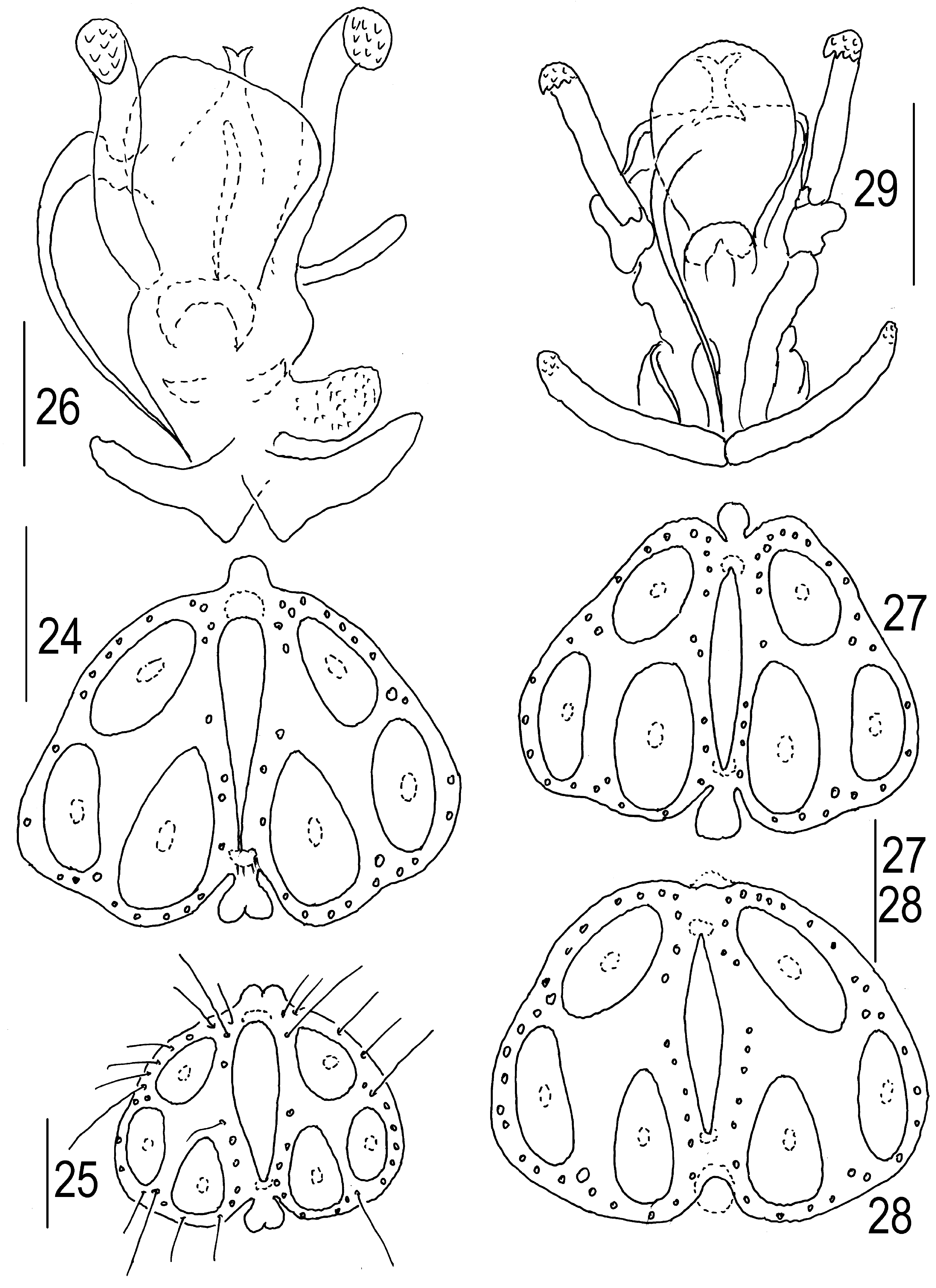
Male. Genital plate wider than long, anterior margin with a small, knob-shaped projection, posterior margin indented, with a short rounded or bifurcated median projection, with 17–28 pairs of setae; acetabula in a triangular position, in most cases ac-3 larger than ac-2 and ac-1 ( Fig. 24 View FIGURES 24 – 26 ) but occasionally all acetabula subequal ( Fig. 25 View FIGURES 24 – 26 ). Ejaculatory complex ( Fig. 26 View FIGURES 24 – 26 ) with long anterior arms and comparatively short posterior ones, proximal chamber large subrectangular, with straight anterior margin, pair of rudimentary proximal horns extending to anterior margin of proximal chamber.
Measurements (n=10). Idiosoma L 800–1150; seta Fch L30–42; coxal plates I + capitulum mL 270–320; genital plate L 150–165, W 190–225, genital acetabula (ac-1–3) L/W 50–60/36–42, 50–80/35–38, 55–85/42–50; cheliceral segments L: base 180–250, chela 90–105; pedipalp segments (P–1–5) L: 30–35, 95–115, 65–85, 125–140, 40–48; legs segments L: I-Leg-1–6: 50–65, 80–100, 100–125, 145–155, 160–175, 145–175; II-Leg-1–6: 50–65, 85– 100, 110–140, 165–215, 190–225, 180–215; III-Leg-1–6: 65–90, 100–115, 130–170, 205–250, 240–290, 200–250; IV-Leg-1–6: 125–150, 120–150, 175–215, 235–315, 265–315, 230–270.
Remarks. The morphology of the larva described by Wainstein (1980) as H. longiporus Thor, 1898 is in perfect agreement with the description of the larva given above. The combination of the dorsal plate broad with convex lateral margin, absent tmas on the coxal plates III, subequal setae Pi and Pe and I-Leg-2/3 with two heavy setae each is diagnostic for the larva H. nigromaculatus . Therefore, the larva described under the name H. longiporus ( Wainstein 1980) probably refer to H. nigromaculatus .
The dorsal plate in larvae H. nigromaculatus from the Netherlands is larger, L 274–321 µm and 292–300 µm, respectively ( Van Hezewijk & Davids 1985, Martin & Davids 2002) than Yaroslavl larvae, others morphometric data without essential differences.
The description of the deutonymph of H. nigromaculatus has been made based on two specimens from the European part of Russia and two specimens from the Asian part of Russia (Kamchatka) ( Tuzovskij 1990). The new description is based only on European specimens. The Asian specimens differ from European specimens by the presence of swimming seta on II–IV-Leg-5 and, probably, belong to another species.
The morphometric data of adults H. nigromaculatus , except of genital plate of male after Gerecke et al. (1960), are without essential differences from the known descriptions ( Martin 2010, Gerecke et al. 2016). See general remarks.


No known copyright restrictions apply. See Agosti, D., Egloff, W., 2009. Taxonomic information exchange and copyright: the Plazi approach. BMC Research Notes 2009, 2:53 for further explanation.