
Leydigia (Neoleydigia) laevis Gurney, 1927
|
publication ID |
https://doi.org/ 10.11646/zootaxa.2082.1.1 |
|
persistent identifier |
https://treatment.plazi.org/id/03BE87A4-4C66-5246-CE97-E69779B2FA64 |
|
treatment provided by |
Felipe |
|
scientific name |
Leydigia (Neoleydigia) laevis Gurney, 1927 |
| status |
|
IX. Leydigia (Neoleydigia) laevis Gurney, 1927 View in CoL
( Figs 262–337 View FIGURES 262–269 View FIGURES 270–284 View FIGURES 285–293 View FIGURES 294–315 View FIGURES 316–321 View FIGURES 322–337 )
Leydigia sp. , unpublished material of Sars from Victoria.
Leydigia laevis Gurney, 1927, p. 73 View in CoL –75, fig. 9; Smirnov 1971, p. 458, figs 567–568 (after Gurney); Smirnov and Timms 1983, p. 55, fig. 65 (after Gurney).
? Leydigia laevis Gurney View in CoL in Shiel and Dickson 1995, p. 33; Smirnov 1995, p. 5; Sanoamuang, 1998, Tab. 1.
? Leydigia australis Sars View in CoL in Chapman and Lewis 1976, p. 69, fig. 419.
Not Leydigia laevis Gurney View in CoL in Biswas 1971, p. 129, fig. 12L.
Type locality. A water body in vicinity of Longreach , Central Queensland, Australia (appr. 23º26'S, 144º14'E) GoogleMaps .
Lectotype (selected here). The adult parthenogenetic ♀, 775 µm, tube in 70% alcohol, NHM 2002.692 .
Paralectotypes (selected here). 8 parth. ♀♀, tube in 70% alcohol, NHM 2002.693 – 700 ., initially from the same sample as the lectotype .
Notes on typification. There was a tube in NHM marked as syntypes of L. laevis Gurney, 1927 with label "from mud, Longreach, R. Gurney Esq."; one of these specimens was selected as the lectotype. Gurney (1927) said that material was from three localities: "Blackboy Lagoon", "Whistling Duck swamp", and "a hole in thick timber (Gidea Scrub)", all in the neighbourhood of Longreach. Because the syntypes from the three localities were mixed in the tube, it is impossible to determine an exact type locality for the lectotype
Other material examined. Australia. New South Wales: Unnamed lake 8, 22 km SW Cooma, coll. 14.05.1975 by B. V. Timms, tubes AAK 2003-031 and NNS 1998-156 (36º20'S, 149º01'E) GoogleMaps ; Maffra Lake , 18 km E Dalgety, near Cooma, coll. 14.05.1975 by B. V. Timms, tube NNS 1997-049 (36º32'S, 149º00'E) GoogleMaps ; Lake Bullenbalong , 22 km NW Berridale, coll. 13.05.1975 by B. V. Timms, tube NNS 1997-013 (36º19'S, 148º42'E) GoogleMaps . Victoria: unknown localities " Victoria " and " Victoria 2", slides GOS F9663, F9667, F9668, tubes GOS F4147, F4148. Western Australia: Lake Caitup, coll. 06.09.1998 by Dept of Conservation and Land Management ( CALM), tube AAK 2002-014 (33º45'S, 121º53'E) GoogleMaps ; Magners Road Melaleuca Swamp, coll. 27.09.1998 by CALM, tube AAK 2002-011 (bad condition) ; Lake Bryde , coll. by CALM, tube AAK 2002- 010 (33º21'S, 118º50'E) GoogleMaps .
Diagnosis. Parthenogenetic female. Body subovoid to triangular-ovoid, height varies from instar to instar, maximum height in middle, dorsal margin uniformly curved from tip of rostrum to smooth posterodorsal angle; posterior margin regularly convex. Coarse striation on valves, clearly visible in adults, less so in juveniles, a fine striation on whole surface of valves and head shield very distinct. In anterior view, body compressed laterally, dorsum like rounded triangle in section. Head small, compound eye small, ocellus of similar size or slightly larger. PP = 6–8 IP, lateral head pores about 0.3–0.4 IP distance from midline, all head pores located on especial plate with finer, obscure reticulation. Labral keel widely-triangular-ovoid, posterior margin with three groups of long setules, anterior margin with fringe of long setules from base to apex, also lateral groups of fine, long setules, sometimes longer than longest marginal setules, and several groups of shorter setules on sides of keel. Setae at middle of ventral margin of valves with sparse, short setulation, located slightly submarginally, with long setules between them. In posterior portion of the margin, these setae setulated asymmetrically, posteriormost seta long. Posterior to the last marginal seta, a row of submarginal setules on inner face of posterior margin, ventrally these long and in continuous row; in middle and dorsal parts located relatively far from the margin, remarkably short. 'Setules' of marginal membrane distinctly larger.
Postabdomen broad, subovoid, robust, preanal margin shorter than anus, with 3–4 relatively large projections in its basal 2/3, preanal and postanal angles well defined, whole postanal margin from anus to base of claws regularly curved, no distal margin or dorso-distal angle. Postanal marginal denticles in numerous clusters, size increasing distally in each cluster. 9–10 fascicles of stout lateral setae, decreasing in size basally, normally 3–4 setae in each fascicle on distal portion, 3 setae in each fascicle in middle, where marginalmost setae of each fascicle significantly larger than rest, adjacent seta in about half as long, innermost seta very fine, 17–19 fascicles of lateral setules on basal half of postanal and anal margin. Postabdominal claw approximately as long as preanal plus anal portion of postabdomen, slightly curved in distal half, 0–2 small setules at claw base, basal spine small, but normally present.
Antenna I elongate, not reaching tip of rostrum, with 4–5 transverse rows of long setules on anterior face and series of shorter, more robust setules at tip. Sensory seta arising about 1/6 way along appendage. Largest aesthetasc shorter than half of antenna I length, projecting behind tip of rostrum. Antenna II with 3–4 stout spine-like setules on first, and 3–5 of these setules on second endopod segment. Apical swimming setae with basal and distal segments bilaterally armed with fine, long setules basally, and with unilaterally armed distal segments, no chitinous insertions within distal segments. Basal and distal lateral setae subequal in size, slender, shorter than apical setae.
Trunk limb I with ODL large, elongated, conically narrowing distally, bearing a row of fine setules and a long seta with distal segment unilaterally armed with short, dense setules, IDL with 3 medial clusters of minute setules, and 3 marginal clusters of setules: in basal and medium clusters setules long and fine, in distalmost cluster setules short and stout; first IDL seta smallest, with numerous short setules distally; second and third IDL setae of unequal length, with differing setulation, endite 3 with long, stiff seta 1, armed with long, fine setules and small receptor near base, endite 2 with two longer soft setae (e–f) subequal in length, seta 2 rudimentary, naked, with sensillum near base, endite 1 with rudimentary seta 3. Two ejector hooks of unequal size. Trunk limb II with exopodite ovoid, small, supplied with a tuft of short, robust setules. Distalmost scraper with naked basal segment, on distal lobe with distal group of short setules and basal cluster of long setules. On limb III, exopodite subquadrabgular, with single, rudimentary lateral seta, and two distal setae (1–2) of unequal size and different armature, seta 2 with distal segment armed bilaterally with setules of different length, filter plate III with distalmost and basalmost setae smaller than rest. Trunk limb IV exopodite with six setae, setae 2 and specially 1 short, but with long, fine setules, in filter plate distalmost seta with inflated basal segment and fully setulated, all setae with inflated tips. Trunk limb V with semi-circular exopodite, ont inner face of limb, two setulated setae of subequal length, but basalmost markedly thinner. Distal armature of gnathobase as a setulated lobe.
Ephippial female. Shape similar to parthenogenetic female, but dorso-distal angle completely smooth, dorsal wall of carapace additionally chitinised; in anterior view, body with a thick dorsal keel, as thick as in parthenogenetic female, although chamber for resting egg expands laterally. Ephippium transparent, slightly pigmented, with single resting egg within a chamber, with additional hexagonal sculpture, no demarcation line visible in female, but it appear during casting off the ephippium.
Adult male. Body low and ovoid and head larger than that in female, rostrum as in female. Postabdomen elongate, distally gradually narrows to inflated base of penis and postabdominal claws, lateral setae short. Penis thick, shorter than half length of claw.
Size. Juvenile and adult parthenogenetic female 560–1200 µm, ephippial female up to 1950 µm.
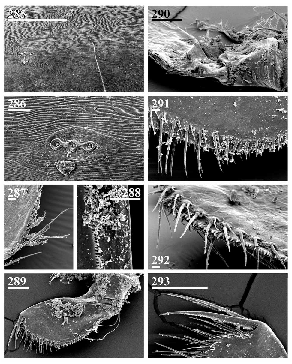
Full redescription. Parthenogenetic female. General. In lateral view body subovoid, height varies from instar to instar (body height/length = 0.58–0.63 in juveniles, 0.63–0.70 in adults), maximum height in middle ( Figs 262, 266 View FIGURES 262–269 , 270–272, 274 View FIGURES 270–284 ). Dorsal margin uniformly curved from tip of rostrum to smooth postero-dorsal angle; posterior margin regularly convex. Postero-ventral angle completely rounded; ventral margin slightly convex. Coarse striation on valves, clearly visible in large adults ( Figs 266 View FIGURES 262–269 , 271–272, 279 View FIGURES 270–284 ), but less so in juveniles ( Fig. 274 View FIGURES 270–284 ) and small adults ( Figs 262 View FIGURES 262–269 , 270 View FIGURES 270–284 ). Fine striation on whole surface of valves and head shield very distinct ( Figs 278–280 View FIGURES 270–284 , 285–286 View FIGURES 285–293 ), small 'dots on valves' are not external structures. In anterior view, body significantly compressed laterally, with maximum width in region of brood pouch, without dorsal keel, but dorsum like rounded triangle in section ( Figs 267 View FIGURES 262–269 , 273 View FIGURES 270–284 ), in dorsal and ventral view body subovoid, elongated ( Figs 268–269 View FIGURES 262–269 ).
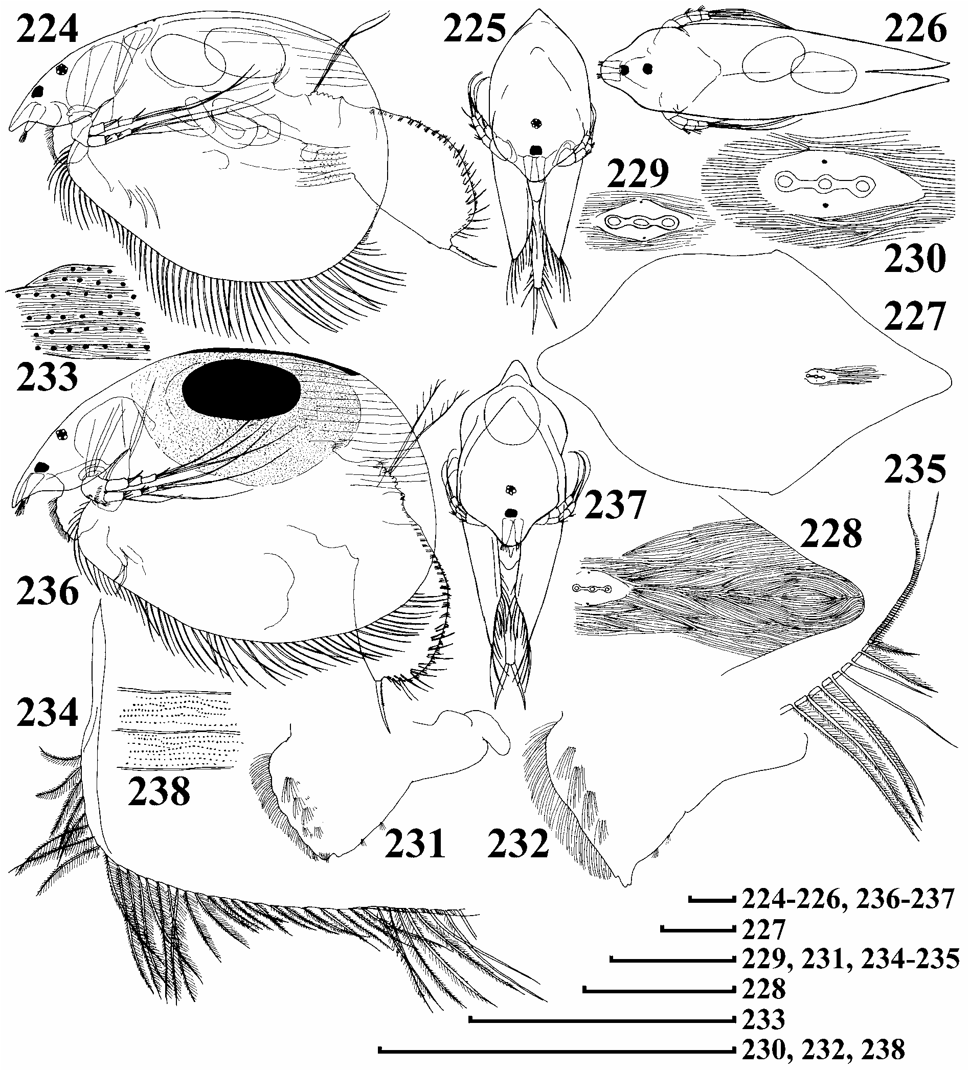
Head relatively small, triangle-round in lateral view, with short, blunt, downward pointing rostrum ( Fig. 263 View FIGURES 262–269 ). Compound eye small, ocellus of similar size or slightly larger, of irregular shape, distance from tip of rostrum to ocellus equal to or slightly more than that between ocellus and eye.
Head shield wide, in posterior portion three major head pores with relatively wide connection between them, PP = 6–8 IP ( Figs 277–278 View FIGURES 270–284 , 285–286 View FIGURES 285–293 ). Central pore of the same size as anterior or posterior, or somewhat narrower, located in middle. Lateral head pores about 0.3–0.4 IP distance from midline, at level of central major head pore. All head pores located on a plate with finer, obscure reticulation.
Labrum with fleshy main body, small distal labral plate, and large medial labral keel ( Figs 294–297 View FIGURES 294–315 ). Distal plate short, setulated. Main body with lateral projections, setulated dorsally, with series of small setules distally. In lateral view, labral keel widely-triangular-ovoid, with well-defined apex. Posterior margin slightly convex, with three tufts of long setules, anterior margin regularly convex, with fringe of long setules from base to apex, also 5–6 lateral groups of fine, long setules, sometimes longer than longest marginal setules, and several groups of shorter setules on sides of keel.
Valves large, subovoid, with numerous setae on ventral margin, longest in posterior half. In middle of margin setae with sparser, shorter setulation, bases located slightly submarginally, with long setules between them ( Fig. 298 View FIGURES 294–315 ). In posterior, thickened, portion these setae asymmetrically setulated (but not inflated), the posteriormost seta long, no setules between their bases ( Figs 287 View FIGURES 285–293 , 299–300 View FIGURES 294–315 ). Posteriorly to the last marginal seta, a row of submarginal setules starts on inner valve face of valve posterior margin ( Figs 300–301 View FIGURES 294–315 ). Ventrally these setules long and not in continuous series; in medium and dorsal parts located far from the margin, and remarkably short ( Figs 288 View FIGURES 285–293 , 302–303 View FIGURES 294–315 ). 'Setules' of marginal membrane distinctly larger.
Thorax relatively long. Abdomen short, distal edges of segments with transverse rows of setules, basal abdominal segment with abdominal projection.
Postabdomen wide, subovoid, robust, with markedly widening postanal portion ( Figs 265 View FIGURES 262–269 , 276 View FIGURES 270–284 , 289 View FIGURES 285–293 , 304–305 View FIGURES 294–315 ).Ventral margin almost straight, specially in middle, with groups of minute setules. Preanal margin ( Fig. 290 View FIGURES 285–293 ) shorter than anus, with 3–4 relatively large projections in basal 2/3, preanal and postanal angles well defined ( Figs 264 View FIGURES 262–269 , 275 View FIGURES 270–284 , 306–307 View FIGURES 294–315 ). Whole postanal margin from anus to basis of claws regularly curved, no distal margin or dorso-distal angle. Each side of postabdomen provided with row of long postanal marginal denticles ( Figs 291–292 View FIGURES 285–293 , 308 View FIGURES 294–315 ), organised in numerous clusters, size increasing distally in each cluster. On postanal margin, about 9–10 fascicles of stout lateral setae, decreasing in size basally, normally 3–4 setae in each fascicle on distal portion, 3 setae in each fascicle in middle, where marginalmost setae of each fascicle significantly larger than rest, second about half as long, next seta very fine. These setae merge with about 17–19 fascicles of lateral setules on basal half of postanal and anal margin. Additional row of fine fascicles in preanal region.
Postabdominal seta relatively short, but longer than anal plus preanal margin; its distal segment shorter than basal, and armed with long, densely located setules ( Fig. 304 View FIGURES 294–315 ).
Postabdominal claw approximately as long as preanal plus anal portion of postabdomen, slightly curved in distal half or straight, with row of small setules along ventral margin ( Figs 293 View FIGURES 285–293 , 309–311 View FIGURES 294–315 ). On lateral side, two successive series of slender setules along the dorsal margin, setules of basal series long, with distalmost setules specially long and thickened, also, 0–2 small setules at claw base. Basal spine small, but normally present ( Figs 316–318 View FIGURES 316–321 ).
Antenna I ( Figs 312 View FIGURES 294–315 , 319 View FIGURES 316–321 ) elongate, not reaching tip of rostrum, with 4–5 transverse rows of long setules on anterior face and series of shorter and more robust setules at tip. Sensory seta long, slender, arising about 1/ 6 way from tip. Nine aesthetascs projecting behind tip of rostrum, of slightly varied size, largest shorter than half appendage length.
Antenna II ( Fig. 313 View FIGURES 294–315 ) relatively short. Coxal part with two sensory setae, and lobe with row of fine, long setules. Basal segment robust, with transverse series of numerous, fine, long setules ( Fig. 314 View FIGURES 294–315 ), rudimentary distal spine and short setules at distal margin. Antennal branches elongated, exopod and endopod subequal in length, all segments cylindrical, with fascicles of setules; 3–4 long and stout spine-like setules on first, and 3- 5 setules on second endopod segment ( Figs 320–321 View FIGURES 316–321 ).
Antennal formula, setae 0–0–3/1–1–3, spines 1–0–1/0–0–1. On both exopod and endopod three long apical swimming setae, all with basal and distal segments bilaterally armed with fine, long setules basally, and with unilaterally armed distal segments, no chitinous insertions within distal segments ( Fig. 315 View FIGURES 294–315 ). Basal and distal lateral setae subequal in size, somewhat shorter than apical setae. Spine on basal segment of exopod long, reaching middle of apical segment. Apical spines of exopod and endopod of subequal length, only a littlet longer than apical segments.
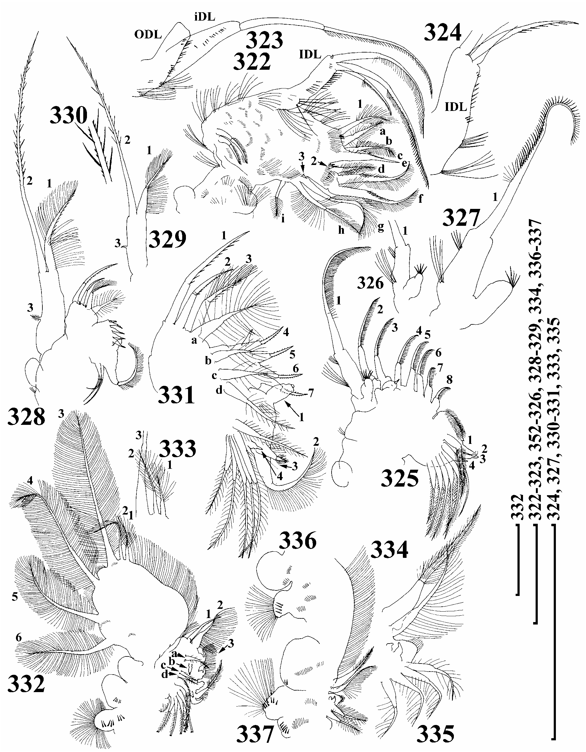
Trunk limb I ( Fig. 322 View FIGURES 322–337 ) with a small, globular epipodite, without accessory seta, ODL large, elongate, narrowing distally, with a row of fine setules and long seta with distal segment unilaterally armed with short, dense setules. IDL with 3 medial clusters of short setules, and 3 marginal clusters of setules: in basal and medial clusters setules long and fine, in distalmost setules short and stout ( Figs 323–324 View FIGURES 322–337 ), and three bisegmented setae; first seta smallest, with numerous short setules distally; second and third setae unequal in length, differing in setulation.
Endite 3 with soft setae a–c of subequal size, and long, stiff seta 1, armed with long, fine setules, a small receptor near its base. On endite 2 densely feathered setae d–f, seta d short, setae e–f subequal in length, a rudimentary, naked seta 2 with sensillum near base. Endite 1 with 2-segmented setae g–i, and a rudimentary seta 3. Fascicles of slender setules on inner face of limb, plus groups of longer, more robust setules on ventral margin. Two ejector hooks of unequal size. On limb base, a maxillar process with single row of long, fine setules.
Trunk limb II ( Fig. 325-327 View FIGURES 322–337 ) with small, globular epipodite. Exopodite ovoid, small, without setae, but with a tuft of short, robust setules. Inner portion of limb ('endopodite') with eight scrapers. Distalmost scraper 1 with long setules distally, naked basal segment, on distal lobe with distal tuft of short setules and basal group of long setules. A series of small projections posterior to scrapers 2–3, plus small sensillum. Portion of gnathobase bordering 'endopodite' somewhat inflated, and densely setose. Distal armature of gnathobase with four elements. Filter plate with seven setae, distalmost somewhat shorter than rest, with inflated basal segment and fully setulated, while others armed bilaterally.
Trunk limb III with sub-globular epipodite ( Fig. 328-331 View FIGURES 322–337 ). Exopodite subquadrabgular, with rudimentary lateral seta 3, and distal setae 1–2, seta 2 with distal segment armed bilaterally with setules of different lengths. Distal endite with three setae, 1 and 2 stout, distal segments with minute setules, 3 asymmetrically armed distally. A small sensillum near each seta 2 and 3. Basal endite with setae 4–7, armed with fine, very short setules distally, bottle-shaped sensillum near seta 4. On posterior limb face, four soft setae, seta a longest, all armed with long, widely separated setules distally.
Distal armature of gnathobase with four elements. Filter plate with six bilaterally setulated setae, basalmost smaller than others, distalmost also smaller, similar in size and armature with soft seta at basal endite.
Trunk limb IV with setulated pre-epipodite and globular epipodite, both with series of robust setules ( Fig. 332 View FIGURES 322–337 ). Exopodite wide, subovoid, with six setae, seta 1 shortest, seta 2 next shortest, seta 3 largest, 4–6 diminishing in size, all with similar armature of long, fine setules ( Fig. 333 View FIGURES 322–337 ).
Inner portion of limb IV with four marginal setae, seta 1 stout, with minute setules distally, 2–4 with inflated basal segments and slender, unilaterally setulated distal segments, in seta 2 basal segment also setulated, a slender sensillum near seta 2. On posterior limb face, three soft setae (a, c–d) and straight, trilobed sensillum (b). Distal armature of gnathobase with four elements. Filter plate with five setae, distalmost with inflated basal segment and fully setulated, all setae with inflated tips.
Trunk limb V with small setose pre-epipodite, and globular epipodite, both with rows of setules, among which some are robust ( Figs 336–337 View FIGURES 322–337 ). Exopodite large, with four setae. Inner limb portion an elongate, flat lobe, with setose inner margin ( Fig. 334 View FIGURES 322–337 ). On inner face, two setulated setae of subequal length, basalmost markedly slender. Distal armature of gnathobase a setulated lobe, with 2 small projections and two setae in 'filter plate' (In a single, atypical female I found 4 setae in filter plate V, a recapitulation to an ancestral state, Fig. 225 View FIGURES 224–238 ).
Ephippial female. In lateral view, shape similar to parthenogenetic female, body height/length = 0.64–0.68, but dorso-distal angle completely smooth, dorsal wall of carapace additionally thickened ( Fig. 281 View FIGURES 270–284 ). In anterior view, body with a thick dorsal keel, thickness as in parthenogenetic female, though chamber for resting egg expands laterally ( Fig. 282 View FIGURES 270–284 ). Ephippium transparent, only slightly brown, with single resting egg within chamber with additional hexagonal sculpture, no demarcation line visible in female, but it appears during casting off of ephippium ( Figs 283–284 View FIGURES 270–284 ). Only a dorsal portion of valves retained in ephippium, rest rejected after shedding.
Male. No original material was accessible. According to Gurney's (1927) illustrations, body lower and more ovoid and head larger than in female, rostrum as in female. Eye and ocellus of subequal size. Postabdomen elongate, with almost straight ventral margin and regularly convex dorsal margin, maximum height in middle, distally postabdomen tapering to inflated base of penis and postabdominal claws, lateral setae short, their arrangement unclear. Penis thick, shorter than half length of claw. Trunk limb I with thin, Ushaped copulatory hook.
Size. Lectotype, relatively young adult female 775 µm; juvenile and adult parthenogenetic females from unnamed lake 8, 22 km SW Cooma 560–1200 µm (n = 100), ephippial females up to 1950 µm (n= 4); females from Victoria, up to 1075 µm (n=20). According to Gurney (1927), female up to 600 µm and adult male 300 µm.
Differential diagnosis. L. laevis is unique in having the 'setules' of the marginal membrane on the posterior valve margin much larger than those of the submarginal row.
Taxonomical notes. This species was obtained by G.O. Sars from Victoria and labelled Leydigia sp. , so, he had doubts about its identity. The results of his study of this species remained unpublished, as did those on the future Neothrix armata Gurney, 1927 and other interesting animals (see Smirnov 1992).
Gurney's (1927) description was brief, but accompanied by realistic illustrations. In his description there were no traits which are helpful for the discrimination of L. laevis from any other acanthocercoides -like species. In addition, Gurney (1927) had only small adult females (only up to 600 µm), due to which he noted the absence of coarse striation, which is not characteristic for smaller females, but is well-defined in larger adults.
Smirnov (1971) and then Smirnov and Timms (1983) discriminated L. laevis from L. ciliata by the punctation pattern of the valves, which is a subjective feature. These authors used the illustrations of Gurney (1927). The description of Biswas (1971) from India suggested that he was dealing with other species (according to postabdomen form). A record from Thailand ( Sanoamuang 1998) must be checked.
Distribution. At this moment, I can confirm L. laevis only in Australia, where it is the most common species of the genus.


| NHM |
University of Nottingham |
| R |
Departamento de Geologia, Universidad de Chile |
| V |
Royal British Columbia Museum - Herbarium |
No known copyright restrictions apply. See Agosti, D., Egloff, W., 2009. Taxonomic information exchange and copyright: the Plazi approach. BMC Research Notes 2009, 2:53 for further explanation.
|
Kingdom |
|
|
Phylum |
|
|
Class |
|
|
Order |
|
|
Genus |
Leydigia (Neoleydigia) laevis Gurney, 1927
| Kotov, Alexey A. 2009 |
Leydigia laevis
| Shiel, R. J. & Dickson, J. A. 1995: 33 |
| Smirnov, N. N. 1995: 5 |
Leydigia australis
| Chapman, A. & Lewis, M. 1976: 69 |
Leydigia laevis
| Biswas, S. 1971: 129 |
Leydigia laevis
| Smirnov, N. N. & Timms, B. V. 1983: 55 |
| Smirnov, N. N. 1971: 458 |
| Gurney, R. 1927: 73 |