
Tritonia, Cuvier, 1797
|
publication ID |
https://doi.org/10.11646/zootaxa.4526.4.1 |
|
publication LSID |
lsid:zoobank.org:pub:3CFFF3AC-C447-4FCE-B6F8-D2B7BAE8B678 |
|
DOI |
https://doi.org/10.5281/zenodo.5971398 |
|
persistent identifier |
https://treatment.plazi.org/id/03BE87CC-FFAF-DE07-9B8E-9FE67C175930 |
|
treatment provided by |
Plazi |
|
scientific name |
Tritonia |
| status |
|
Tritonia View in CoL sp.
( Figs. 2B View FIGURE 2 , 3 View FIGURE 3 C–D, 7–8)
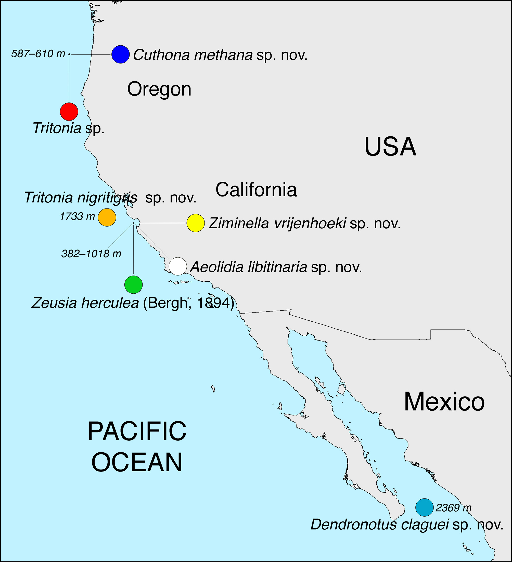
Material examined. Hydrate Ridge, Oregon, Northeast Pacific Ocean ( 44.67° N, 125.098° W), 587 m depth, ROV Jason II (dive 593), 6 Sep 2011, 1 specimen 17 mm preserved length (SIO-BIC M12395), GenBank accession numbers: MH756139 View Materials ( COI), MH756134 View Materials ( 16S), MH 756145 View Materials ( H3).
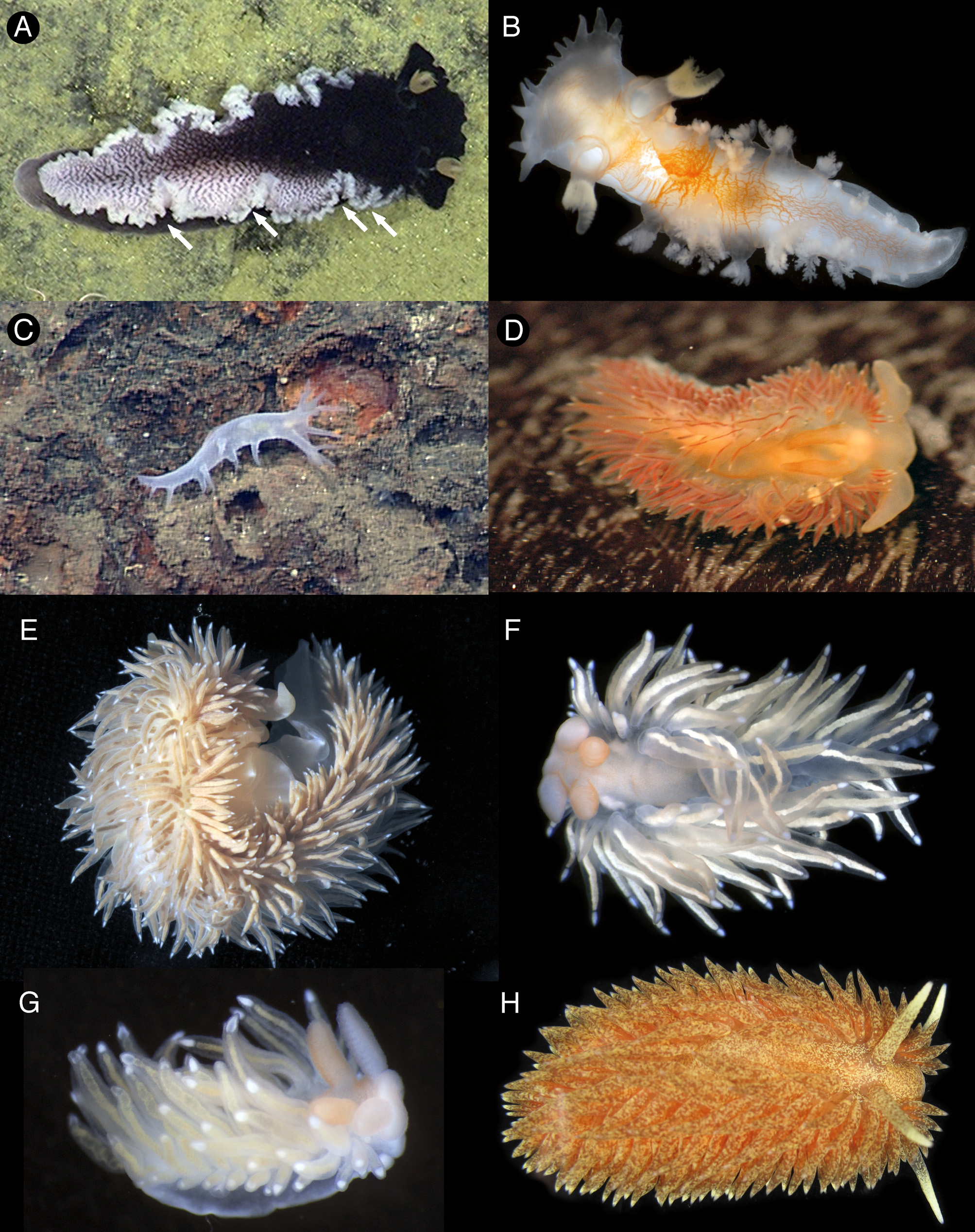
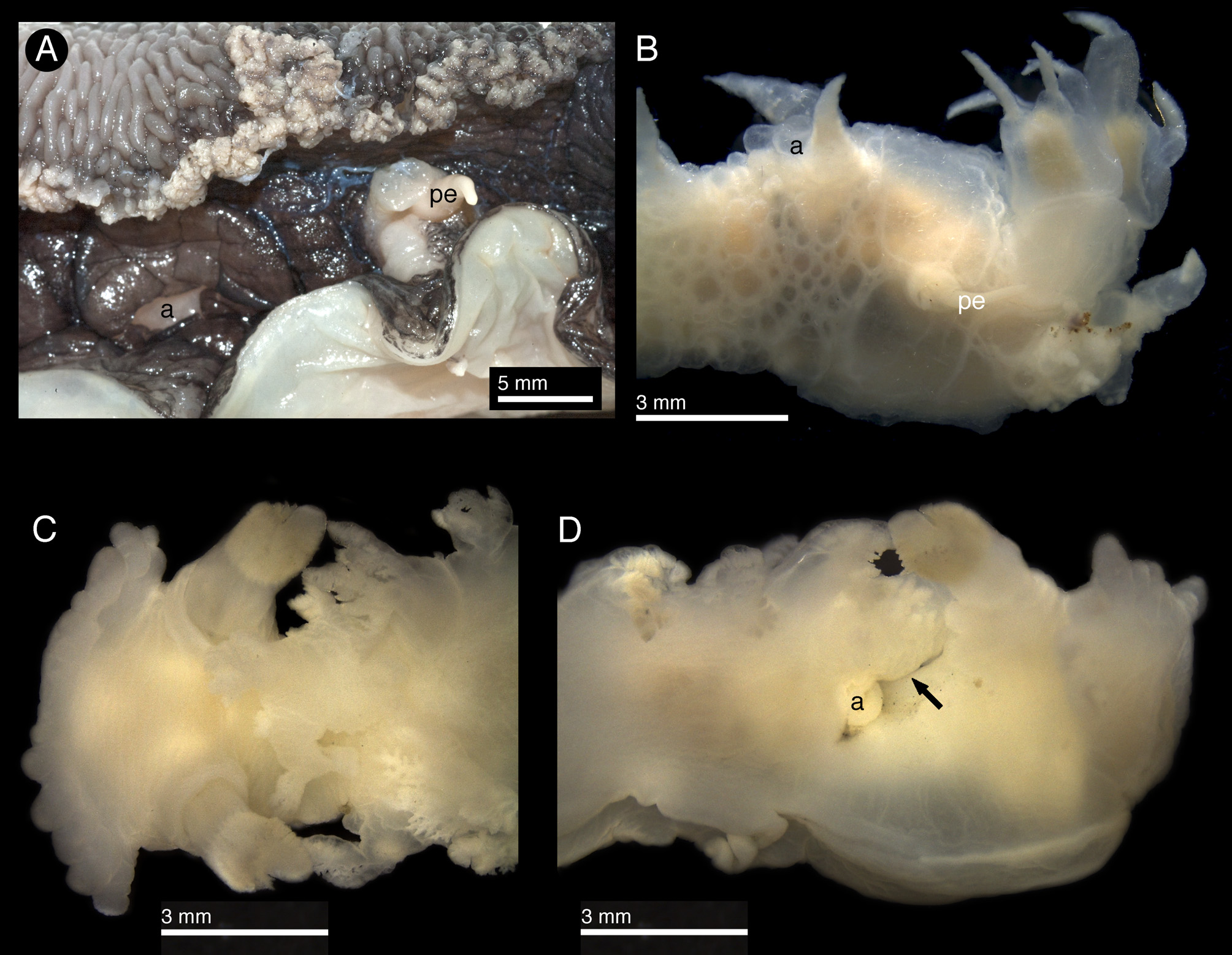
Description. Body elongate, narrow, tapering towards the posterior end ( Fig. 2B View FIGURE 2 ). Lateral margins with 8 branched appendages (cerata), tripinnate, decreasing in size towards the posterior end ( Fig. 2A View FIGURE 2 ). Dorsum smooth, lacking tubercles or projections. Velum wide, triangular, with 14 conical, simple projections. Rhinophores elongate, narrow, each with a smooth, long stalk. Rhinophoral clubs with 5 tripinnate papillae, forming a circle closed posteriorly by a much more elongate papilla, covered with lamellae on its posterior end ( Fig. 3C View FIGURE 3 ), located on anterior end of notum. Rhinophoral sheaths low, with smooth edges. Foot smooth, elongate posteriorly, visible dorsally at posterior end and in notum invaginations. Anal opening on right side of body ( Fig. 3D View FIGURE 3 ). Background color translucent white, with a concentration of orange pigment on the central dorsum. Body contractions concentrate the pigment, forming an irregular network of red lines. Notum edge translucent white with the lateral appendages (cerata) opaque white to cream. Velum and velar appendages translucent white. Rhinophores with translucent white stalks and beige sheath clubs. Foot uniform translucent white.
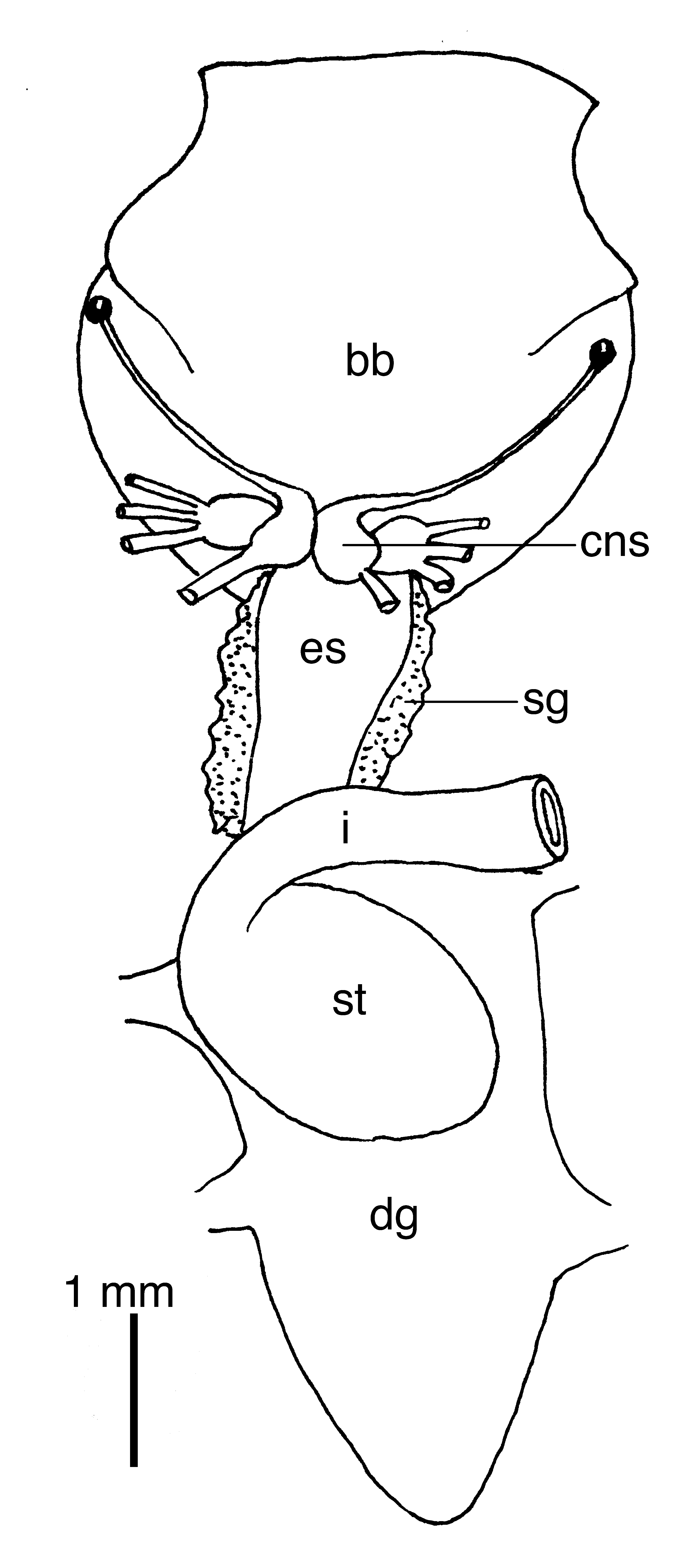
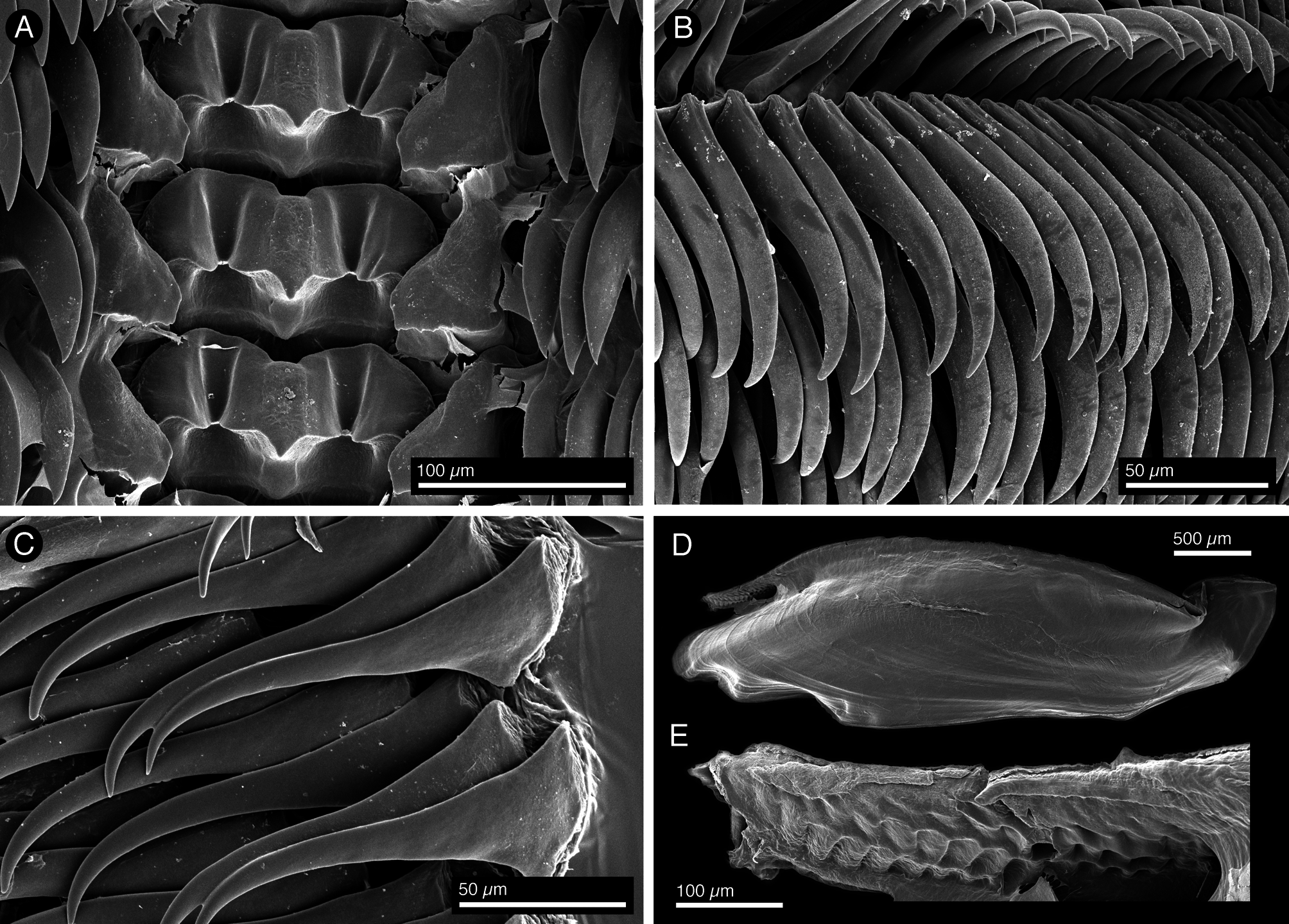
Digestive system with large, muscular buccal bulb ( Fig. 7 View FIGURE 7 ). Esophagus narrow, connecting anteriorly into buccal bulb, where two large, flat salivary glands connect. Intestine emerging dorsally from the stomach, forming a loop towards the right side of body where it opens into the anus ( Fig. 3D View FIGURE 3 ). Radular formula 44 × 49.1. 49 in the single specimen examined. Rachidian teeth broad, with a short, wide central cusp, shorter than tooth, and 1–2 denticles or folds on each side of central cusp ( Fig. 8A View FIGURE 8 ). Lateral teeth hook-shaped, with sharp, elongate cusps lacking denticles ( Fig. 8B View FIGURE 8 ). Outer lateral teeth very elongate with reduced bases ( Fig. 8C View FIGURE 8 ). Jaws elongate ( Fig. 8D View FIGURE 8 ), with a differentiated masticatory border ( Fig. 8E View FIGURE 8 ).
Reproductive system absent, specimen was probably castrated as suggested by damage on the right side of the body visible in the live ( Fig. 2B View FIGURE 2 ) and preserved ( Fig. 3D View FIGURE 3 ) specimen.
Biology. This species was collected at 587 m, from Hydrate Ridge, an area of methane seeps about 44 nautical miles off Newport, Oregon ( Fig. 1 View FIGURE 1 ). It came from an inactive, experimental rock (Levin Lab E12, rock F), upon which an unidentified soft coral (SIO-BIC Co2330) was growing. Other organisms collected from the soft coral were an unidentified amphipod (SIO-BIC C11265) and the polychaete worm Eusyllis nuchalata ( SIO-BIC A2679 ) .
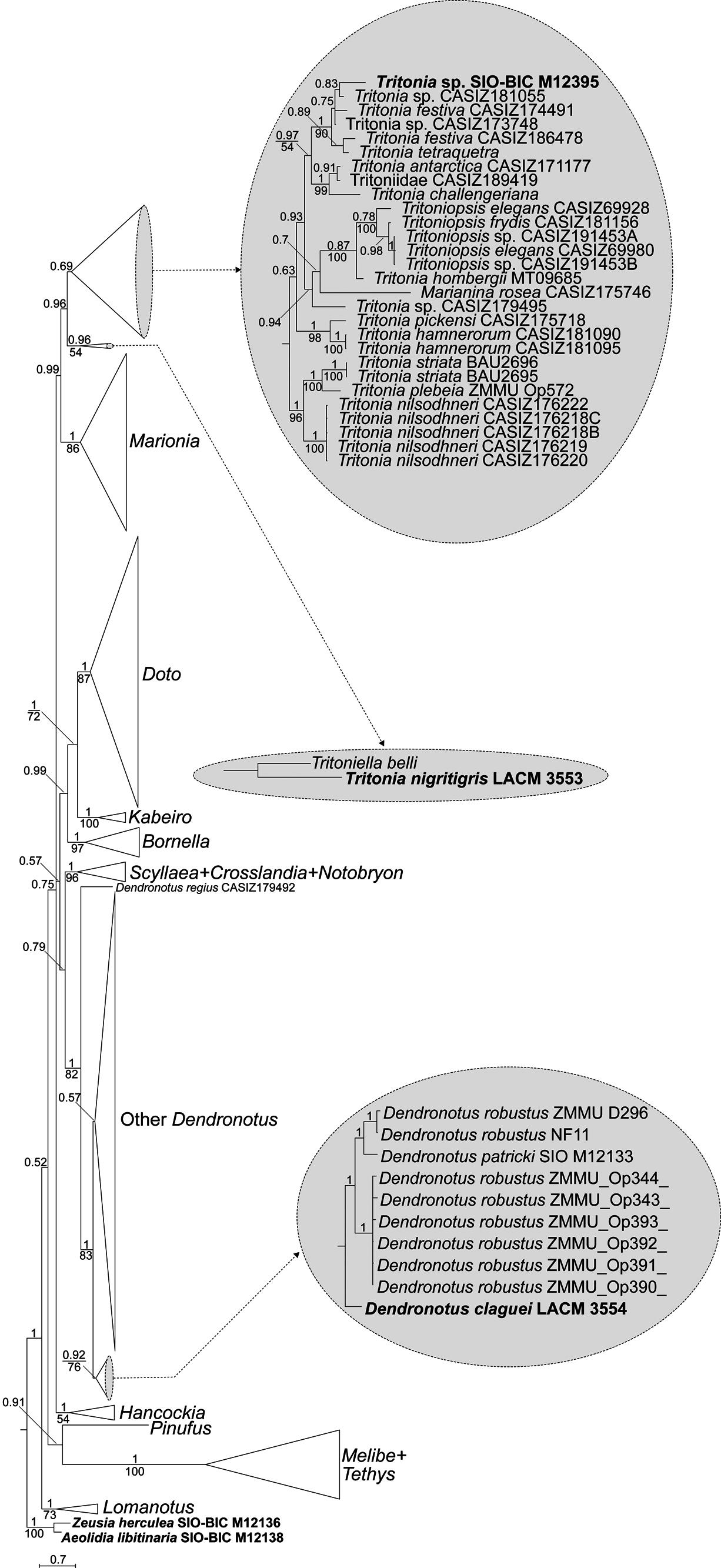
Phylogenetic position. Tritonia sp. is placed in a clade including Eastern Pacific species of Tritonia such as T. festiva (Stearns, 1873) and Tritonia tetraquetra ( Pallas, 1788) , as well as two other unidentified specimens ( Fig. 6 View FIGURE 6 ).
Remarks. Tritonia sp. appears to be different from other species of Tritonia described to date. However, the single specimen examined lacked a reproductive system. Damage on the right side of the body may suggest the animal was either harmed during collection or castrated by a parasite.
The external morphology of Tritonia sp. resembles that of Tritonia myrakeenae Bertsch & Mozqueira Osuna, 1986 , also found in the Eastern Pacific, but the latter lacks red pigment on the dorsum and often has two white pigment patches posterior to the head ( Bertsch & Mozqueira Osuna, 1986), absent in Tritonia sp. The radula of T. myrakeenae is also different from that of Tritonia sp. The former has a rachidian tooth with an elongate central cusp and two strong lateral denticles ( Bertsch & Mozqueira Osuna, 1986), whereas Tritonia sp. has a much broader rachidian with a shorter central cusp. Unfortunately, no molecular data for T. myrakeenae is available for comparison.
| MH |
Naturhistorisches Museum, Basel |
No known copyright restrictions apply. See Agosti, D., Egloff, W., 2009. Taxonomic information exchange and copyright: the Plazi approach. BMC Research Notes 2009, 2:53 for further explanation.
|
Kingdom |
|
|
Phylum |
|
|
Class |
|
|
SubClass |
Heterobranchia |
|
Order |
|
|
SubOrder |
Cladobranchia |
|
Family |