
Ricanula unica, Ren, Lan-Lan, Stroiński, Adam & Qin, Dao-Zheng, 2016
|
publication ID |
https://doi.org/ 10.11646/zootaxa.4168.3.7 |
|
publication LSID |
lsid:zoobank.org:pub:CA4E98CE-740F-4C21-8141-60FFEFF7860A |
|
DOI |
https://doi.org/10.5281/zenodo.6062978 |
|
persistent identifier |
https://treatment.plazi.org/id/03BF5105-391D-6D75-FF69-78AB52DC0E9E |
|
treatment provided by |
Plazi |
|
scientific name |
Ricanula unica |
| status |
sp. nov. |
Ricanula unica View in CoL sp. nov.
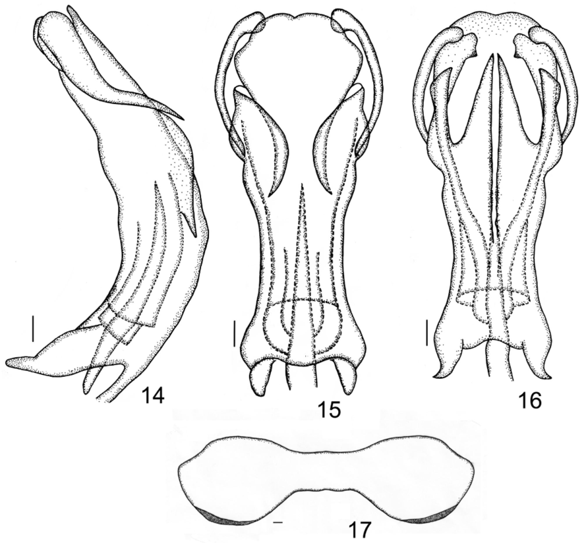
( Figs 1–17 View FIGURES 1 – 6 View FIGURES 7 – 13 View FIGURES 14 – 17 )
Etymology. The name is derived from the Latin word “unicus” (single), referring to only one pair of apical processes of the aedeagus.
Diagnosis. Ricanula unica sp. nov. is similar to Ricanula pulverosa (Stål) , but differs from the latter in having only one pair of apical spinose processes of aedeagus (with two pairs of apical spinose processes of aedeagus in R. pulverosa ).
Description. Length (inclu. teg.): male 9.3–10.5 mm, female 9.7–11.3 mm.
Head. Head with compound eyes (in dorsal view) a little narrower than the widest part of mesonotum ( Fig. 3 View FIGURES 1 – 6 ). Vertex short, 7.3 times wider at the anterior margin than long in midline, anterior and posterior margins arcuate, posterior margin arcuate more than anterior margin; disc of vertex without median carina, from anterior to posterior margins downward sloped ( Fig. 3 View FIGURES 1 – 6 ).
Frons at upper margin as wide as high in midline, 1.29 times wider at widest part (about the level of lower margin of compound eyes) than long in midline; upper margin slightly convex, lateral margins arcuate, not incised near ocelli, in lower part slightly curved to frontoclypeal suture; frontal disc with 3 carinae separated basally, lateral carinae arcuate, almost parallel to lateral margins and finishing basally at the same level than the median carina, reaching the level of antennae ( Fig. 4 View FIGURES 1 – 6 ); frons not in the same plane, the apical part below the level of antenna strongly sloped downward, oriented postero-ventral ( Fig. 4 View FIGURES 1 – 6 ).
Compound eyes oval, with callus at lower margin ( Figs 1–4 View FIGURES 1 – 6 ). Pedicel elongate, barrel-shaped ( Fig. 2 View FIGURES 1 – 6 ). Ocelli present ( Figs 2, 4 View FIGURES 1 – 6 ).
Frontoclypeal suture arcuate ( Fig. 4 View FIGURES 1 – 6 ). Clypeus ecarinate, distinctly narrower than frons, disc with median portion convex longitudinally ( Fig. 4 View FIGURES 1 – 6 ).
Rostrum reaching mesotrochanters, apical segment distinctly shorter than subapical.
Thorax. Pronotum distinctly longer in midline than vertex; anterior and posterior margins arcuate, almost parallel in median portion ( Fig. 3 View FIGURES 1 – 6 ).
Mesonotum elongate, distinctly longer than cumulative length of vertex and pronotum in midline; median carina keel-shaped, almost reaching scutellum; lateral carinae not connecting with anterior margin, almost reaching posterior margins; anterolateral carinae connected with anterior margin, not connected with lateral carinae ( Fig. 3 View FIGURES 1 – 6 ).
Tegmina ( Figs 1, 2, 5 View FIGURES 1 – 6 ) membranous, elongately-triangular; costal margin weakly arcuate, apical angle broadly rounded, placed distad to claval angle, posterior margin arcuate. Costal area of tegmina with sparse transverse veinlets, a little wider than postcostal cell and widened apically, postcostal cell narrower than costal area, without transverse veinlets; basal cell small, widely rounded; veins ScP+R, MP1+2, MP3+4 and CuA leaving basal cell separated. ScP+R vein forked just after leaving basal cell, CuA forked before middle of clavus. Tegmen without lines of transverse veinlets. Cubital cell with transverse veinlets, icu veinlets present. Claval veins Pcu and A1 fused about midlength of clavus. Transverse veinlets present in the median portion of tegmen closing inner margin.
Wings small, costal area present, small; postcostal cell distinctly longer than wide, and 2 transverse veinlets rm and m-cu present ( Fig. 6 View FIGURES 1 – 6 ).
Pro- and mesofemora as long as pro- and mesotibia tibiae, both square in cross section; metafemur square, shorter than metatibiae. Metatibia with 2 lateral spines and 6 apical teeth; basitarsomere a little longer than cumulative length of second and hind tarsomere, with 5–7 apical teeth. Metatibiotarsal formula 2/6/5–7.
Coloration. General color of body dark brown to dark ( Figs 1–2 View FIGURES 1 – 6 ). Lateral margins of vertex creamy ( Fig. 3 View FIGURES 1 – 6 ); apical margin of frons near frontoclypeal suture with brown yellowish band, median portion of clypeus and rostrum brown ( Fig. 4 View FIGURES 1 – 6 ). Eyes sordid brown, ornamented with irregular black patches ( Figs 3–4 View FIGURES 1 – 6 ). Gena light brown with three pale yellowish spot. Tegmina piceous-brown, costal margin with about 14 transverse black stripes from base to a little beyond middle, between the transverse black stripes filled with greyish-white stripes, near middle a large pale flavescent or greyish-white spot marked by two central transverse back lines, disc of tegmina near posterior margin with pale yellowish transverse veinlets ( Fig. 5 View FIGURES 1 – 6 ). Wings brown, each side of A2 with a grayish narrowed band longitudinally ( Fig. 6 View FIGURES 1 – 6 ). Pro- and mesofemora brown, both tibiae brown yellowish. Metafemora and metatibiae brown yellowish. Abdomen dark brown.
Male terminalia. Anal tube elongate; basal margin, in dorsal view, about twice shorter than posterior margin, posterior margin strongly concave, basal margin slightly convex, lateral margins straight, anus placed a bit after midlength, paraproct surpassing the posterior margin ( Fig. 12 View FIGURES 7 – 13 ). Anal tube, in lateral view, strongly extending the end of the pygofer, ventral margin slightly concave ( Fig. 7 View FIGURES 7 – 13 ).
Pygofer in lateral view higher than wide; dorsally narrower than ventrally, posterior margin almost straight, posterior-dorsal angle with process, caudodorsal angle angulate ( Fig. 7 View FIGURES 7 – 13 ).
Genital styles, in lateral view, obviously longer than wide and bearing spine-like process at the end of dorsal margin; both lower and upper margins convex; ventral margin in caudo-dorsal angle widely rounded and surpassing the posterior margin of process, hind margin straight ( Fig. 7 View FIGURES 7 – 13 ).
Phallic complex. Phallic complex slender, arcuate in lateral view ( Figs 7 View FIGURES 7 – 13 , 14 View FIGURES 14 – 17 ). In apical part, periandrium shorter than aedeagus, upper margin of dorsal periandrium “w” shaped with split in middle line, surpassing the half length of periandrium; lateral margin of dorsal periandrium, in ventral view, bending ventral and slightly inside-out forming two small crescented lobes. Basal part of periandrium without any additional structures, dorsal periandrium shorter than the ventral one, middle part of upper margin of ventral periandrium slightly concave ( Figs 14–16 View FIGURES 14 – 17 ).
Aedeagus longer than periandrium, with pair of well sclerotized, smooth and spinose processes; each process with a single apex; processes bending from dorsal to ventral side, about one third of phallic complex, oriented ventrally ( Figs 14–16 View FIGURES 14 – 17 ).
Female terminalia. Pregenital sternite with lateral lobes well developed, median portion distinctly narrower than lateral lobes; anterior margin weakly convex medially; posterior margin almost straight with middle portion slightly incised ( Fig. 17 View FIGURES 14 – 17 ).
Anal tube in lateral view elongate, reaching a half of the upper margin of gonoplac, ventral margin convex ( Fig. 8 View FIGURES 7 – 13 ).
Anal tube, in dorsal view, 1.87 times longer in midline than wide at the widest part, the widest near median portion, lateral margins convex, basal margin slightly concave, posterior margin arcuate, anus placed after midlength, paraproct distinctly surpassing the posterior margin of anal tube ( Fig. 13 View FIGURES 7 – 13 ).
Gonoplac with posterior margin bearing 2–3 well visible rows of blunt and short teeth; posterior ventral part partly membranous ( Fig. 9 View FIGURES 7 – 13 ).
Gonapophysis VIII partly laterally flattened, tapering apicad, dorsal margin slightly concave with sharp apex and well visible teeth at the posterior-dorsal margin, with spiniferous microsculpture near apex; endogonocoxal process narrower and shorter than gonaphophysis VIII, smooth ( Fig. 11 View FIGURES 7 – 13 ).
Gonapophysis IX as in Fig 10 View FIGURES 7 – 13 , ventral portion membraneous, dorsal portion sclerotized; gonospiculum bridge flatted caudo-dorsally, needle-like ventro-dorsally.
Bursa copulatrix with two widely connected white, circular and partly wrinkled pouches; the first pouch with well visible cells and sclerotized ornamentation, the second one without cells but with well visible numerous surface pores ( Fig. 8 View FIGURES 7 – 13 ).
Spermatheca well developed; ductus receptaculi wrinkled, longer than diverticulum ductus.
Type materials. Holotype, male: [ China: Guangxi, Guilin, Huaping National Nature Reserve , 1000 m, coll. Yinfeng Meng, 26 Jul. 2014] . Paratypes: 1♂ and 3♀♀, same data as holotype ; 4♂♂ and 6♀♀: [ China: Guangxi, Langping, Cenwanglao mountain , coll. Yinfeng Meng and Ye Xu, 11 Aug. 2014] ; 4♂♂ and 12♀♀: [ China: Guangxi, Guilin, Huaping National Nature Reserve , coll. Lanlan Ren, 18 Jul. 2015] ; 9♂♂ and 18♀♀: [ China: Guizhou, Duyun, Dongpeng mountain , coll. Lanlan Ren, 3 Jul. 2015] .
Distribution. China (Guangxi, Guizhou).


No known copyright restrictions apply. See Agosti, D., Egloff, W., 2009. Taxonomic information exchange and copyright: the Plazi approach. BMC Research Notes 2009, 2:53 for further explanation.
|
Kingdom |
|
|
Phylum |
|
|
Class |
|
|
Order |
|
|
Family |
|
|
Genus |