
Oreodytes Seidlitz, 1887
|
publication ID |
https://doi.org/ 10.11646/zootaxa.4820.1.1 |
|
publication LSID |
lsid:zoobank.org:pub:AED07F7B-6F71-4CDE-A923-9AB3FD84FC7C |
|
DOI |
https://doi.org/10.5281/zenodo.4397181 |
|
persistent identifier |
https://treatment.plazi.org/id/03BF87FF-354C-FF80-76BA-F929B1BAFD83 |
|
treatment provided by |
Plazi |
|
scientific name |
Oreodytes Seidlitz, 1887 |
| status |
|
Oreodytes Seidlitz, 1887 View in CoL (sensu novo)
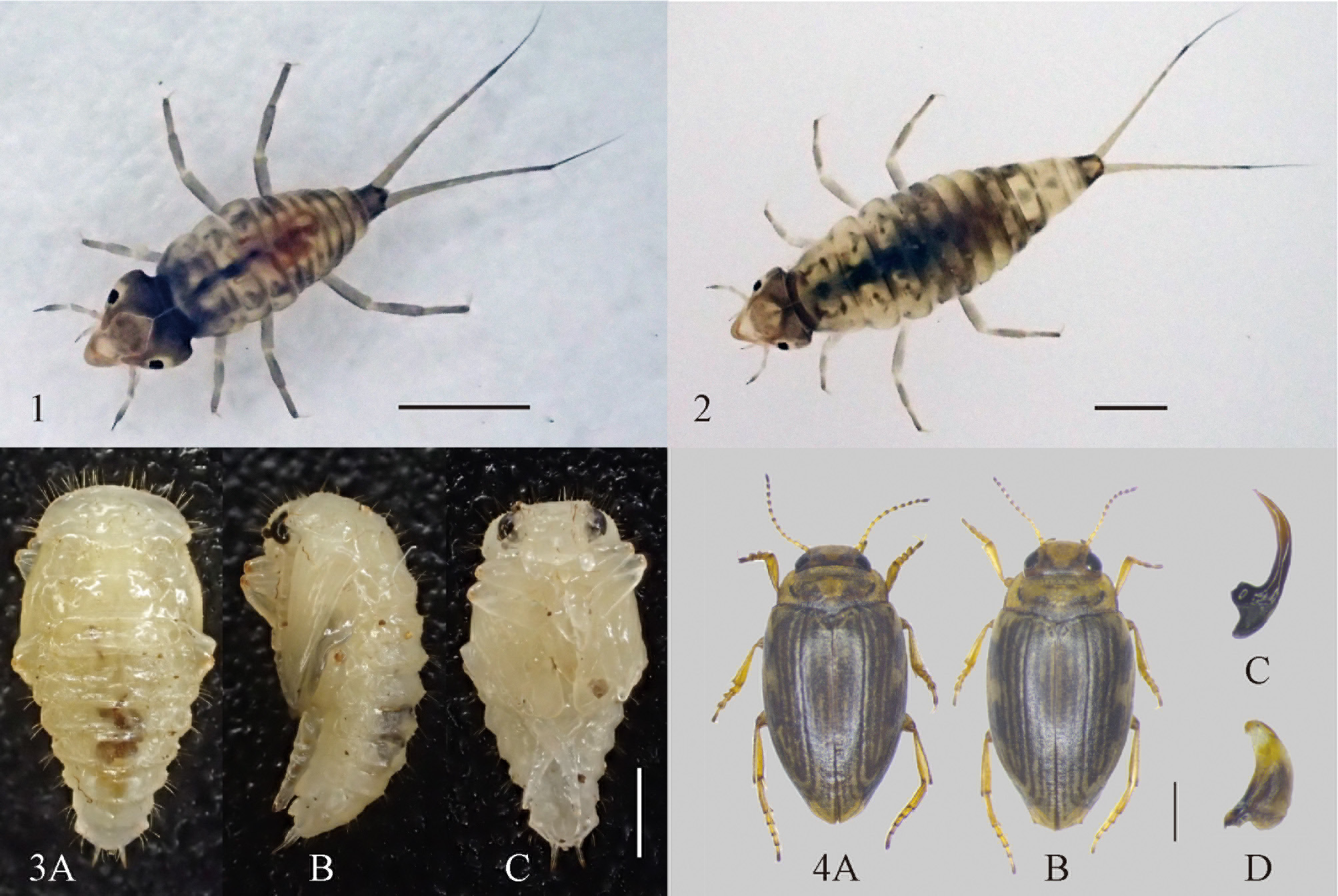
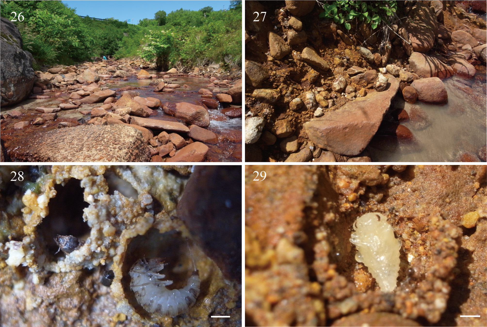
( Figs. 1–25 View FIGURES 1–4 View FIGURES 5–6 View FIGURES 7–13 View FIGURES 14–18 View FIGURES 19–23 View FIGURES 24–25 , 28–29 View FIGURES 26–29 )
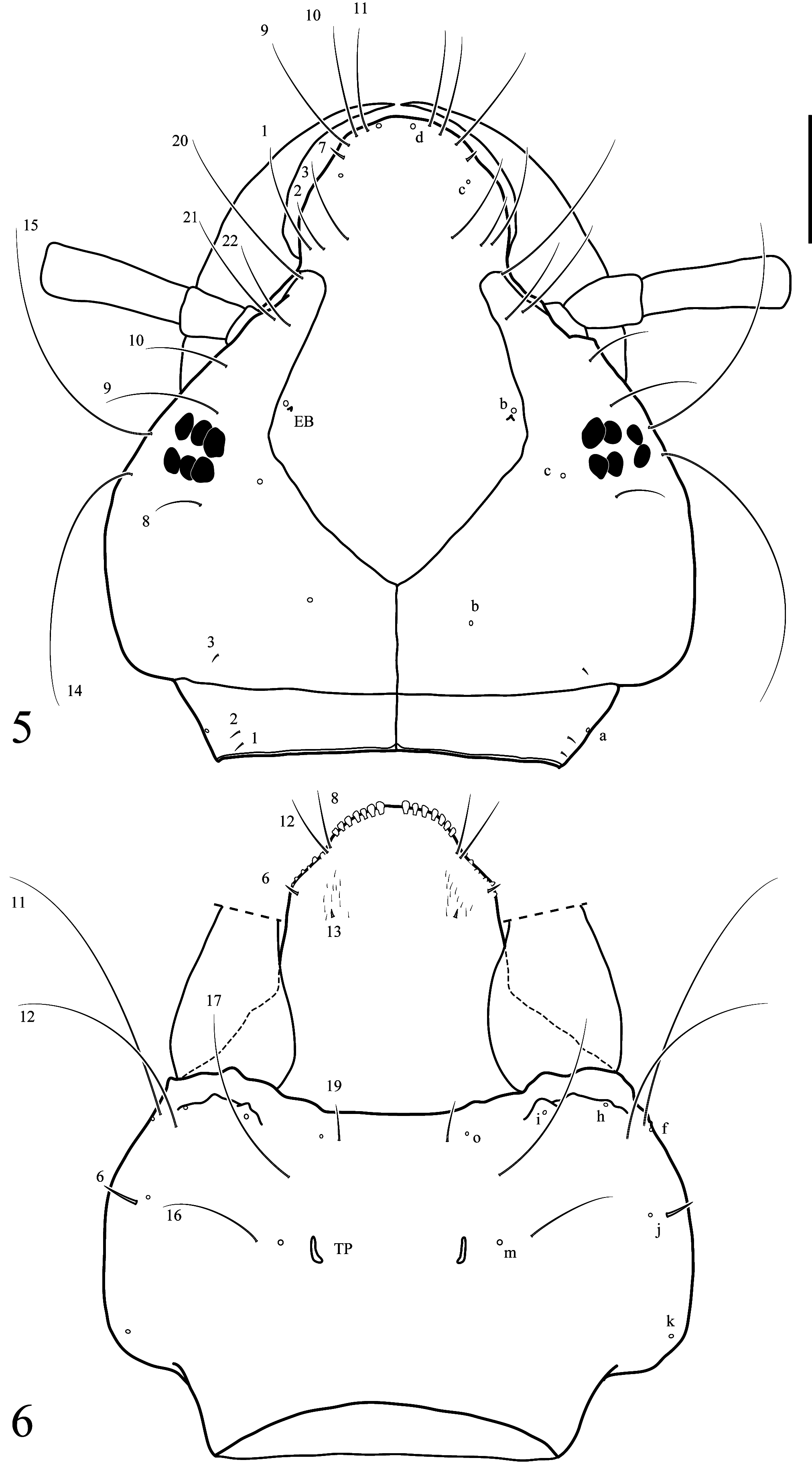
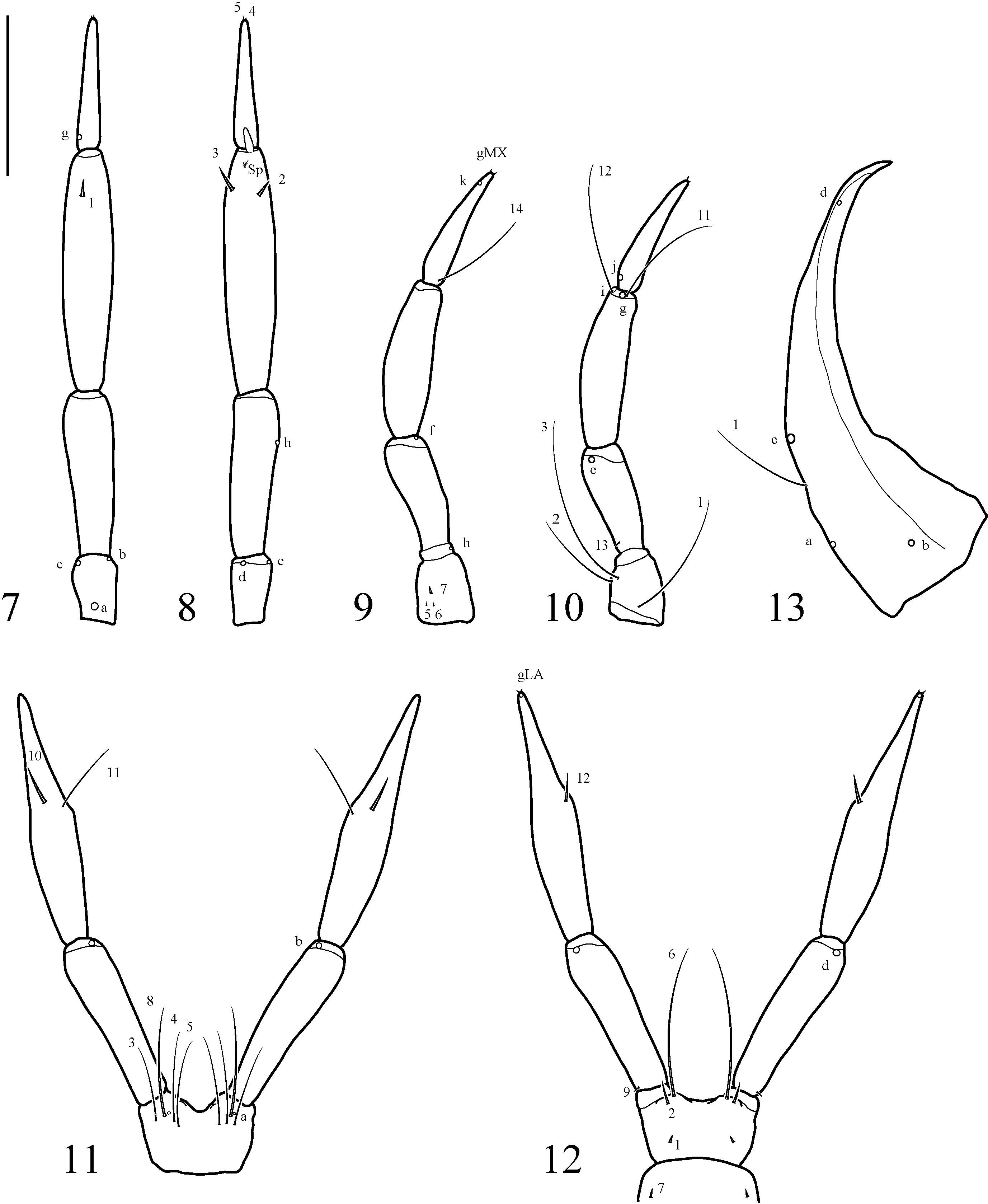
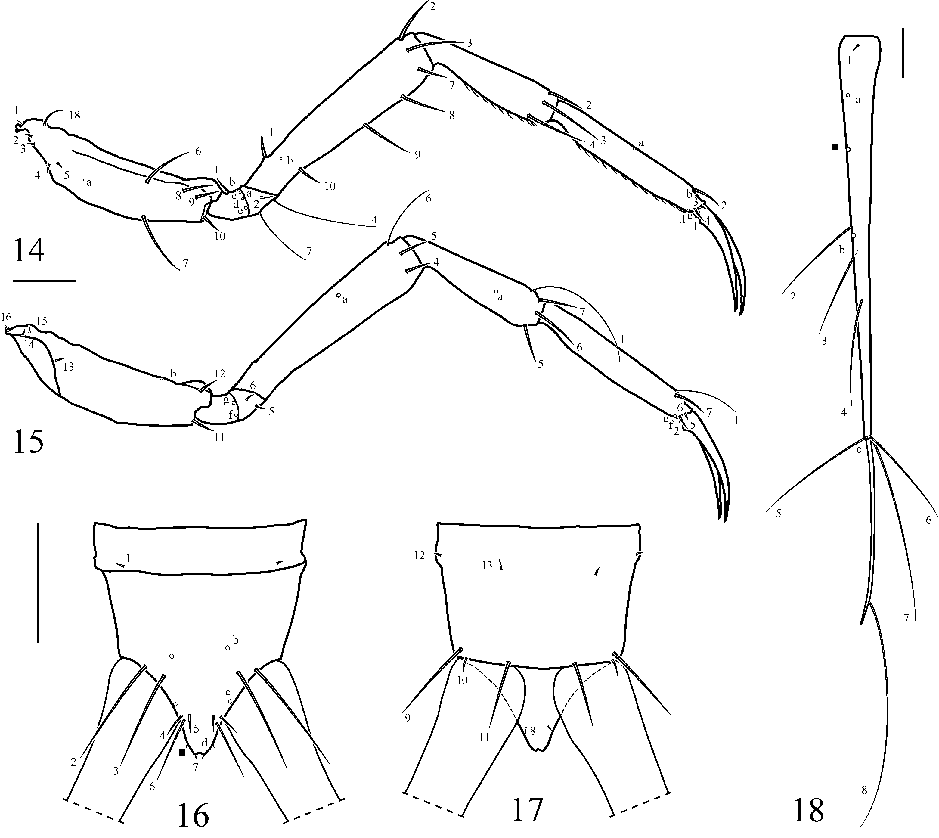
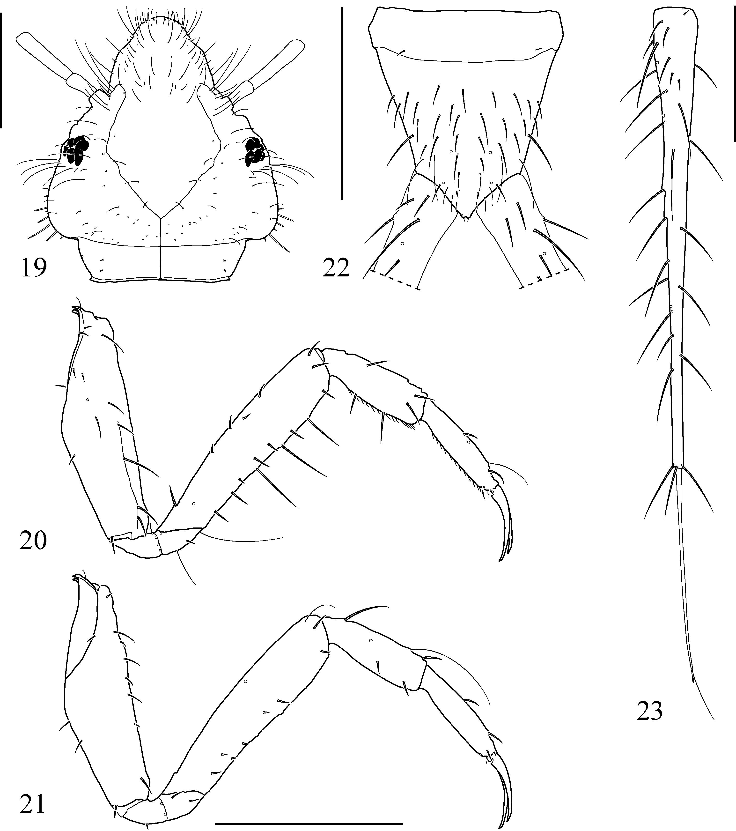
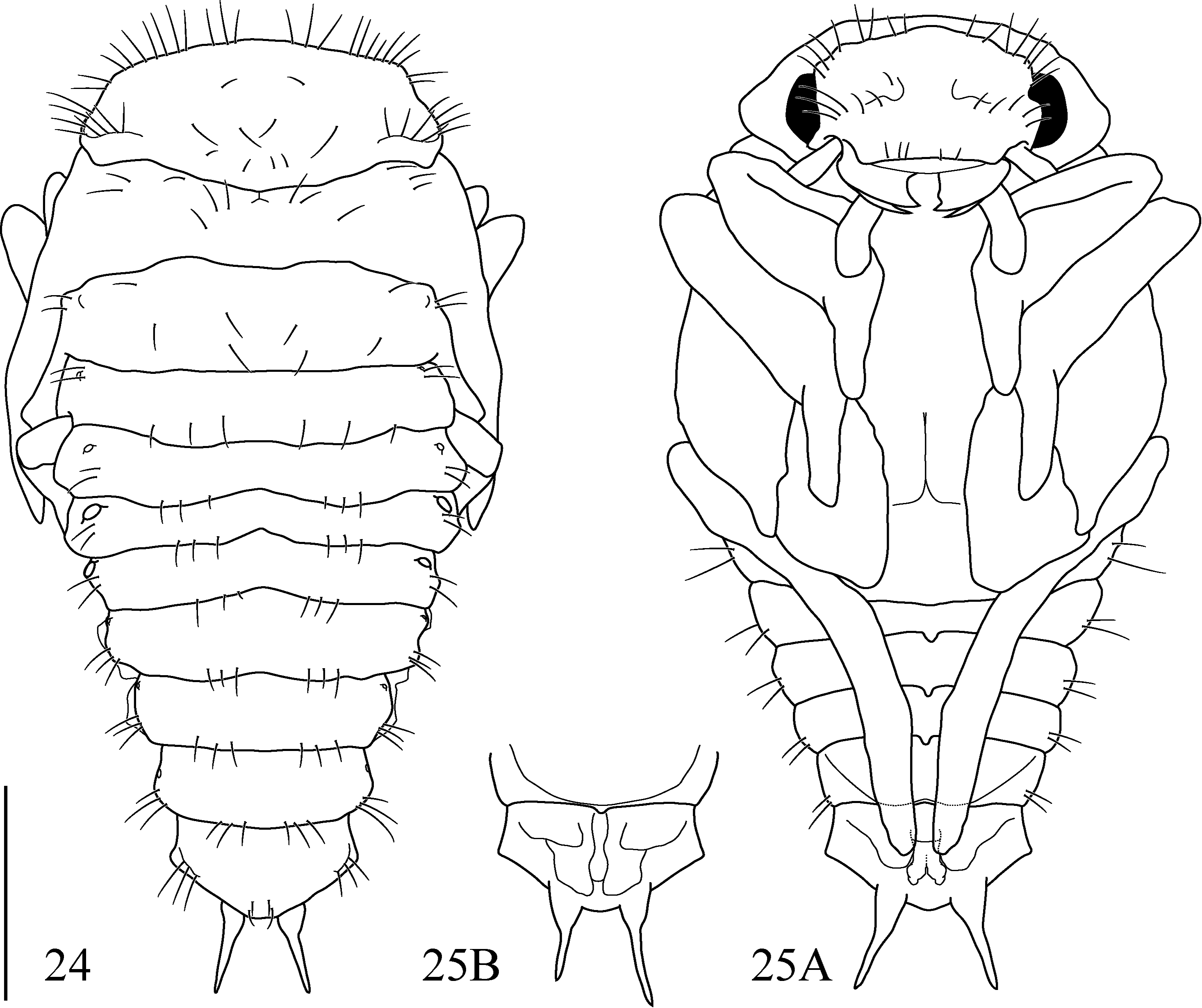
Diagnosis. Larvae of Oreodytes can readily be distinguished from those of other species of Deronectina described in detail (i.e., Deuteronectes Guignot, 1945 ; Hornectes Fery & Ribera, 2018 ; Nectoporus Guignot, 1950 ; Neonectes J. Balfour-Browne, 1944 ) (cf. Alarie & Nilsson 1996; Alarie et al. 1996; Alarie 1997) by the following combination of characters: body fusiform, narrow ( Figs 1–2 View FIGURES 1–4 ); cephalic capsule distinctly constricted posterior to occipital suture; occipital suture present from instar I ( Fig 5 View FIGURES 5–6 ); parietale lacking pores PAd and PAe ( Figs 5–6 View FIGURES 5–6 ); temporal spines acute apically and strongly developed ( Fig 19 View FIGURES 19–23 ) (instars II and III); lamellae clypeales interrupted medially for a short distance ( Fig 6 View FIGURES 5–6 ) (instar I); antennomere 2 lacking secondary setae; antennomere 3 with a ventroapical spinula and pore ANf absent ( Figs 7–8 View FIGURES 7–13 ); primary seta AN2 inserted subapically, either lower or at about level of ventral spinula ( Fig 8 View FIGURES 7–13 ); maxillary cardo fused to stipes ( Fig 10 View FIGURES 7–13 ); primary seta MX 1 inserted on maxillary stipes ( Fig 10 View FIGURES 7–13 ); primary seta MX 5 present; prementum lacking lateral spinulae ( Figs 11–12 View FIGURES 7–13 ); legs lacking natatory setae ( Figs 20–21 View FIGURES 19–23 ); primary seta TR2 of trochanters present ( Fig 14 View FIGURES 14–18 ); spinulae present on ventral margin of tibiae and tarsi ( Figs 14 View FIGURES 14–18 , 20 View FIGURES 19–23 ); abdomen segments VII and VIII lacking bluntly rounded secondary setae ( Fig 22 View FIGURES 19–23 ); abdomen segment VIII strongly constricted posterior to insertion of urogomphi, and short, less than 0.20 times LLAS in instar III ( Figs 16 View FIGURES 14–18 , 22 View FIGURES 19–23 ); primary seta AB5 short, not extending beyond apex of siphon ( Fig 16 View FIGURES 14–18 ); urogomphus elongate, more than 1.9 times HW (urogomphomere 1 more than 1.1 times longer than HW) ( Figs 18 View FIGURES 14–18 , 23 View FIGURES 19–23 ); urogomphomere 1 at least 1.7 times longer than urogomphomere 2, with an additional pore and primary setae UR2, UR3, and UR4 about subequally distant ( Fig 18 View FIGURES 14–18 ), with secondary spine-like setae ( Fig 23 View FIGURES 19–23 ).
No known copyright restrictions apply. See Agosti, D., Egloff, W., 2009. Taxonomic information exchange and copyright: the Plazi approach. BMC Research Notes 2009, 2:53 for further explanation.