
Styraconyx turbinarium, Bartels, Paul J., Fontoura, Paulo & Nelson, Diane R., 2015
|
publication ID |
https://doi.org/10.11646/zootaxa.3955.3.6 |
|
publication LSID |
lsid:zoobank.org:pub:055C37C7-7DBD-4239-A22F-64488D47D8C9 |
|
DOI |
https://doi.org/10.5281/zenodo.5678912 |
|
persistent identifier |
https://treatment.plazi.org/id/03BF8F73-FF93-EA58-FF28-FF64FD1C7C80 |
|
treatment provided by |
Plazi |
|
scientific name |
Styraconyx turbinarium |
| status |
sp. nov. |
Styraconyx turbinarium sp. nov.
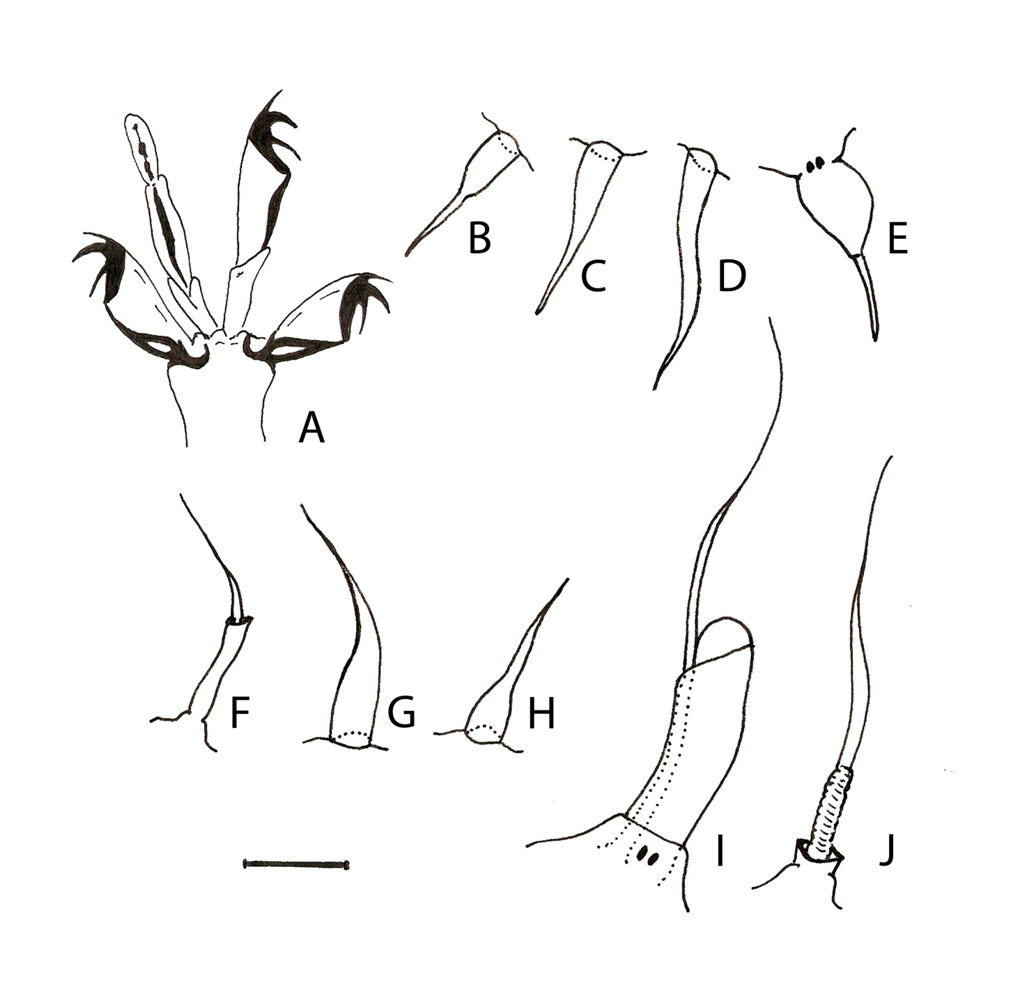
Figs. 3 View FIGURE 3 A–D; Fig. 4 View FIGURE 4 A–J; Table 1 View TABLE 1
Material examined. Holotype (female) and two paratypes (one female and one male) extracted from drifting Turbinaria ornata in Opunohu Bay ( 17° 30.5' S; 149° 51.2' W).
Type repository. The holotype (slide FP, 14.9.1) and the two paratypes (slides FP, 14.9.2 and 14.9.3*) are deposited in the collection of P. Fontoura at the Department of Biology, Faculty of Sciences, University of Porto, Portugal. [* this is a poor preparation]
Specific diagnosis. Styraconyx with subterminal mouth cone, eyespots not visible in specimens mounted in PVA. Elongate primary clavae and lateral cirri A situated on a common pedestal and enveloped by a common membrane extending almost to claval tip. Indistinct secondary clavae. Peduncles present on all digits: external, sinuous with two small lateral expansions; internal, rod-shaped. Internal digits with heart-shaped proximal pads. Three pointed claws, with strong basal hook and thin accessory spine. Sense organs present on all legs, spines on legs I–III and club-shaped papilla with terminal spine on leg IV. Cuticle coarsely punctated but otherwise smooth.
Holotype description. Female with slight green colour before mounting, 164 µm long and 78 µm wide (in lateral view) between leg pairs II and III ( Fig. 3 View FIGURE 3 A). Eye spots not visible in our specimens mounted in PVA. The cuticle coarsely punctated ( Fig. 3 View FIGURE 3 B), consisting of small pillars ( ca. 20 pillars/10 µm). The punctation also extends to the ventral cuticle and proximal part of the legs. The dorsal cuticle smooth, without any ridges, folds or other ornamental patterns. Subterminal protruded mouth opening, formed of dome-shaped cuticular annular fold without buccal papillae. Complete set of cephalic sense organs, present. Primary clava elongate (14.4 µm long and 2.2 µm wide); lateral cirrus A (27.8 µm); both arise from common dorso-lateral pedestal (about 3.5 µm); both are enveloped by transparent membranous sheath extending almost to the claval tip ( Fig. 3 View FIGURE 3 C; 4I). The primary clava/ lateral cirrus A ratio, 0.52. Van der Land’s body present in the base of the primary clavae. The lateral cirri A unsegmented, i.e. no visible line separating the scapus and the flagellum. Secondary clavae, indistinct. Dorsofrontal internal cephalic cirri (12.9 µm long), located on short cirrophores ( ca. 1.8 µm), comprising stout scapus (6.1 µm long) followed by short flagellum ( ca. 6.8 µm long) telescopically inserted in the scapus ( Fig. 3 View FIGURE 3 C; 4F). Both ventrally located external cephalic cirri (13.0 µm long) and dorsal medial cirrus (9.9 µm long) lack cirrophores, scapus and flagellum indistinct, however proximal portion of cirri (probably corresponding to scapus) much thicker than sharpened distal portion (probably flagellum) ( Fig. 4 View FIGURE 4 G; 4H). Dorsal medial cirrus inserted well back on head, not extending beyond front edge of head.
Sense organs on all legs, present. Leg I segmented spine (8.5 µm long) with differentiation of scapus and flagellum ( Fig. 4 View FIGURE 4 B); legs II ( Fig. 4 View FIGURE 4 C) and III ( Fig. 4 View FIGURE 4 D), unsegmented spines, 9.5 µm long and 12.5 µm long, respectively. Leg IV, sense organ (10.1 µm long) consists of smooth papilla (5.3 µm) and terminal spine (4.8 µm), arising on short cirrophore, with basal diffractive structure ( Fig. 3 View FIGURE 3 A; 4E). This papilla seems to be asymmetrical, having the posterior portion less curved than the anterior, but this character must be evaluated with care because the shape of the structure can be deformed by the slide mounting process.
Cirrus E (24.3 µm long) consists of short (3.0 µm long) bell-shaped cirrophore, scapus ( ca. 7 µm long) with accordion-like appearance, and flagellum ( ca. 14.3 µm long) ( Fig. 4 View FIGURE 4 J).
Legs consist of coxa, femur, tibia and tarsus. Four digits on each leg. First three pairs of legs, digits of similar length (leg I: internal digits 4.8 µm long, external digits 3.8 µm long). Leg IV, digits longer (internal digits 7.8 µm long, external digits 4.9 µm long). Heart-shaped proximal pads on internal digits, present. Peduncles present on both internal and external digits. The external hook-shaped peduncles ( ca. 6.5 µm long on legs IV) attached to the base of the claws and have two small basal lateral processes ( ca. 4.7 µm distant to the tip). The external peduncles, strongly curved at base and enlarged (about 1.5 µm wide) distal to the lateral processes.
Distal part of external peduncles, hollow; hollow area not extending to peduncle base. The very thin internal rod-shaped peduncles extend from the proximal pads to the claw base ( Fig. 3 View FIGURE 3 D; 4A).
Claws attached to the peduncle at almost a right angle, not crescent-shaped, with claw sheath. Claw sheath not covering tip of claw hooks, however this character needs to be confirmed. Claws on all legs, internal and external digits of similar size ( ca. 3.3 µm long). Each claw with three points, comprising thin dorsal spine-like accessory hook, clearly shorter than primary claw hook, and downward directed secondary hook, similar or slightly longer than primary hook ( Fig. 3 View FIGURE 3 D; 4A).
The gonopore consists of a six-cell rosette (9.7 µm from the anus). Seminal receptacles and ducts are not observable in our specimens.
Differential diagnosis. To clearly differentiate S. turbinarium sp. nov. from similar species we used characters of major taxonomic and phylogenetic value suggested by Kristensen & Higgins (1984) and highlighted by D’Adabbo Gallo et al. (1989): namely the type of peduncles and claws and the type of leg sense organs. In addition, the shape of primary clavae ( Renaud-Mornant 1982a) and particular characteristics of some species ( e.g. cuticular characters) were also considered.
Excluding the new species described in this paper, 12 Styraconyx species are known as of now. They can be divided into two major groups according the number of peduncles on each foot: seven species constituting the S. sargassi group have four peduncles on each foot, and five species of the Styraconyx hallasi Kristensen, 1977 group have two peduncles on external digits only. Our new species, S. turbinarium sp. nov., has four peduncles on each foot corresponding to the species of the first group: Styraconyx craticulus ; Styraconyx craticuliformis Chang & Rho, 1998 ; S. haploceros ; Styraconyx paulae Robotti, 1971 ; S. sardiniae ; S. sargassi ; and Styraconyx tyrrhenus D’Addabbo Gallo et al., 1989 . From this group, Styraconyx haploceros with reduced sense organs, and S. paulae characterized by having a spine-like primary clavae, can be easily distinguished from all the other species including the new species.
The new species shares with S. craticulus and S. sargassi the long sausage-shaped primary clavae, whereas all the other species have a club-shaped or a lanceolate primary clavae. However, unlike S. sargassi , the primary clavae and the lateral cirri A have a common cirrophore and are enveloped by a common membrane extending beyond the base in both the new species and S. craticulus . Additionally, the relative size of claw hooks and especially the external peduncles are very different in S. turbinarium sp. nov. compared to S. sargassi . In the new species, the accessory hook of the claws is thinner and shorter and the external peduncles are much more robust and differently shaped (see figure 45D, page 39 in Kristensen & Higgins 1984, and Fig. 3 View FIGURE 3 D). In addition, the lateral cirri A are divided in S. sargassi and undivided in the new species. The different algal associations of the two species are also important. According to Kristensen & Higgins (1984), S. sargassi was found in Sargassum (and once on benthic Dictyota ) whereas our S. turbinarium was found in drifting Turbinaria .
By having a cuticle without the characteristic grid-like pattern S. turbinarium sp. nov. differs from S. craticulus . In addition, in the new species eye spots were not observed and the sense organs on leg IV have a smooth surface (they are granular, with small tubercles, in S. craticulus ). The claws of the new species, when compared with the claws of S. craticulus , are less curved, the secondary hook is directed downwards, and the accessory hook is smaller and thinner than the other claw hooks. In S. craticulus all the cephalic cirri are segmented; in contrast in the new species only the internal cephalic cirrus has a distinct scapus and flagellum. Another very important difference between these two species concerns the shape of the peduncles on the external digits; they are thinner and with the internal hollow area located in the proximal portion in S. craticulus . In contrast, in S. turbinarium sp. nov. the hollow area of the peduncles is restricted to the distal portion following the lateral processes ( Fig. 3 View FIGURE 3 D, 4A).
Within the S. sargassi group, in addition to S. craticulus and the new species, only S. tyrrhenus has the primary clavae and the lateral cirri A situated on common cirrophores and enveloped by a common membrane extending beyond the base. However, in comparison with S. turbinarium sp. nov., in S. tyrrhenus the primary clavae are shorter and club-shaped as shown by the primary clava/lateral cirrus A ratio ( Tables 1 View TABLE 1 , 2). Excluding the shape and size of clavae and the presence of segmented external cephalic cirri in S. tyrrhenus , all the other features exhibited by the two species are very similar, including the shape of peduncles. Morphologically overall, this makes S. tyrrhenus the most similar species to S. turbinarium sp. nov. Despite the high variability in morphometric data mentioned by Kristensen & Higgins (1984) and the limited number of specimens examined, the membranes enveloping the clavae and lateral cirri A and cirri E seem to be relatively longer in S. turbinarium sp. nov.
Etymology. The new species is named after the alga Turbinaria with which it is associated. Styraconyx turbinarium translates to Styraconyx of Turbinaria .

No known copyright restrictions apply. See Agosti, D., Egloff, W., 2009. Taxonomic information exchange and copyright: the Plazi approach. BMC Research Notes 2009, 2:53 for further explanation.
|
Kingdom |
|
|
Phylum |
|
|
Class |
|
|
Order |
|
|
Family |
|
|
SubFamily |
Halechiniscinae |
|
Genus |