
Stegana, Meigen, 1830
|
publication ID |
https://doi.org/ 10.1111/j.1096-3642.2009.00575.x |
|
DOI |
https://doi.org/10.5281/zenodo.10545499 |
|
persistent identifier |
https://treatment.plazi.org/id/03BFAB37-AC0E-FFCE-FC23-34E6FDCFFD97 |
|
treatment provided by |
Valdenar |
|
scientific name |
Stegana |
| status |
|
STEGANA View in CoL (S.S.) MEIGEN, 1830
Stegana Meigen, 1830: 79 View in CoL .
Type species: Stegana nigra Meigen, 1830 .
Diagnosis: Main axis of eye vertical; face yellow, unicolorous; palpus large and black, with numerous small setae (modified from Bächli et al., 2004).
Description: Male and female: Eyes red. Ocellar triangle black, with a pair of small setae above ocellar setae. Postvertical setae slightly behind vertex ridge. Frons with several minute setulae medially. Proclinate orbital setae nearer to ptilinal fissure than to inner vertical setae. Pedicel yellow; arista plumose, with pubescence between dorsal and ventral branches. Lunule brown to black. Facial carina absent; gena yellowish brown, black at vibrissa corners. Vibrissa prominent; other orals small. Occiput glossy, yellow, black around occipital foramen. Thorax yellow to dark brown. Mesopleuron with a black longitudinal stripe above (running from propleuron to base of halter). Postpronotal lobe with one long and a few small setae. Acrostichal setulae in approximately ten irregular rows. One pair of prescutellar setae. Scutellum with basal setae divergent, apical setae cruciate. Wing dark brown anteriorly, paler posteriorly, yellowish at tip; curved downward on distal part. Basal medial–cubital cross-vein present; vein C 1 with two setae, less differentiated. Costal vein with six to eight minute spinules on ventral surface before vein R 4+5. Vein R 2+3 markedly curved to costa at tip; veins R 4+5 and M 1 convergent distally. Halters white. Fore femur with one row (four to six) of setae on ventral surface. Apical seta present on mid tibia. Preapical dorsal setae present on all tibiae. Mid tibia with five to six strong setae on basal part of dorsal surface. Mid and hind tarsomeres with two and one row(s) of minute cuneiform setulae on ventral surface, respectively; fore and hind tarsomeres I slightly shorter than the rest combined; mid tarsomere I longer than the rest combined. Abdominal sternites broadened, wider than long.
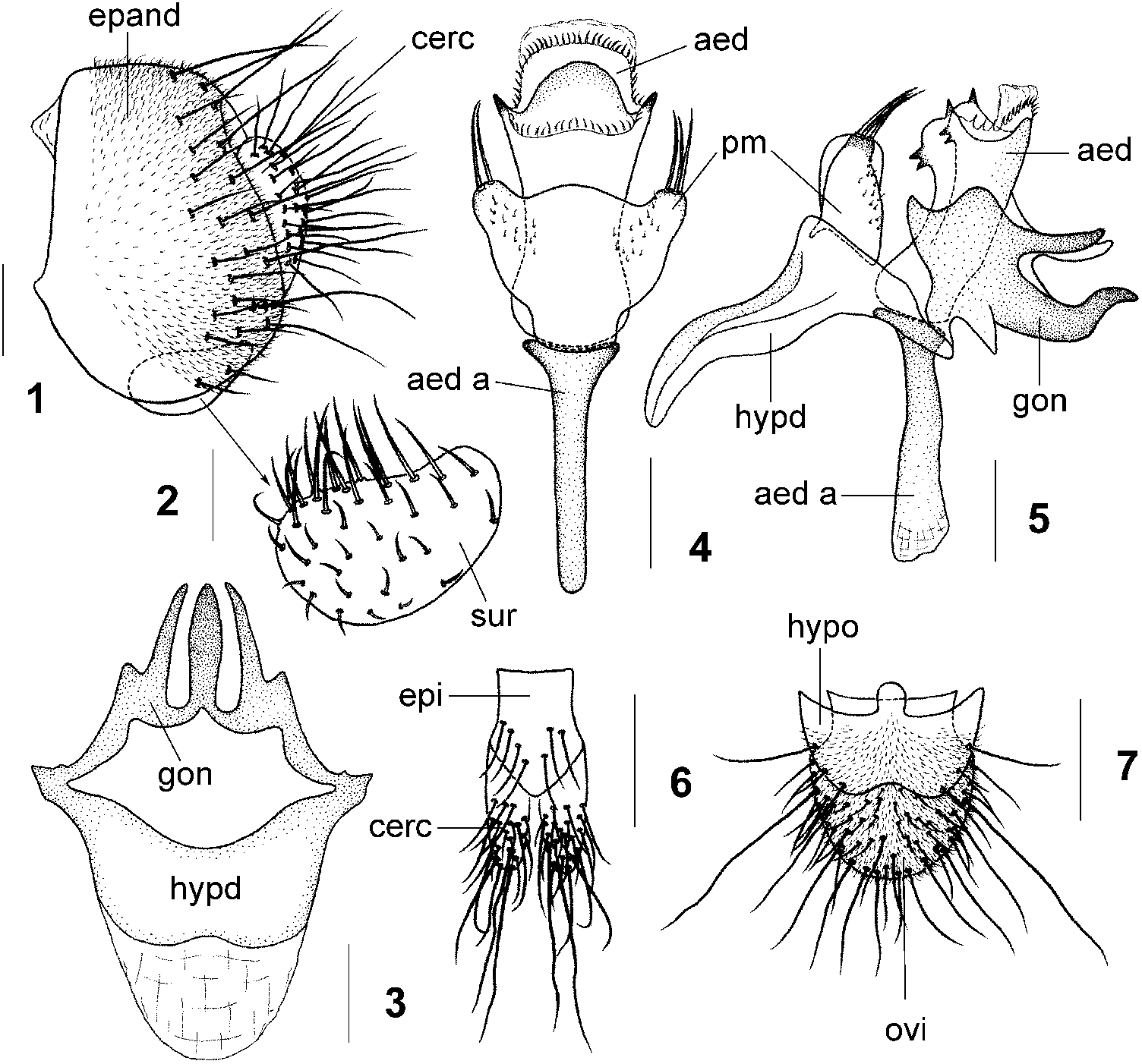
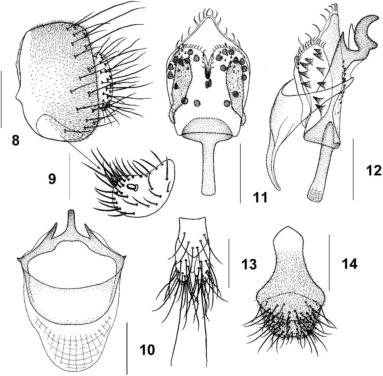
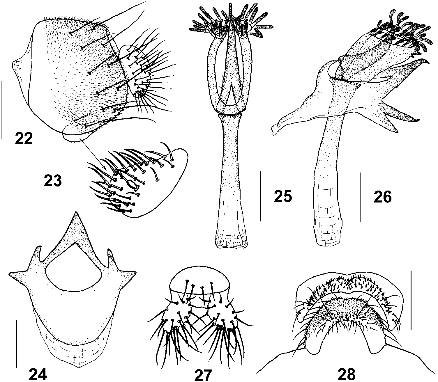
Male terminalia. Epandrium pubescent except for anterior margin, with numerous setae on dorsal to posterolateral portion on each side ( Figs 1 View Figures 1–7 , 8 View Figures 8–14 , 15 View Figures 15–21 , 22 View Figures 22–28 , 29, 34 View Figures 29–35 ). Surstylus anterodorsally fused with epandrium, with several setae on outer surface ( Figs 2 View Figures 1–7 , 9 View Figures 8–14 , 16 View Figures 15–21 , 23 View Figures 22–28 , 30, 35 View Figures 29–35 ). Cercus separated from epandrium, setigerous, mostly lacking pubescence ( Figs 1 View Figures 1–7 , 8 View Figures 8–14 , 15 View Figures 15–21 , 22 View Figures 22–28 , 29, 34 View Figures 29–35 ). Hypandrium anteromedially protruded and broadened ( Figs 6 View Figures 1–7 , 10 View Figures 8–14 , 17 View Figures 15–21 , 24 View Figures 22–28 , 31 View Figures 29–35 , 36 View Figures 36–40 ). Gonopods fused to each other, forming posteromedian lobe, strongly sclerotized, posterolaterally contiguous to posterior ends of hypandrium ( Figs 6 View Figures 1–7 , 10 View Figures 8–14 , 17 View Figures 15–21 , 24 View Figures 22–28 , 31 View Figures 29–35 , 36 View Figures 36–40 ). Parameres developed, sometimes fused with each other ( Figs 7 View Figures 1–7 , 8, 11, 12 View Figures 8–14 , 18, 19 View Figures 15–21 , 25, 26 View Figures 22–28 , 32, 33 View Figures 29–35 , 37, 38 View Figures 36–40 ). Aedeagus mostly with tentacle-like setae distally ( Figs 7 View Figures 1–7 , 8, 11, 12 View Figures 8–14 , 18, 19 View Figures 15–21 , 25, 26 View Figures 22–28 , 32, 33 View Figures 29–35 , 37, 38 View Figures 36–40 ). Aedeagal apodeme strong, contiguous to base of aedeagus ( Figs 7 View Figures 1–7 , 8, 11, 12 View Figures 8–14 , 18, 19 View Figures 15–21 , 25, 26 View Figures 22–28 , 32, 33 View Figures 29–35 , 37, 38 View Figures 36–40 ).
Female terminalia. Epiproct and cercus not pubescent ( Figs 6 View Figures 1–7 , 13 View Figures 8–14 , 20 View Figures 15–21 , 27 View Figures 22–28 , 39 View Figures 36–40 ); oviscapt (sternite VIII) and hypoproct mostly with numerous setae and pubescence ( Figs 7 View Figures 1–7 , 14 View Figures 8–14 , 21 View Figures 15–21 , 28 View Figures 22–28 , 40 View Figures 36–40 ).


| R |
Departamento de Geologia, Universidad de Chile |
No known copyright restrictions apply. See Agosti, D., Egloff, W., 2009. Taxonomic information exchange and copyright: the Plazi approach. BMC Research Notes 2009, 2:53 for further explanation.
|
Kingdom |
|
|
Phylum |
|
|
Class |
|
|
Order |
|
|
Family |
Stegana
| Li, Tong, Cao, Huazhi, Gao, Jianjun & Chen, Hongwei 2010 |
Stegana
| Meigen JW 1830: 79 |