
Notohymena australis Foissner
|
publication ID |
https://doi.org/10.11646/zootaxa.4000.4.4 |
|
publication LSID |
lsid:zoobank.org:pub:7FB26592-09EC-4980-A5CE-3E9C662D1F5B |
|
DOI |
https://doi.org/10.5281/zenodo.6101950 |
|
persistent identifier |
https://treatment.plazi.org/id/03C06549-F731-9A26-FF08-FBBDFADBE9B6 |
|
treatment provided by |
Plazi |
|
scientific name |
Notohymena australis Foissner |
| status |
|
Notohymena australis Foissner & O’ Donoghue, 1990
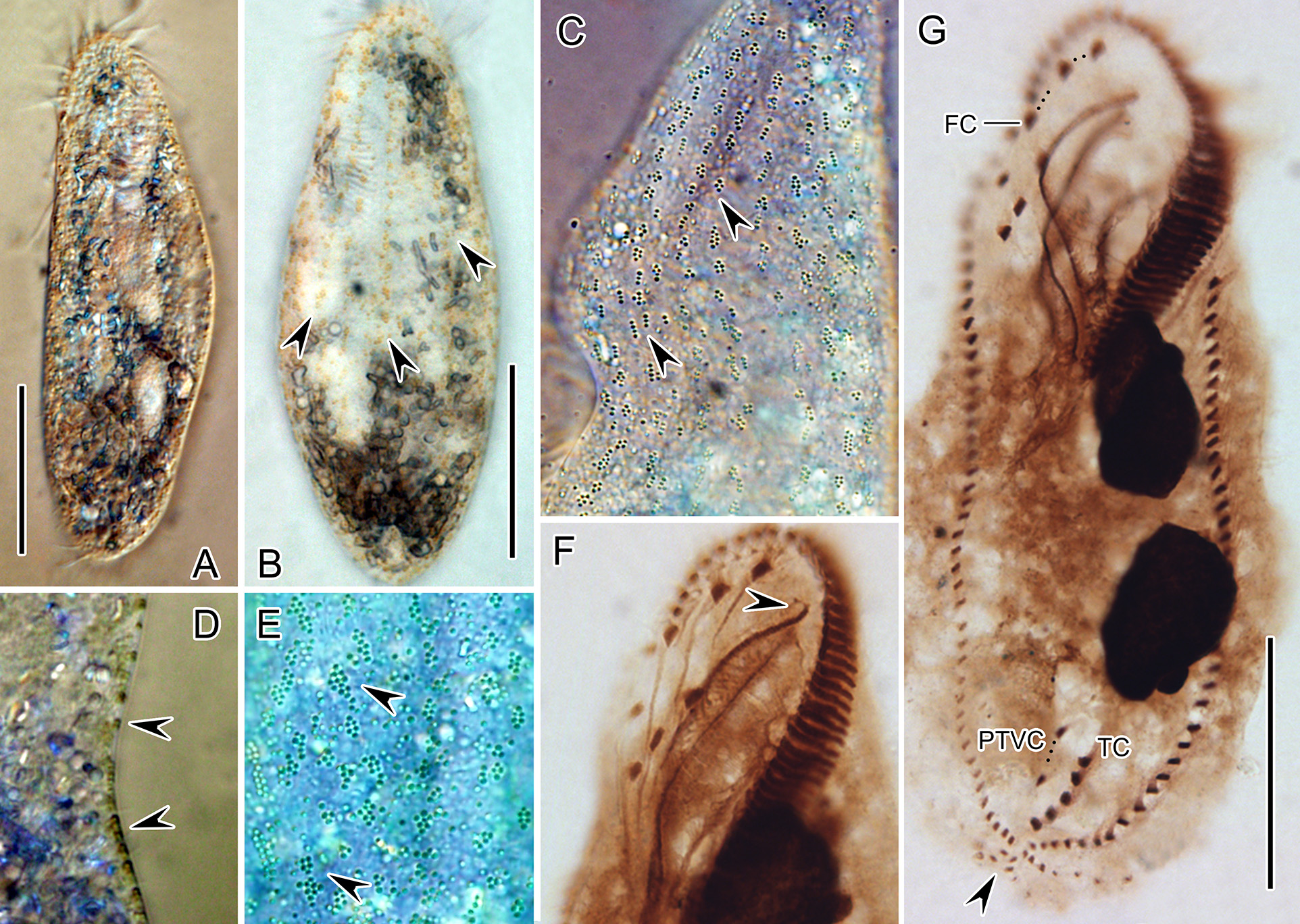
( Figure 3 View FIGURE 3 A–G; Table 1)
This species was well described by original report, thus only brief redescription based on new populations was supplied.
Morphological description of Chinese population: Body 110-130 × 30-50 µm, long elliptical in outline, with length: width ratio approximately 4: 1 in vivo ( Fig. 3 View FIGURE 3 A, B). Body flexible but not contractile. Cortical granules yellow or orange, spherical, about 0.4 µm across ( Fig. 3 View FIGURE 3 C, D), and they arranged mainly along cirral rows and dorsal kineties ( Fig. 3 View FIGURE 3 B, C, E). Buccal field about 46% of body length ( Fig. 3 View FIGURE 3 F). Bases of largest membranes in life about 10 µm. Two ellipsoidal macronuclear nodules, 15–33 × 8–11 µm in size. One contractile vacuole about 20 µm across, located left of cell mid-line, slightly in front of mid-body. Cytoplasm usually containing small lipid droplets with size about 1–7 µm in diameter ( Fig. 3 View FIGURE 3 A, B). Locomotion by swimming rotating about its longitudinal axis moderately fast or relatively slow crawling on bottom of Petri dishes or on debris.
Paroral bends to left and has a hooked distal ( Fig. 3 View FIGURE 3 F, G). Cirri on ventral side arranged in typical 8: 5: 5 pattern. Frontal cirri is about 12–15 µm long in life, transverse cirri, about 18 µm. Two marginal rows converging posterior ( Fig. 3 View FIGURE 3 G). Six dorsal kineties, cilia about 5 µm long; leftmost 3 (dorsal kineties 1, 2, and 3) of body length; fourth and fifth dorsal kineties terminate below the mid-body respectively; rightmost dorsal kinety (dorsal kinety 6), very short, only composed of 2–5 dikinetids. 7–10 caudal cirri located at posterior body margin and arranged in three rows, located at the posterior end of dorsal kineties 1, 2, and 4 respectively ( Fig. 3 View FIGURE 3 G).
Remarks: This species was recorded successively from Australia, Bavaria, Korea and Japan. Our population corresponds well with previous reports (Foissner & O’ Donoghue, 1990; Choon & Shin, 2010; Hu & Yasushi, 2015) in terms of cyst, locomotion, cortical granules and marginal cirri, and differs from them in larger adoral zone about 46% (vs. 38% in Australian population, 36% in Korean population, 37% in Japanese population) and bases of largest membranes in life about 10 µm (vs. 7 µm in Australian population, 8 µm in Japanese population).
No known copyright restrictions apply. See Agosti, D., Egloff, W., 2009. Taxonomic information exchange and copyright: the Plazi approach. BMC Research Notes 2009, 2:53 for further explanation.
|
Kingdom |
|
|
Phylum |
|
|
Class |
|
|
Order |
|
|
SubOrder |
Sporadotrichina |
|
Family |
|
|
Genus |