
Pattersoniella vitiphila Foissner, 1987
|
publication ID |
https://doi.org/10.11646/zootaxa.4000.4.4 |
|
publication LSID |
lsid:zoobank.org:pub:7FB26592-09EC-4980-A5CE-3E9C662D1F5B |
|
DOI |
https://doi.org/10.5281/zenodo.6101948 |
|
persistent identifier |
https://treatment.plazi.org/id/03C06549-F732-9A25-FF08-FB56FB14E9BF |
|
treatment provided by |
Plazi |
|
scientific name |
Pattersoniella vitiphila Foissner, 1987 |
| status |
|
Pattersoniella vitiphila Foissner, 1987
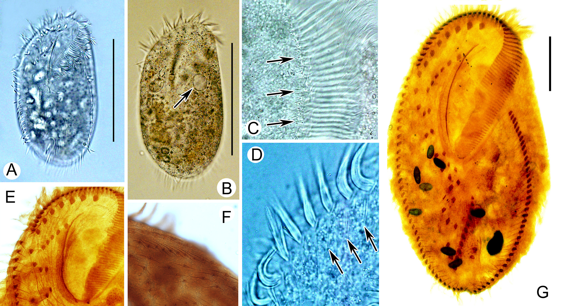
( Figure 2 View FIGURE 2 A–G; Table 1)
This species was originally described in terms of both living morphology and infraciliature by Foissner (1987). Chinese population shows some variation in morphometric data, thus a redescription was supplied here.
Morphological description of Chinese population: Cells in vivo 310-360 × 140-180 µm, broadly elliptical in outline, with ratio of length to width approximately 2: 1 ( Fig. 2 View FIGURE 2 A, B). Body inflexible, but very fragile when observed under microscope on a slide. Cortical granules lacking. Cell dark brown in color, different sized food vacuoles containing algae or small ciliates. Buccal field about 2/5–1/2 of body length. Basal fibers of cirri and membranelles of adoral zone detectable in vivo ( Fig. 2 View FIGURE 2 C, D) and after staining ( Fig. 2 View FIGURE 2 E). 21–35 ellipsoidal macronuclear nodules, each about 20 × 15 µm in size, located in the cell mid-line irregularly. One contractile vacuole detectable at anterior third and near dorsal side, with diameter about 27 µm ( Fig. 2 View FIGURE 2 B). A forming cyst observed, spherical, about 100 µm across. Locomotion by swimming moderately fast or crawling relatively slow on bottom of Petri dishes or on debris.
Ventral infraciliature as shown in Fig. 2 View FIGURE 2 G. Frontal cirri and buccal cirri distinctly enlarged, about 37 µm long. Transverse cirri, about 35 µm in length, arranged roughly in a J-shaped row in posterior region of cell, only posterior-most reaching posterior end of body. Length of marginal cirri about 20–24 µm. Invariably three caudal cirri. Dorsal cilia arranged altogether about 15 kineties on average, only three or four of them about body length. Two associated fibers of each dorsal dikinetid visible after staining, about 14 µm long ( Fig. 2 View FIGURE 2 F).
Remarks: The original population from Fiji Island has 13–18 (14.6 on average) ventral cirri in normal specimens and 18–20 (18.7 on average) cirri in giant specimens that present after culture. Our population has 18– 27 (21.3 on average) ventral cirri and larger size which shows the similarities with the giant specimens of original population ( Foissner 1987). The number of cirri might be closely related to the body size.
No known copyright restrictions apply. See Agosti, D., Egloff, W., 2009. Taxonomic information exchange and copyright: the Plazi approach. BMC Research Notes 2009, 2:53 for further explanation.
|
Kingdom |
|
|
Phylum |
|
|
Class |
|
|
Order |
|
|
SubOrder |
Sporadotrichina |
|
Family |
|
|
Genus |