
Sphingius nilgiriensis Gravely, 1931
|
publication ID |
https://doi.org/ 10.11646/zootaxa.4896.4.3 |
|
publication LSID |
lsid:zoobank.org:pub:0824AFA4-4E8B-419B-972C-0FA0A88538FF |
|
DOI |
https://doi.org/10.5281/zenodo.4387709 |
|
persistent identifier |
https://treatment.plazi.org/id/03C08789-C535-4F64-FF62-D01B8913FEF5 |
|
treatment provided by |
Plazi |
|
scientific name |
Sphingius nilgiriensis Gravely, 1931 |
| status |
|
Sphingius nilgiriensis Gravely, 1931 View in CoL
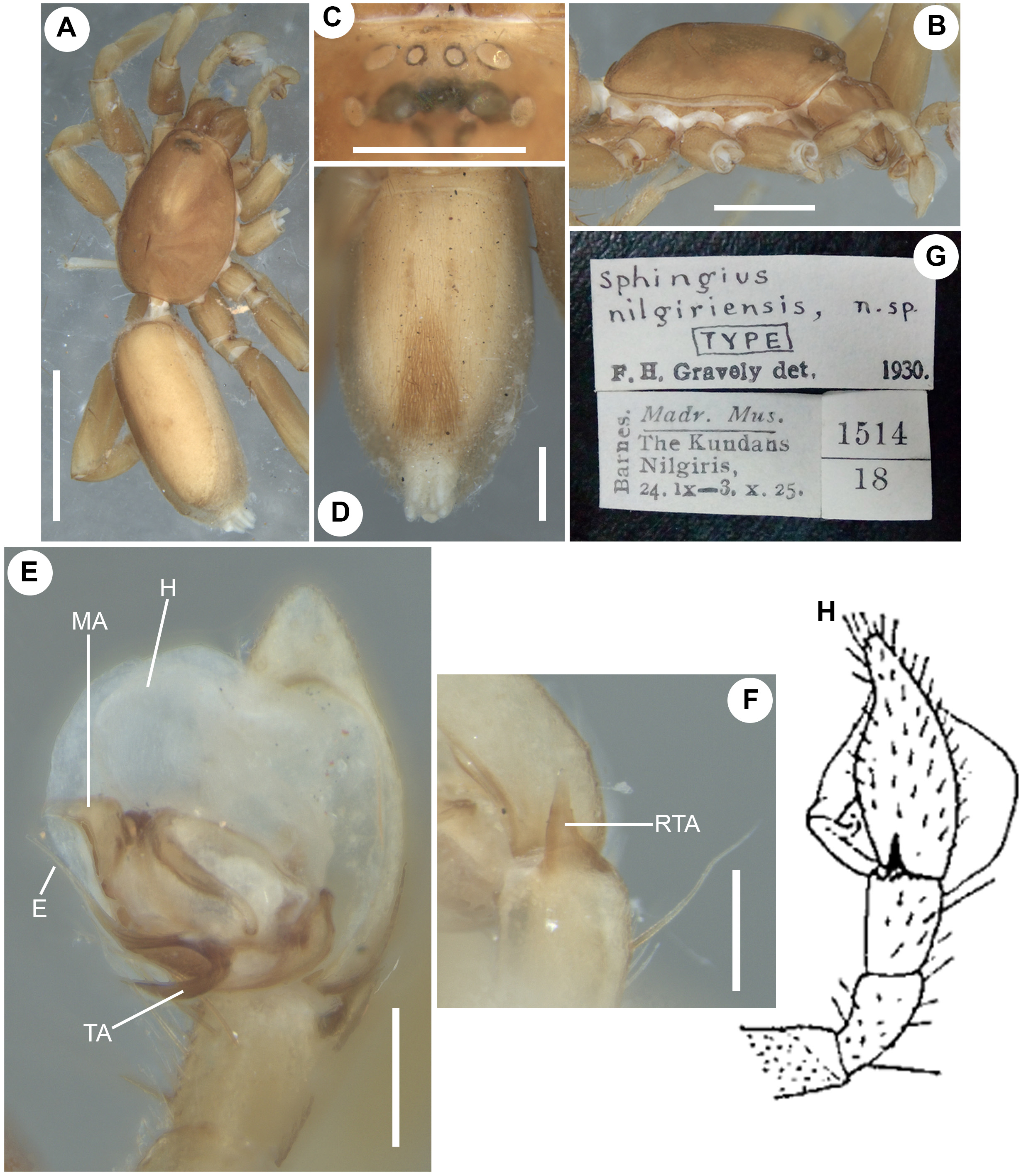
Fig. 6 View FIGURE 6
Sphingius nilgiriensis Gravely, 1931: 271 View in CoL , fig. 19C; Majumder & Tikader, 1991: 155, figs 330–333.
Type material. Syntype Ƌ from INDIA: Tamil Nadu: Nilgiris: Kundah (not Kundahs): (11°15’46.41’’N, 76°37’59.98’’E), 1856 m a.s.l., leg. Barnes, 24 September–3 October 1925, deposited in NZC-ZSI (no register number), examined ( Fig. 6G View FIGURE 6 ) GoogleMaps .
Diagnosis. Males of S. nilgiriensis seem closely related to the males of S. superbus Dankittipakul, Tavano & Singtripop, 2011 , but can easily be separated from the latter by group of bristles located on the proximal part of ventral scutum which are restricted to the rear end of the posterior margin of ventral scutum in S. superbus (compare Fig. 6D View FIGURE 6 with Dankittipakul et al. 2011: fig. 5).
Supplementary description. Male ( Fig. 6 View FIGURE 6 A–D). Dorsal scutum covering almost entire length of opisthosoma; ventral scutum smaller than dorsal scutum, less sclerotized; posterior half of venter medially bears group of bristles ( Fig. 6D View FIGURE 6 ). Body length 5.26. Carapace length 2.33, width 1.85. Opisthosoma length 2.93, width 1.34. Eye diameters: ALE 0.11, AME 0.06, PLE 0.09, PME 0.08. Eye interdistances: ALE–PLE 0.09, AME–ALE 0.03, AME–AME 0.05, AME–PME 0.10, PME–PLE 0.08, PME–PME 0.05. Chelicerae length 0.79. Clypeus height at ALEs 0.07, at AMEs 0.08. Measurements of pedipalp and legs. Pedipalp 1.88 [0.73, 0.32, 0.29, 0.54], I 5.74 [1.59, 0.85, 1.30, 1.10, 0.90], II 4.88 [1.50, 0.78, 1.07, 0.97, 0.56], III 4.56 [1.23, 0.69, 0.84, 1.06, 0.74], IV 7.32 [2.03, 0.96, 1.58, 1.83, 0.92]. Leg formula: 4123. Pedipalp ( Fig. 6 View FIGURE 6 E–F, H): retrolateral tibial apophysis short, broad at base, gradually narrowing towards apex ( Fig. 6F View FIGURE 6 : RTA). Accessory tegular apophysis short, flat, angular in retrolateral view ( Fig. 6E View FIGURE 6 : TA). Embolus moderately long, thin, with blunt tip ( Fig. 6E View FIGURE 6 : E). Median tegular apophysis large, with retrolateral process with truncated apex ( Fig. 6E View FIGURE 6 : MA). Conductor apparently absent.
Female. Unknown.
Note. In his original description, Gravely (1931) mentioned that the most conspicuous feature of S. nilgiriensis was a white membraneous structure in its pedipalp. It is, in fact, the haematodocha of the expanded pedipalp ( Fig. 6E View FIGURE 6 : H).
Remarks. The ZSI collection has one glass bottle for this species labelled as “ Type ” (1514/18), containing a male specimen in fairly good condition, with broken legs and with intact pedipalps.
No known copyright restrictions apply. See Agosti, D., Egloff, W., 2009. Taxonomic information exchange and copyright: the Plazi approach. BMC Research Notes 2009, 2:53 for further explanation.
|
Kingdom |
|
|
Phylum |
|
|
Class |
|
|
Order |
|
|
Family |
|
|
Genus |
Sphingius nilgiriensis Gravely, 1931
| Sankaran, Pradeep M., Caleb, John T. D. & Sebastian, Pothalil A. 2020 |
Sphingius nilgiriensis
| Majumder, S. C. & Tikader, B. K. 1991: 155 |
| Gravely, F. H. 1931: 271 |