
Anthicimimus Franz
|
publication ID |
https://doi.org/ 10.11646/zootaxa.4033.3.10 |
|
publication LSID |
lsid:zoobank.org:pub:9183A8D0-C65F-4F88-9B4F-ED315BCF4F25 |
|
DOI |
https://doi.org/10.5281/zenodo.6094589 |
|
persistent identifier |
https://treatment.plazi.org/id/03C087C8-CC22-9B60-77A3-B0E7FE1DB988 |
|
treatment provided by |
Plazi |
|
scientific name |
Anthicimimus Franz |
| status |
|
Anthicimimus Franz , new status
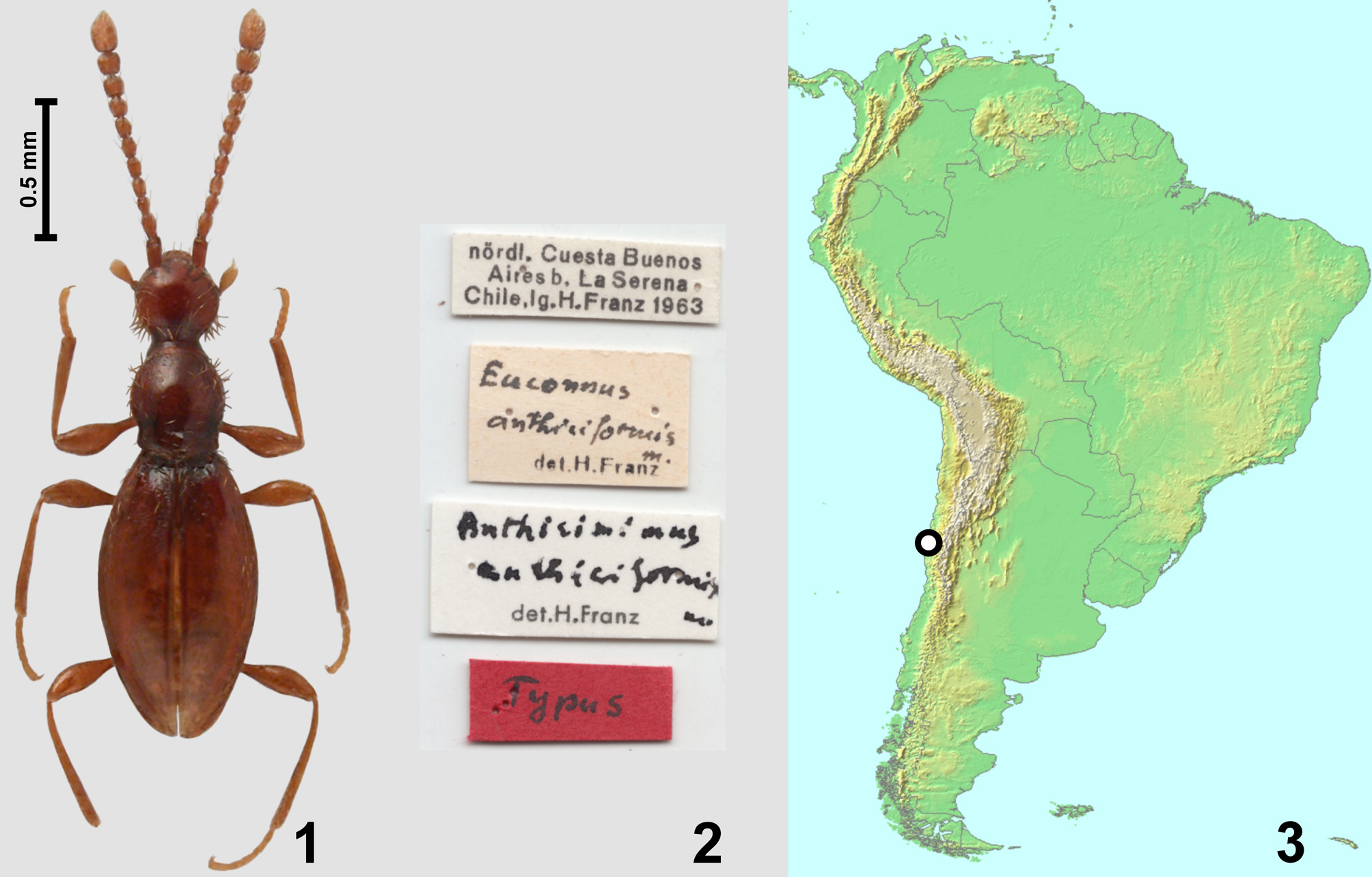
Anthicimimus Franz, 1993: 100 (as subgenus of Sciacharis View in CoL ). Type species: Euconnus anthiciformis Franz, 1967 (des. orig.).
Revised diagnosis. Head with anteriorly located eyes and long tempora, with distinct frontoclypeal groove, occipital constriction slightly broader than half HW; tempora, genae and sides of pronotum with thick bristles; submentum lacking lateral sutures; hypostomal ridges complete and connecting slightly in front of posterior tentorial pits; antennae gradually thickened distally; pronotum with transverse antebasal groove and without pits, sublateral and lateral carinae; prosternum with long basisternal part, nearly complete notosternal sutures (obliterated posteriorly), complete hypomeral ridges, broadly open procoxal sockets and distinct, subtriangular and narrow prosternal process; mesoventral intercoxal process carinate with median portion projecting ventrally more than anterior and posterior portions; ventrolateral foveae present; metaventrite with distinct, elongate anterior metaventral process and short, subtriangular metaventral intercoxal process with median notch; metacoxae contiguous; each elytron with one very small but deep and asetose basal fovea.
General body shape of female ( Fig. 1 View FIGURES 1 – 3 ) strongly elongate and slender, body deeply constricted between head and pronotum but weakly so between pronotum and elytra, flattened; appendages long and slender, vestiture of setae sparse, short and suberect, in addition to thin setae thick bristles are present on various body parts.
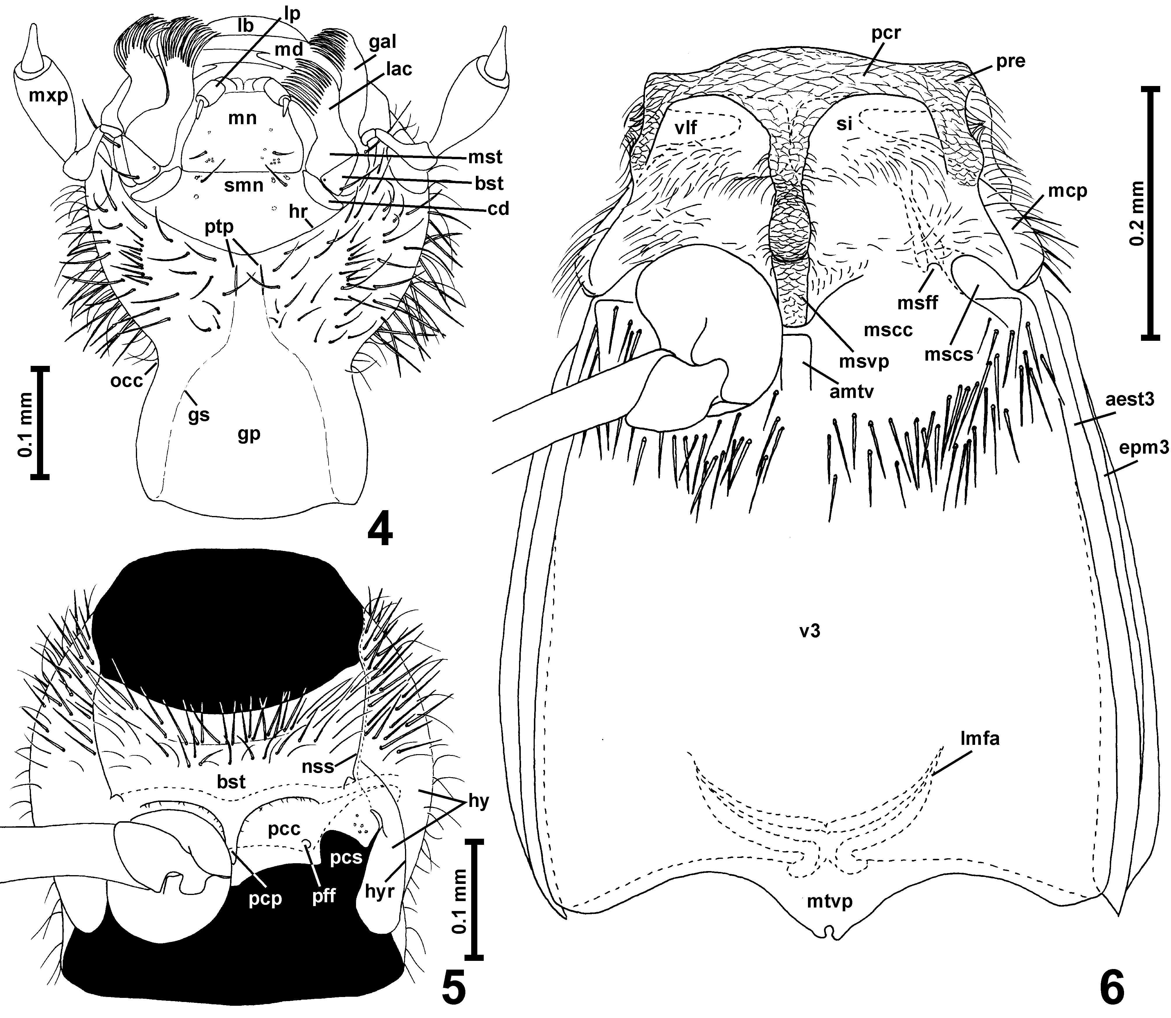
Head capsule ( Figs 1 View FIGURES 1 – 3 , 4 View FIGURES 4 – 6 ) large in relation to pronotum; divided by occipital constriction into large anterior and small posterior part ('neck' region), posterior part retracted into pronotum. 'Neck' region much narrower than anterior part of head, short and weakly broadening toward foramen occipitale; narrowest place of occipital constriction ( Fig. 4 View FIGURES 4 – 6 ; occ) slightly broader than half HW. Anterior part of head capsule strongly convex dorsally and flattened ventrally, round in dorsal view, with small round eyes located anteriorly. Tempora much longer than compound eyes; vertex transverse and strongly convex, its posterior portion not bulging posterodorsally, anteriorly vertex confluent with subtrapezoidal and strongly convex posterior portion of frons, which is abruptly and steeply declining anteriorly in front of weakly marked supraantennal tubercles; genae elongate, weakly convex. Clypeus short and broad, demarcated from frons by distinct transverse frontoclypeal groove. Vestiture of head capsule composed mostly of thin setae, but tempora and genae with thick bristles. Gular plate ( Fig. 4 View FIGURES 4 – 6 ; gp) in posterior part broad and with indistinctly marked gular sutures ( Fig. 4 View FIGURES 4 – 6 ; gs), anterior part adjacent to posterior tentorial pits narrow and nearly parallel-sided; posterior tentorial pits ( Fig. 4 View FIGURES 4 – 6 ; ptp) strongly elongate, slot-shaped and located anteriorly to transverse impression delimiting 'neck' region from anterior part of head.
Mouthparts ( Fig. 4 View FIGURES 4 – 6 ) only partly exposed in the studied specimen. Labrum strongly transverse, with rounded anterior margin. Mandibles ( Fig. 4 View FIGURES 4 – 6 ; md), subtriangular and strongly elongate, only slender and curved distal portions visible, each with large subapical mesal tooth. Maxilla elongate, with transverse cardo ( Fig. 4 View FIGURES 4 – 6 ; cd); subtriangular and elongated basistipes ( Fig. 4 View FIGURES 4 – 6 ; bst); short and broad mediostipes ( Fig. 4 View FIGURES 4 – 6 ; mst); elongate galea ( Fig. 4 View FIGURES 4 – 6 ; gal) and lacinia ( Fig. 4 View FIGURES 4 – 6 ; lac), each with long and dense setae along distal and mesal margin; maxillary palp ( Fig. 4 View FIGURES 4 – 6 ; mxp) long, palpomere I minute, elongate, palpomere II strongly elongate, curved and distinctly thickening distally, palpomere III longer than II and strongly broadened, broadest near distal third; palpomere IV small, subconical and slender, with elongate distal portion. Labium with transverse submentum ( Fig. 4 View FIGURES 4 – 6 ; smn) laterally not demarcated by sutures; mentum ( Fig. 4 View FIGURES 4 – 6 ; mn) subtrapezoidal; prementum short, its median region not visible; labial palps ( Fig. 4 View FIGURES 4 – 6 ; lp) shorter than mentum. Posteriorly and laterally mouthparts demarcated by distinct and only weakly arcuate hypostomal ridges ( Fig. 4 View FIGURES 4 – 6 ; hr) which are complete and connected in front of posterior tentorial pits.
Antennae ( Fig. 1 View FIGURES 1 – 3 ) slender and long, gradually but weakly thickening distally; antennomere I strikingly elongate, about three times as long as broad and in lateral view slightly curved.
Prothorax ( Figs 1 View FIGURES 1 – 3 , 5 View FIGURES 4 – 6 ) moderately convex, elongate, broadest in front of middle, in dorsal view bell-shaped; anterior and lateral margins in anterior half rounded; sides in sub-basal region shallowly constricted; anterior pronotal corners indistinctly marked; posterior corners obtuse-angled and blunt; posterior pronotal margin weakly arcuate. Pronotum without sharp lateral edges and without sublateral carinae. Base of pronotum with distinct transverse groove, without pits. Sides of pronotum and external parts of hypomera with thick bristles. Prosternum ( Fig. 5 View FIGURES 4 – 6 ) with basisternal part ( Fig. 4 View FIGURES 4 – 6 ; bst) longer than coxal part and partly sharply demarcated from procoxal cavities ( Fig. 5 View FIGURES 4 – 6 ; pcc) by carina mesally continuous with lateral margins of prosternal intercoxal process ( Fig. 5 View FIGURES 4 – 6 ; pcp), which is elongate subtriangular with sharply marked margins; procoxal sockets ( Fig. 5 View FIGURES 4 – 6 ; pcs) broadly open; profurcal foveae ( Fig. 5 View FIGURES 4 – 6 ; pff) large and distinct. Hypomera ( Fig. 5 View FIGURES 4 – 6 ; hy) elongate, each divided by entire hypomeral ridge ( Fig. 5 View FIGURES 4 – 6 ; hyr) into narrow and long subtriangular inner (adcoxal) part and large external part confluent laterally with side of pronotum; notosternal sutures ( Fig. 5 View FIGURES 4 – 6 ; nss) nearly complete, obliterated only posteriorly, in front of procoxae, so that inner part of each hypomeron is mesally fused with coxal part of sternum.
Mesothorax. Mesonotum subtrapezoidal in shape; mesoscutum strongly transverse, with several thick lateral bristles; scutoscutellar suture well-marked on the surface as transverse ridge; mesoscutellum not visible between bases of elytra in intact specimens, triangular with rounded posterior margin.
Mesoventrite ( Fig. 6 View FIGURES 4 – 6 ) much broader than long, without demarcated anterior ridge; area functioning as procoxal rest ( Fig. 6 View FIGURES 4 – 6 ; pcr) not impressed, asetose and covered with distinct microsculpture composed of polygonal cells; setose impressions ( Fig. 6 View FIGURES 4 – 6 ; si) large but weakly impressed, posteriorly not demarcated by carinae and with setae only in posterior portion; mesoventral intercoxal process ( Fig. 6 View FIGURES 4 – 6 ; msvp) long and keel-like, strongly projecting ventrally, especially its median portion in front of mesocoxae in lateral view forming rounded ventral projection, whereas anterior and posterior portions are lower (i.e., less projecting ventrally); mesocoxal sockets ( Fig. 6 View FIGURES 4 – 6 ; mscs) located lateromesally on mesocoxal projections ( Fig. 6 View FIGURES 4 – 6 ; mcp) and in ventral view exposed; mesofurcal foveae ( Fig. 6 View FIGURES 4 – 6 ; msff) large and located near anterior margin of each mesocoxal cavity ( Fig. 6 View FIGURES 4 – 6 ; mscc). Prepectus ( Fig. 6 View FIGURES 4 – 6 ; pre) short, posterior part of mesanepisternum only partly visible in ventral view. Mesothorax with deep ventrolateral foveae ( Fig. 6 View FIGURES 4 – 6 ; vlf) with setose openings.
Metathorax. Metaventrite ( Fig. 6 View FIGURES 4 – 6 ; v3) much longer than mesoventrite, subtrapezoidal, distinctly broadening posteriorly, anteriorly fused with mesoventrite, lateral margins slightly rounded, lateral (admetacoxal) parts of posterior margin weakly concave, intermetacoxal area weakly projecting posteriorly and forming short subtriangular metaventral intercoxal process ( Fig. 6 View FIGURES 4 – 6 ; mtvp) with deep and rounded median notch; metacoxae contiguous. Metanepisterna ( Fig. 6 View FIGURES 4 – 6 ; aest3) and metepimera ( Fig. 6 View FIGURES 4 – 6 ; epm3) narrow.
Metafurca (metendosternite) ( Fig. 6 View FIGURES 4 – 6 ) with short stem and strongly divergent lateral furcal arms ( Fig. 6 View FIGURES 4 – 6 ; lmfa).
Elytra ( Fig. 1 View FIGURES 1 – 3 ) oval, with rounded apices; humeral callus and basal impression weakly developed; subhumeral line present and carinate; each elytron with one very small but deep asetose basal fovea (visible only in transparent mount).
Metathoracic wings not studied.
Legs ( Fig. 1 View FIGURES 1 – 3 ) long ad slender, unmodified.
Abdomen in the studied specimen partly damaged, terminal portion missing.
Remarks. Anthicimimus does not have the diagnostic characters of Sciacharis , as the closed procoxal sockets and indistinct, diffused prosternal process, and has features not known in the latter genus, as the large anterior metaventral process. The broadly open procoxal sockets are unique among Cyrtoscydmini ; narrowly open procoxal sockets can be found in several genera that occur in Australia ( Palaeoscydmaenus Franz, 1975 ; Leascydmus Jałoszyński, 2014c), Neotropical (Obesoconnus Jałoszyński, 2014d), Oriental ( Siamites Franz, 1989 ) and Palaearctic (Rutaraphes Jałoszyński, 2015) regions. All of them, except for Leascydmus, have the submentum with lateral sutures, which are absent in Anthicimimus . Leascydmus has rudimentary hypostomal ridges, lacks the prosternal process, and has broadly separated metacoxae, characters clearly different than those in Anthicimimus . For the above reasons, Anthicimimus is here elevated to the genus rank.
No known copyright restrictions apply. See Agosti, D., Egloff, W., 2009. Taxonomic information exchange and copyright: the Plazi approach. BMC Research Notes 2009, 2:53 for further explanation.
|
Kingdom |
|
|
Phylum |
|
|
Class |
|
|
Order |
|
|
Family |
Anthicimimus Franz
| Jałoszyński, Paweł 2015 |
Anthicimimus
| Franz 1993: 100 |