
Parvanachis pygmaea ( Sowerby I, 1832 )
|
publication ID |
https://doi.org/ 10.11646/zootaxa.3753.3.1 |
|
publication LSID |
lsid:zoobank.org:pub:DE234954-1829-4277-9E17-78C4E5C18142 |
|
DOI |
https://doi.org/10.5281/zenodo.6141036 |
|
persistent identifier |
https://treatment.plazi.org/id/03C0B65E-FFD6-FF99-FF0F-54B1FD4DFE1C |
|
treatment provided by |
Plazi |
|
scientific name |
Parvanachis pygmaea ( Sowerby I, 1832 ) |
| status |
|
Parvanachis pygmaea ( Sowerby I, 1832) View in CoL
Figure 6 View FIGURE 6 A–D, 7A, 7E
Columbella pygmaea Sowerby, 1832 View in CoL : p. 119, not figured. 1844a: p. 141, pl. 40 fig. 163. Duclos in Chenu, 1848: pl. 25 figs. 1– 2. Adams, C.B., 1852: p. 97. Reeve, 1858: sp. 128, pl. 22, figs. 128–129.
Anachis pygmaea ( Sowerby, 1832) View in CoL . Carpenter, 1857: p. 510; 1863: p. 344. Baker, Hanna & Strong, 1938: p. 250, not figured. Strong & Hertlein, 1939: p. 184, not figured. Keen, 1958, p. 384, sp. 448 (illustration from Sowerby, Thes. Conch.).
Columbella (Seminella) pygmaea (Sowerby) View in CoL . Tryon, 1883: p. 166, pl. 56 figs. 91, 92.
Columbella (Anachis) pygmaea Sowerby. Kobelt, 1897 View in CoL : p. 158, taf. 22, figs. 1, 2.
Anachis dalli Bartsch, 1931 View in CoL : pp. 2–3, pl. 1 fig. 2.
Anachis (Parvanachis) pygmaea ( Sowerby, 1832) View in CoL . Keen, 1971: p. 585, sp. 1209. Abbott, 1974: p. 197, sp. 2068 (not figured).
Parvanachis pygmaea ( Sowerby, 1832) View in CoL . Skoglund, 1992: p. 89.
Types. Four probable syntypes marked by Keen in 1964, NHMUK 1966389, collected by Cuming at Monte Cristo (not St. Elena), Ecuador (in Guayaquil province, according to C.B. Adams, 1852) on dead shells.
Taxonomic history. Sowerby (1832) reports Cuming collecting this species on dead shells in sandy mud at Santa Elena, at 10 fms. Adams (1852) reports finding them under stones at the low water mark. Keen (1971) figures one of Cuming’s specimens from Monte Cristo, as well as Sowerby’s illustration; no material from St.
Elena was found at NHMUK. Sowerby’s (1844) illustration is not specific enough to match a particular specimen, but is consistent with the Monte Cristo specimens. Carpenter (1857) refers to Anachis pygmaea as being variable in color, from ‘light horn with a few purple-brown patches to nearly uniform black brown’; he probably has this species confused with P. adamsi (described below), which is similar in size and whose markings sometimes resemble those of P. pygmaea . In 1864 he refers to his and Adams’ material as Anachis pygmaea var. auriflua , but no formal description or illustration has been found to determine an identity for that name, so it must remain a nomen nudum. One lot of Carpenter’s specimens ( USNM 716231, Carpenter Tablet 2429), is cited in Carpenter (1857) as showing P. pygmaea in different stages of growth, but is not this species; however it has fewer specimens than reported (6 juveniles rather than 7 juveniles and 3 adults) so perhaps the lot was split. Bartsch (1931) named Anachis dalli (holotype USNM 368144) from Taboga Island, Panama. Photos of the likely syntypes of C. pygmaea and the type specimen of A. dalli were both examined, and though Bartsch briefly notes that his species was previously misidentified as A. pygmaea , he does not specify how they differ, and no differences between the types are apparent.
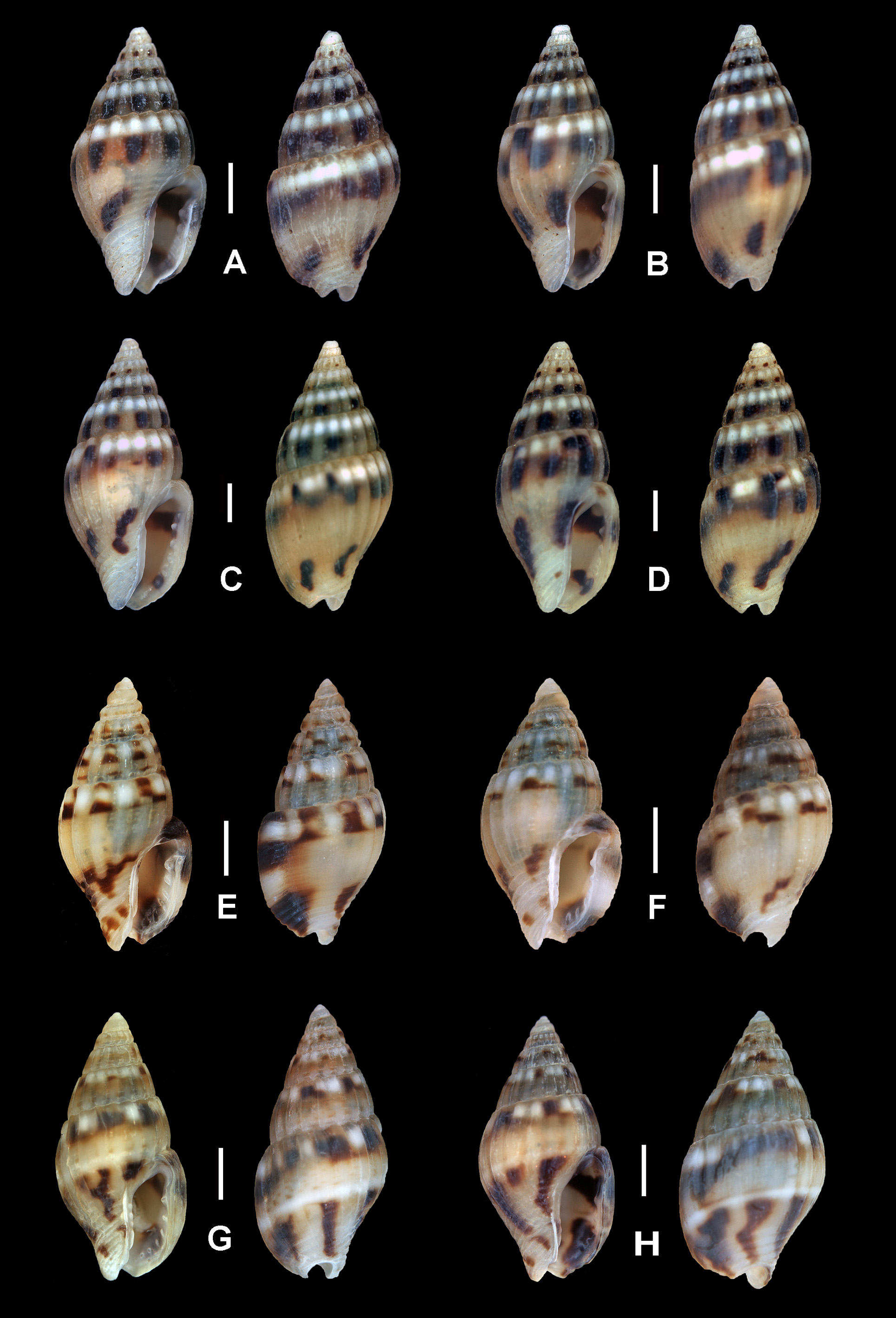
Diagnosis. Small (4 to 7 mm) species with a biconic, axially ribbed shell, straw colored with a white band immediately below the suture and a spiral row of dark markings overlying or slightly below the white band. A second band of dark markings is present below the periphery.
Material. Parvanachis pygmaea was common at most localities (6 of 11, Table 1) though not in high numbers. 45 specimens were collected on and under rocks in muddy sand or sand; most were large juveniles or adults. USNM has these from Baja California to Ecuador. Three were sectioned, three dissected.
Shell ( Figs. 6 View FIGURE 6 A–D): Shell biconic, 4.2 to 6.8 mm long (avg. 5.41 mm) and 2.05 to 2.85 mm wide (avg. 2.40 mm) in 26 specimens measured. Adult shells with 4.5 to 5.5 teleoconch whorls (avg. 4.8). Protoconch smooth, offwhite, with 3 to 3.25 (avg. 3.17 in eight specimens) whorls. Shell sculptured primarily with axial ridges (15 to 18 on the penultimate whorl), weak, evenly spaced spiral grooves between axial ridges on the spire whorls. No welldeveloped subsutural cord except on the first two whorls, unlike P. adamsi , which is the same size and very similar otherwise. Apex usually eroded, unlike similar species. Shell easily recognized by its striking and relatively invariant color pattern. Shell glossy white or yellowish, with a row of purplish brown blotches on the anterior half of each whorl, usually strongest on every other axial ridge, and a white band between the dark spots and the suture. Body whorl with a second row of axially elongated dark blotches below the periphery. Aperture edge thickened, with a few denticles internally. Aperture edge with a shallow posterior sinus. Parietal callus with a weakly denticulate ridge. Aperture color reflects that of the shell exterior, anterior end of shell paler.
Body coloration: Body cream colored, with dense black mottling and white speckles. Front corners of the foot without markings. Siphon mottled, with a black band; tentacles cream colored, with black bands on the base and middle.
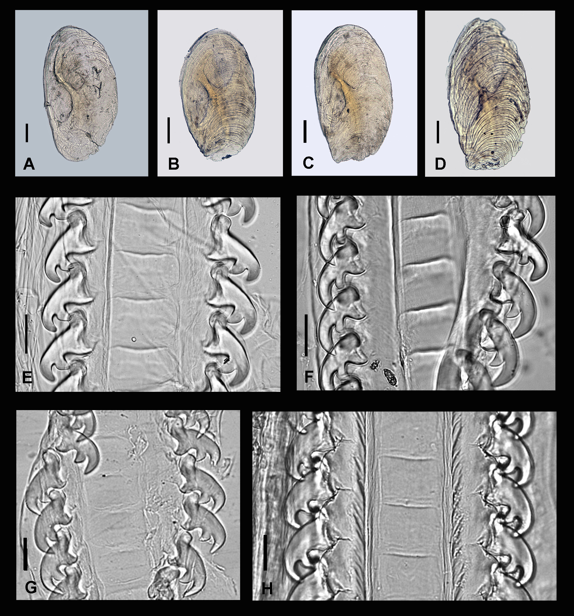
Operculum ( Fig. 7 View FIGURE 7 A): Operculum oblong, with a keel and bilobed muscle scar, and usually a terminal nucleus (one photographed appeared to be damaged, nucleus was sub-terminal). Operculum center is darker tan.
Radula ( Fig. 7 View FIGURE 7 E): Radula similar to those of P. adamsi ( Fig. 7 View FIGURE 7 G) and P. nigricans ( Fig. 7 View FIGURE 7 H). Lateral teeth about 40 µm long in adults, with three pointed secondary cusps; the basal cusp is pointed down and embedded in radular membranes. Center plates are rectangular, about 30 to 40 µm wide by 10 to 25 µm deep. The three specimens (two adult males, one adult female) dissected had radulae with 125 to 132 tooth rows.
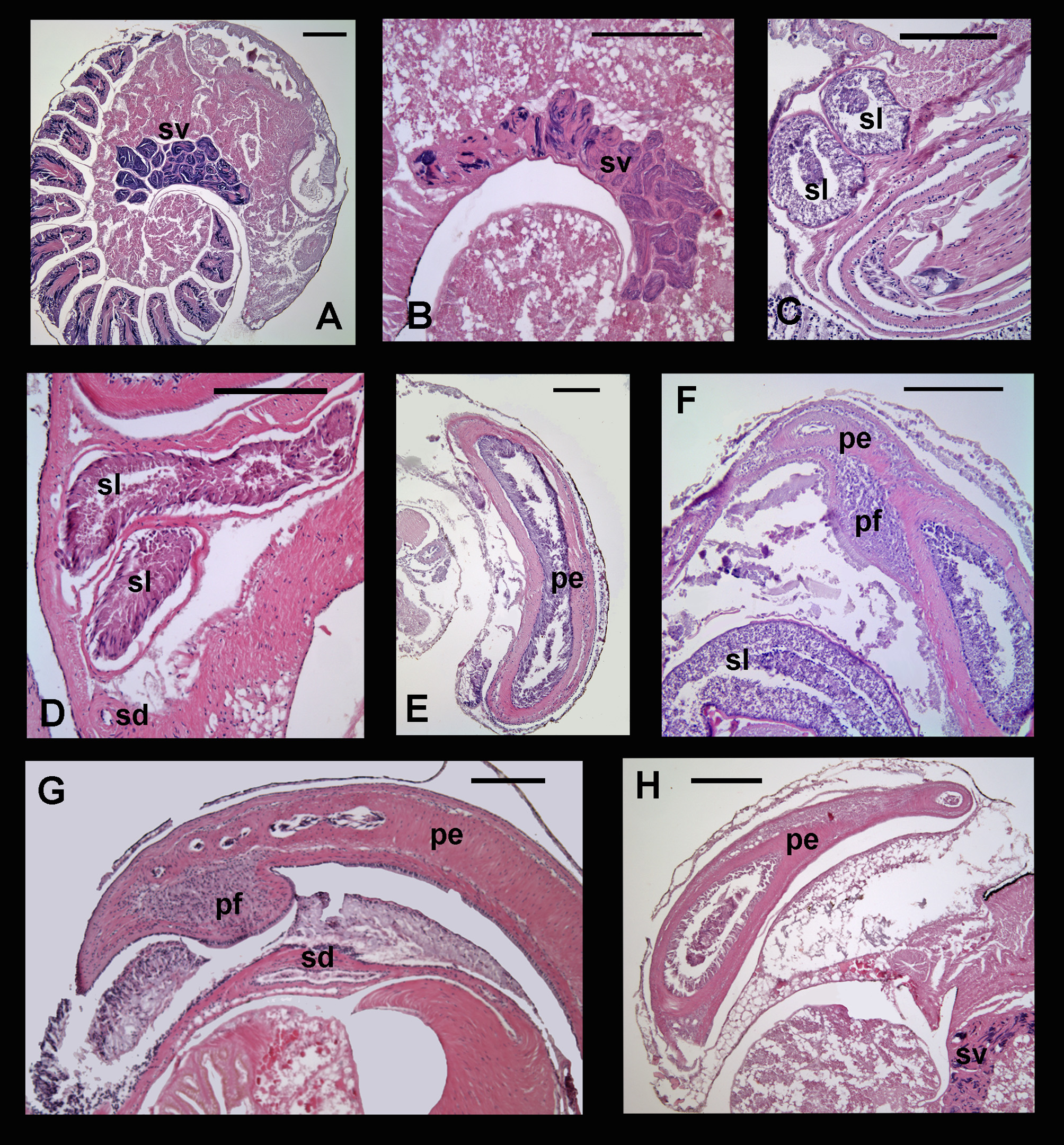
Reproductive anatomy: Spermiduct typical for Parvanachis , with the addition of a flap-like projection at the penis near the base and a penial pouch. Coiled seminal vesicle with a long posterior portion having normal darkstaining sperm, anterior portion with mixed dark-staining and pink-staining sperm cells. Epithelia in both portions flat. Short mantle cavity duct present adjacent to kidney. Spermiduct from mantle Cavity duct to penis base encased in body wall muscle. At penis base, spermiduct leaves body wall and forms a long loop adjacent to the proboscis. Spermiduct loop epithelium ( Fig. 4 View FIGURE 4 C) tall and mucoidal, becomes non-secretory at penis base. Penis long and narrow, with a short filament tip and a flap of tissue protruding to one side near the base ( Fig. 4 View FIGURE 4 F). A semilongitudinal section of the penis where it narrows is shown in Fig. 4 View FIGURE 4 F; at this point the duct also narrows and becomes non-secretory. Penial spermiduct straight. Tissue flap at base does not involve duct or any modified epithelia, so ostensibly has a more mechanical function. Distal portion of penis at rest stored in a pouch in the mantle cavity roof.
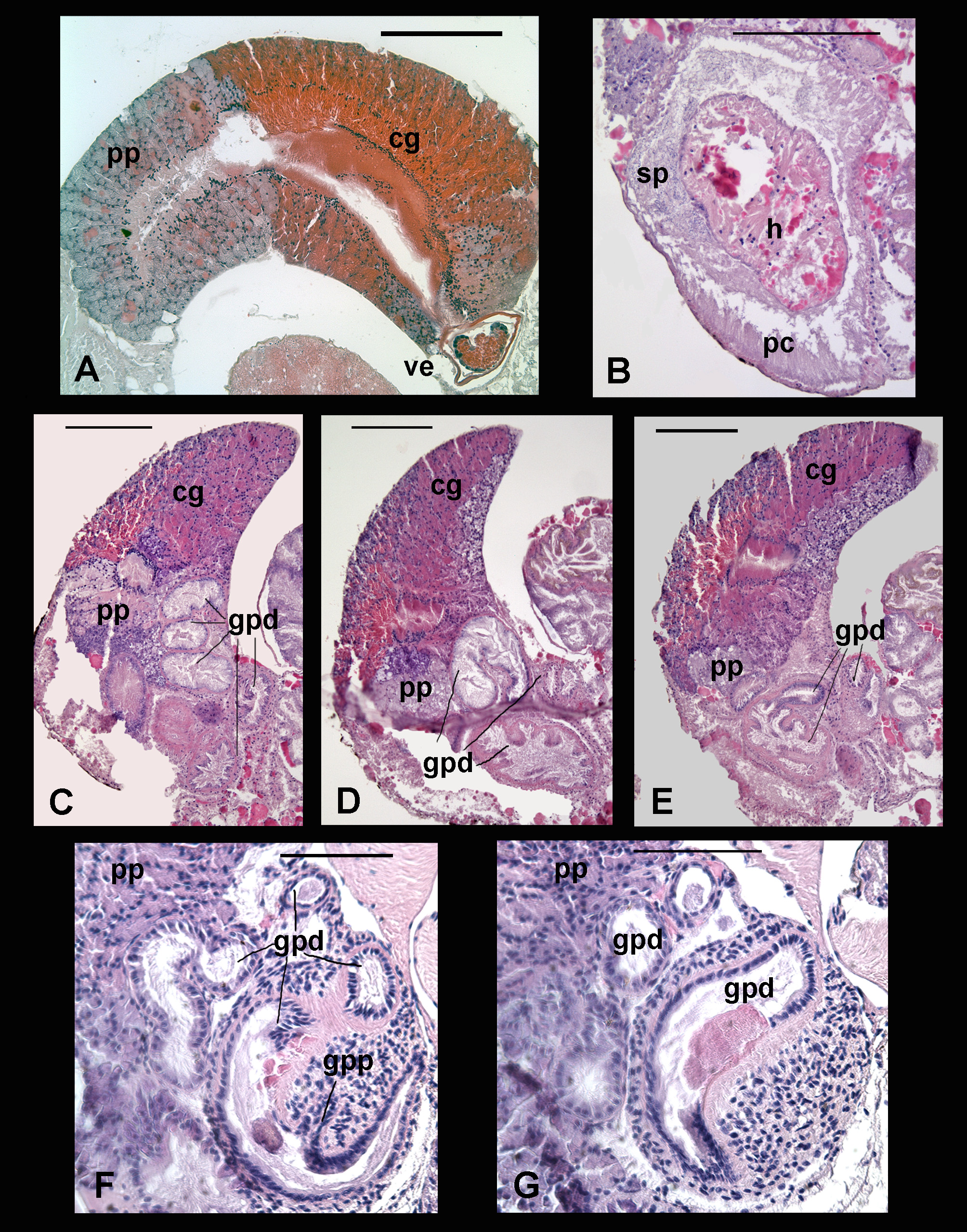
Female reproductive system typical for Parvanachis ; bursa copulatrix absent, coiled gonopericardial duct present. Glands comprise a single mass, which stains red anteriorly and darker purple posteriorly, with a pale purple band across the middle closer to the posterior end. Gonopericardial duct splits from oviduct just inside posterior end of gland mass. Gonopericardial duct muscular and coiled on the oviduct side, with a tall secretory epithelium; then widens to become almost as wide as the glandular mass itself, with a pouched, mucoid wall ( Figs. 3 View FIGURE 3 C–E). Duct narrows before entering the pericardium, which in the specimen sectioned was full of sperm and cellular material. Gonopericardial-pallial duct splits from pericardium adjacent to where gonopericardial duct enters. Vestibule short, non-muscular. The specimen sectioned had an egg capsule in the vestibule, so details of the vestibule epithelium and form could not be ascertained with certainty.
Remarks. The 12 year gap between description of this species and its illustration, combined with lack of specimens from the reported type locality and the obvious similarity between several of these species, creates some uncertainty in this species’ original identity. It is possible, given the similarity between P. pygmaea , P. mullineri and P. adamsi (discussed below), to suggest that the unillustrated Columbella pygmaea Sowerby, 1832 , is actually what I identify here as P. mullineri or P. adamsi . Reeve’s (1858) figure, interestingly enough, shows the dark band adjacent to the suture, consistent with P. mullineri . However, Sowerby’s illustration from 1844, consistent with the existing syntypes, sets the identity of the species.
No known copyright restrictions apply. See Agosti, D., Egloff, W., 2009. Taxonomic information exchange and copyright: the Plazi approach. BMC Research Notes 2009, 2:53 for further explanation.
|
Kingdom |
|
|
Phylum |
|
|
Class |
|
|
Order |
|
|
Family |
|
|
Genus |
Parvanachis pygmaea ( Sowerby I, 1832 )
| Maintenon, Marta J. 2014 |
Anachis dalli
| Bartsch 1931 |
Columbella (Anachis) pygmaea
| Sowerby. Kobelt 1897 |
Columbella pygmaea
| Sowerby 1832 |
Anachis pygmaea (
| Sowerby 1832 |
Anachis (Parvanachis) pygmaea (
| Sowerby 1832 |
Parvanachis pygmaea (
| Sowerby 1832 |