
Utivarachna trisula Dhiya’ulhaq & Dupérré, 2024
|
publication ID |
https://doi.org/ 10.11646/zootaxa.5418.5.6 |
|
publication LSID |
lsid:zoobank.org:pub:8681F446-C4A0-466F-A763-57F426B14523 |
|
DOI |
https://doi.org/10.5281/zenodo.10794114 |
|
persistent identifier |
https://treatment.plazi.org/id/03C0D450-D16B-9539-41AD-FF011FB4D117 |
|
treatment provided by |
Plazi |
|
scientific name |
Utivarachna trisula Dhiya’ulhaq & Dupérré |
| status |
sp. nov. |
Utivarachna trisula Dhiya’ulhaq & Dupérré , sp. nov.
Figures 14–17 View FIGURE 14 View FIGURE 15 View FIGURE 16 View FIGURE 17
Type material. Holotype ♂: Sumatra: Jambi Province: Bungku, Bajubang , Batang Hari (2013_HJ1.1_ AraTrac003N_001), canopy fogging in jungle rubber plantation, 01⁰55’41.6”S, 103⁰15’34.2”E, altitude 48 m, 9.V.2013, leg. J. Drescher ( MZB). GoogleMaps
Paratypes: Sumatra: Jambi Province: 1♀ (2013_HJ1.1_AraTrac003N_002), with the same data as holotype ( MZB) GoogleMaps ; 1♀, Dusun Baru , Air Hitam, Sarolangun, (2013_BJ4.1_AraTrac003N_001), canopy fogging in jungle rubber plantation, 02 ⁰00’56.8”S, 102 ⁰45’12.6”E, altitude 64 m, 14.VII.2013, leg. J. Drescher (ZMH-A0023868).
Etymology. The specific name is taken from Indonesian trisula , meaning “trident”, referring to the trifid RTA. Noun in apposition.
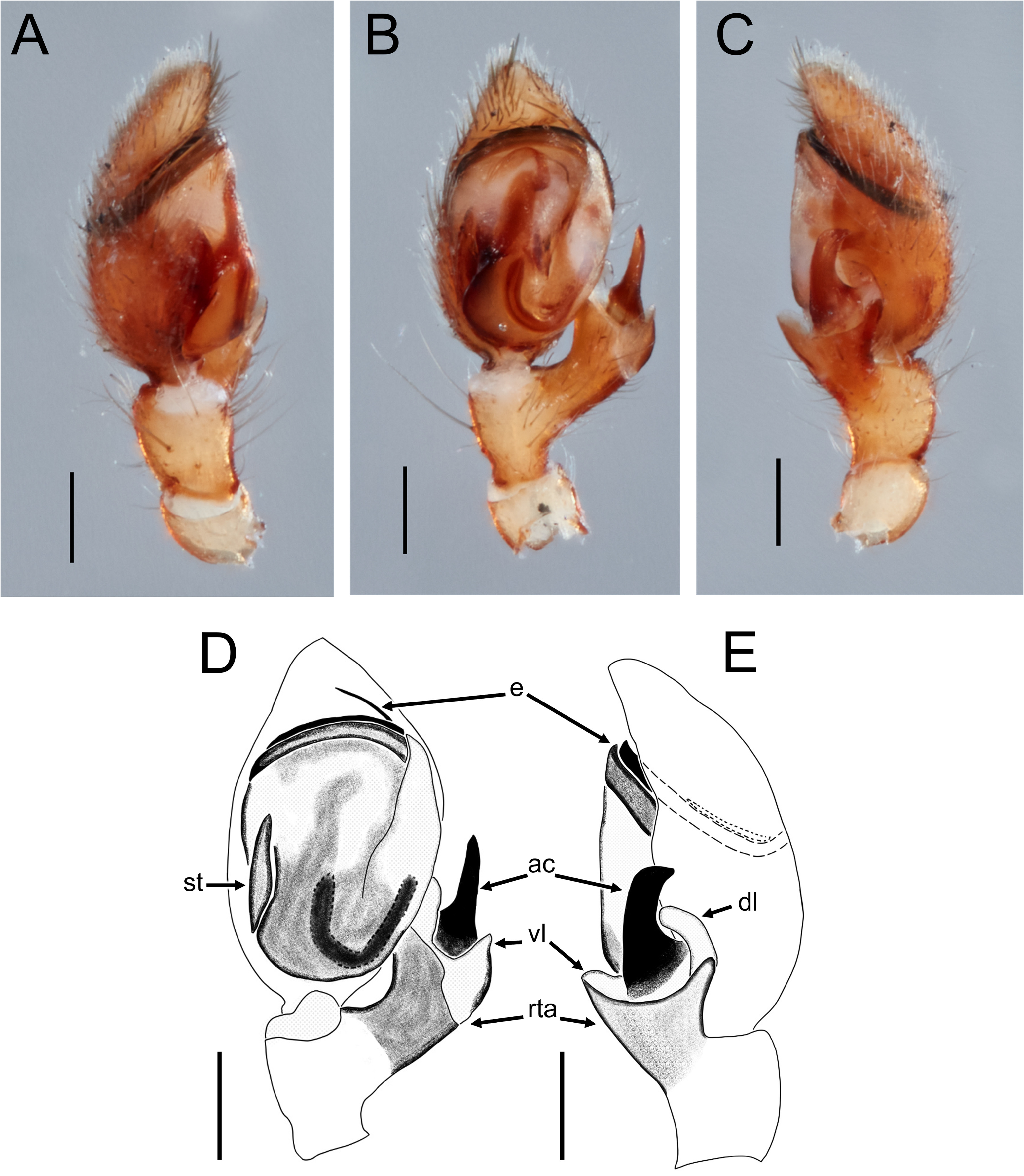
Diagnosis. This species belongs to the kinabaluensis -group based on the following characters: carapace wedge-shaped with undulating lateral margins, posterior end of carapace not produced into an elongated stalk, PER strongly recurved and much longer than AER, palpal tegulum not enlarged and bulbous, bursae curved backwards towards spermathecae. Among the kinabaluensis -group, males of this species are most similar to those of U. kinabaluensis by the RTA ending in a wide arch and an apical claw separated by a membranous area, but can be distinguished by the thumb-shaped RTA-claw with a broad tip ( Fig. 15B, E View FIGURE 15 ) versus curved and with a sickle-shaped tip in U. kinabaluensis ( Deeleman-Reinhold 2001: figs 594–595; Yamasaki 2023: figs 25A, B, D), and the arch of the RTA extending into a thinner, membranous lobe on the dorsal side. Females of this species are most similar to those of U. kinabaluensis in having the copulatory ducts produced into a horizontal helical coil, but can be distinguished by the posteriorly wider bursae versus anteriorly wider in U. kinabaluensis ( Yamasaki et al. 2023: fig. 27C), and the thick, posteriorly broadening connecting ducts ending in not clearly distinguishable spermatheca ( Figs 17B, C View FIGURE 17 ) versus clearly distinguishable, oval-shaped spermatheca in U. kinabaluensis ( Yamasaki et al. 2023: fig. 27C).
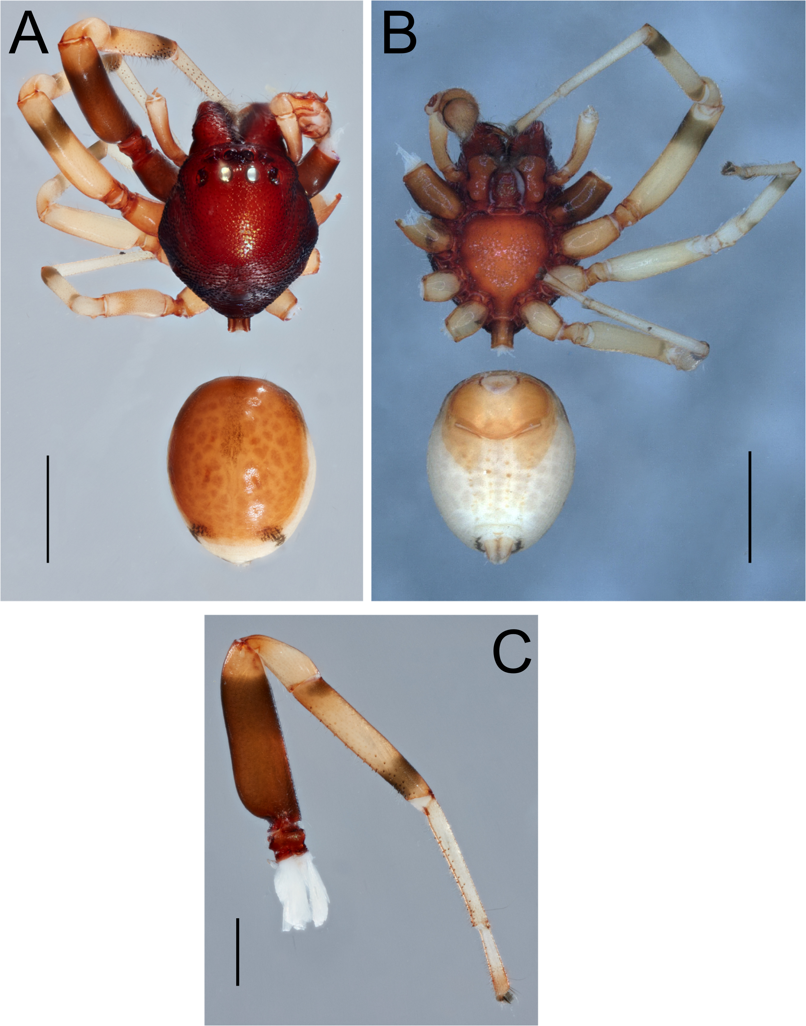
Description. Male (Holotype). Total length 3.41. Carapace length 1.72; width 1.72. Carapace maroon, almost wedge-shaped, with narrowly truncate posterior end, strongly sclerotized, surface granulated, with each granule ending in long white seta, lateral margins undulating, fovea short but distinct; PER longer than AER, both slightly recurved ( Fig. 14A View FIGURE 14 ); clypeus short, with slight projection between chelicerae. Eye diameters: AME 0.09; ALE 0.12; PLE 0.11; PME 0.11. Eye interdistances: AME–AME 0.07; AME–ALE 0.08; ALE–ALE 0.38; PME–PME 0.14; PME–PLE 0.16; ALE–PLE 0.11; AME–PME 0.08; PLE–PLE 0.69. MOA: length 0.28; anterior width 0.25; posterior width 0.35. Clypeus height 0.21. Sternum heart-shaped, colored as carapace, surface granulated ( Fig. 14B View FIGURE 14 ).
Abdomen length 1.69; width 1.30. Abdomen oval, pale-colored, with two pairs of dark patches on lateral margin, one anteriorly and one posteriorly, as well as darker-shaded cardiac pattern and pair of small dark patches directly anterior to spinnerets; most of dorsal surface of abdomen covered with orange scutum ( Fig. 14A View FIGURE 14 ); ventral side of abdomen sclerotized on area anterior to epigastric furrow, projecting a short distance posterior to it on sides, four longitudinal rows of rather faintly sclerotized dots present between epigastric furrow and spinnerets ( Fig. 14B View FIGURE 14 ).
Legs yellow, covered with long white setae, especially on ventral surface, striated with black bands distally and proximally on tibiae I–IV, while on femora I, II only with distal bands. Anterior legs stouter and longer than posterior legs, ventrally filled with leg cuspules from tarsus to tibiae ( Fig. 14C View FIGURE 14 ), metatarsi III and IV distally with comb-like structure followed by brush of setae. Leg measurements: leg I 4.31 (1.26, 0.45, 1.15, 0.96, 0.49); leg II 3.94 (1.18, 0.35, 1.05, 0.9, 0.46); leg III 2.97 (0.88, 0.28, 0.69, 0.78, 0.34); leg IV 3.67 (1.00, 0.31, 0.86, 1.06, 0.44).
Male palp ( Fig. 15 View FIGURE 15 ): Cymbium and bulb oval, sperm duct U-shaped with rather broad turn. Embolus long, coiled horizontally, looping twice, visible dorsally through cymbium. RTA rather large, extending retrolaterally, ending in wide arch and apical claw, lobes of arch thumb-shaped, dorsal lobe continuing into longer, thinner part; apical claw stout, thumb-shaped with wide base, positioned between arch-lobes, separated by narrow membranous area.
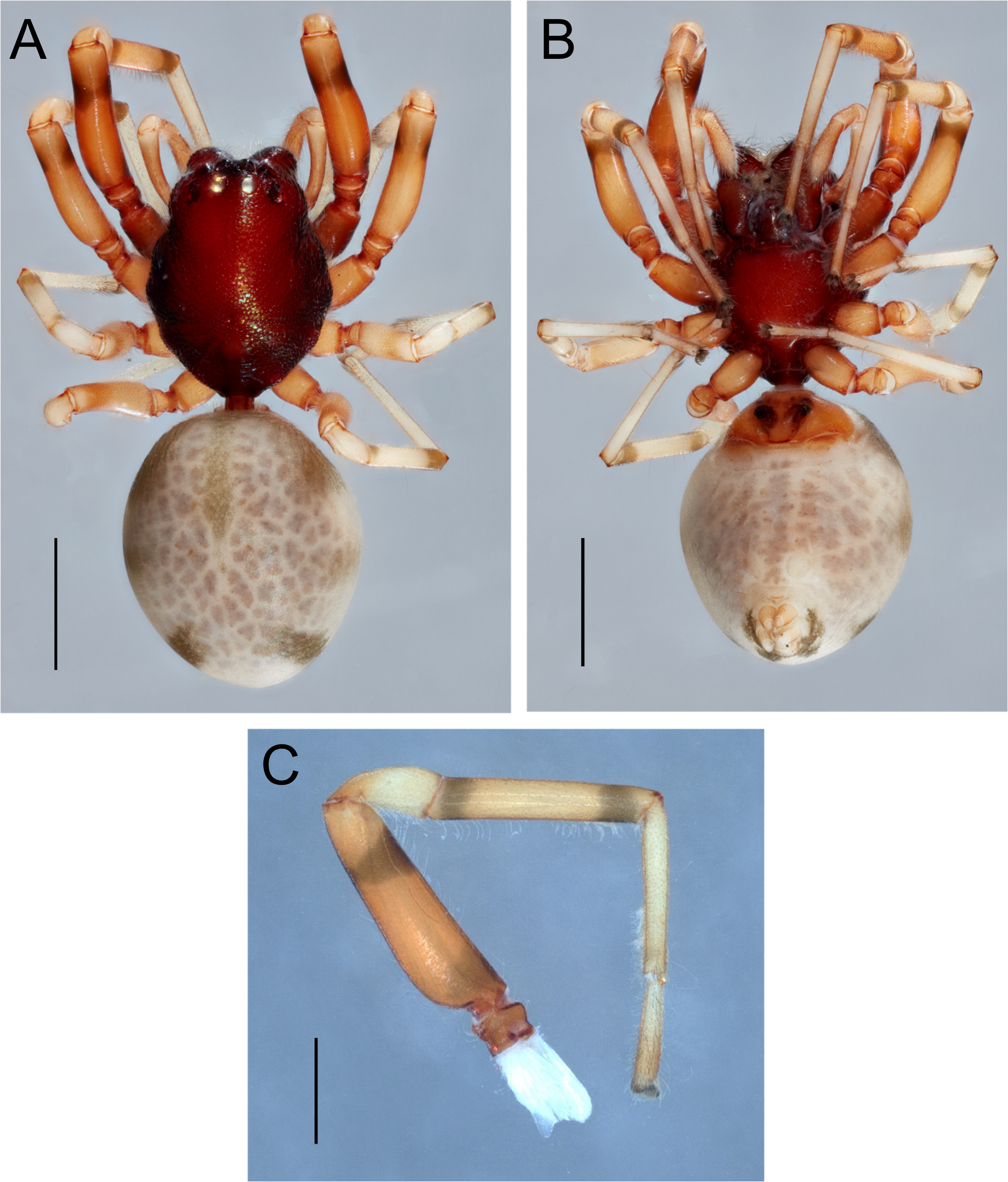
Female (Paratype). Total length 3.88. Carapace length 1.70; width 1.35. Eye diameters: AME 0.10; ALE 0.12; PLE 0.11; PME 0.11. Eye interdistances: AME–AME 0.06; AME–ALE 0.06; ALE–ALE 0.34; PME–PME 0.15; PME–PLE 0.15; ALE–PLE 0.09; AME–PME 0.06; PLE–PLE 0.63. MOA: length 0.27; anterior width 0.26; posterior width 0.34. Clypeus height 0.18. Abdomen length 2.18; width 1.71. General appearance as in male except abdomen not covered in dorsal scutum, with three pairs of dark lateral patches ( Fig. 16A View FIGURE 16 ), longitudinal rows of sclerotized dots absent on venter ( Fig. 16B View FIGURE 16 ). Leg cuspules absent ( Fig. 16C View FIGURE 16 ). Leg measurements: leg I 3.98 (1.14, 0.46, 1.04, 0.83, 0.51); leg II (3.90; 1.18, 0.43, 0.99, 0.91); leg III 2.85 (0.79, 0.26, 0.68, 0.76, 0.36); leg IV 3.50 (0.89, 0.26, 0.84, 1.06, 0.45).
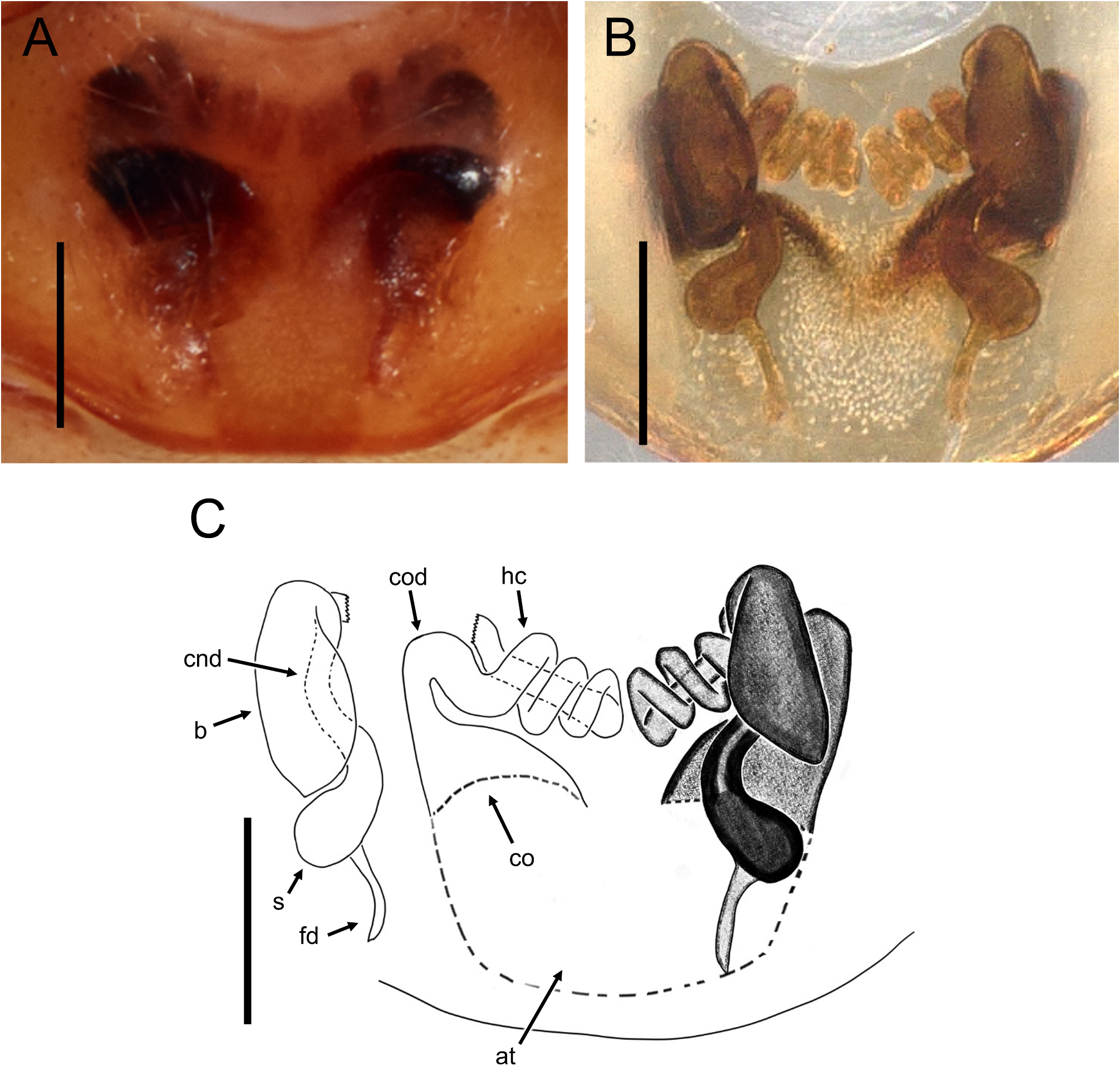
Copulatory organs ( Fig. 17 View FIGURE 17 ): Atrium wider than long. Copulatory openings located on anterior corners of atrium, roughly midway between pedicel and epigastric furrow. Copulatory ducts initially thick, narrowing anteriorly, extending into broad horizontal coil and then into three smaller helical coils, finally turning into straight tube towards middle of coils. Bursae slightly inflated, pear-shaped, anterior part curved. Connecting ducts thick, positioned in between the copulatory openings and bursae, broadening posteriorly, ending into bulbous, not clearly distinguishable spermathecae, together forming an S-shaped structure. Fertilization ducts extending from bulbous posterior end of spermathecae, as long as the latter, slightly curved.
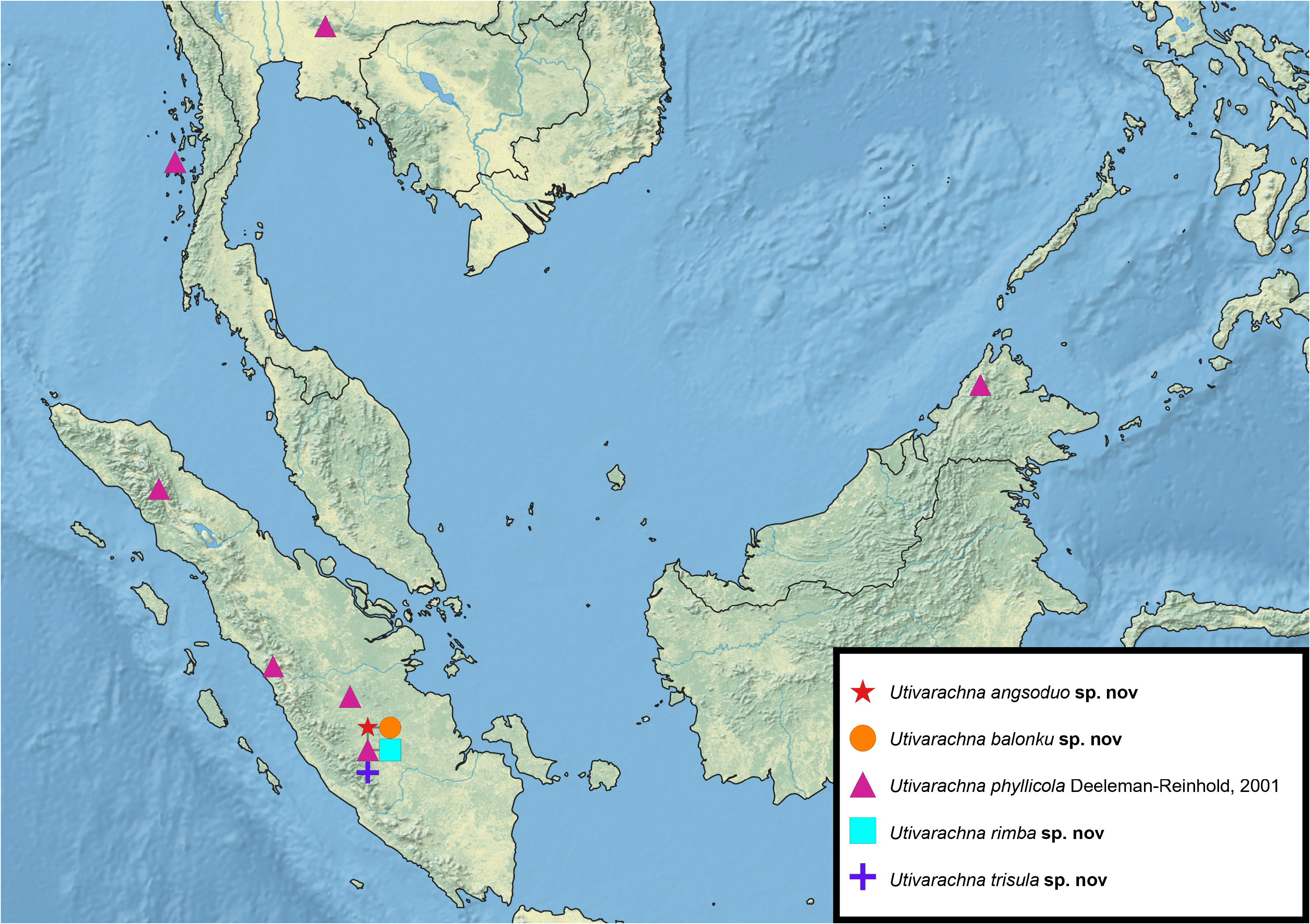
Distribution. Jambi Province, Sumatra ( Maps 1 View MAP 1 and 2 View MAP 2 ).
Remarks. All specimens were collected by fogging in jungle rubber plantations, and are considered arboreal. The genitalic similarity of U. trisula and U. kinabaluensis to members of the fukasawana -group (particularly in regards to the RTA-claw and coiling of the copulatory ducts) might suggests a closer relationship with them than towards other members of the kinabaluensis -group. Currently, the kinabaluensis -group is only distinguished from the fukasawana -group by a single character, which is the absence of an elongation of the carapace posterior end. Thus, a revision of the grouping of Utivarachna species is likely needed.

| MZB |
MZB |
No known copyright restrictions apply. See Agosti, D., Egloff, W., 2009. Taxonomic information exchange and copyright: the Plazi approach. BMC Research Notes 2009, 2:53 for further explanation.
|
Kingdom |
|
|
Phylum |
|
|
Class |
|
|
Order |
|
|
Family |
|
|
Genus |