
Laccobius (Yateberosus)
|
publication ID |
https://doi.org/ 10.2478/aemnp-2018-0017 |
|
publication LSID |
lsid:zoobank.org:pub:D8FC2D46-DB1D-422C-8403-614A7D29C157 |
|
DOI |
https://doi.org/10.5281/zenodo.3705502 |
|
persistent identifier |
https://treatment.plazi.org/id/03C1257D-FFEE-415F-8825-FF664885FD8E |
|
treatment provided by |
Tatiana |
|
scientific name |
Laccobius (Yateberosus) |
| status |
|
Laccobius (Yateberosus) View in CoL sp.
Material examined. 3 larvae of second instar, 5 larvae of third instar ( NMPC, KMNH): “ NEW CALEDONIA / Grande Terre (N-Prov.) / Koniambo / 05.01.05 (CONF-015) / leg. C. Pöllabauer / Confiance bassin / 479.822 mE / 7.673.494 mN / 45 m a.s.l.”. All larvae were collected in the ultramafic gravel of riffles in an unshaded part of the middle reach of Confiance River, 21°2’26”S / 164°48’10”E (Koniambo Mountain, Koné Municipality, North Province, New Caledonia).
Additional material available. 9 larvae ( NHMW): same label data as above. These larvae were not examined in detail in the present study.
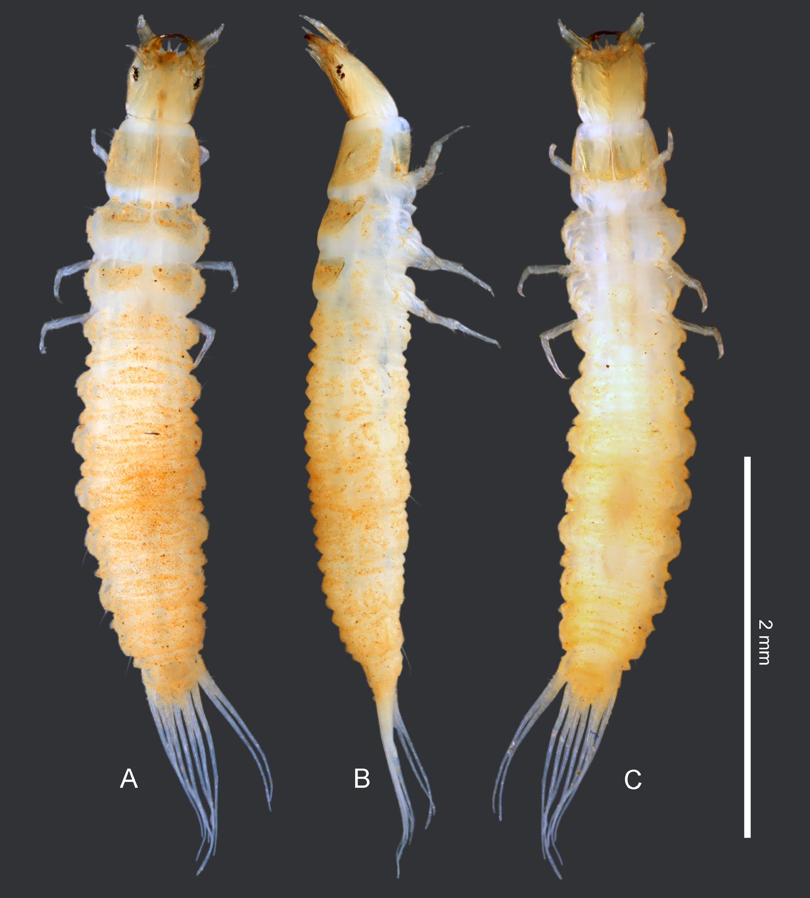
General morphology. Third instar. Colour. Head and sclerotized body parts yellowish to reddish brown (reddish
brown colour probably stems from very high content of iron oxide in the habitat). Membranous parts yellowish to white ( Figs 1A–C View Fig ).
Head ( Figs 1–2 View Fig ). Head superprognathous, slightly longer than wide, subquadrate, widest in anterior third, slightly narrowing posteriad. Frontal lines absent. Surface of head capsule smooth. Each side of head with a group of six stemmata, two anterodorsal ones larger than remaining ones. Clypeolabrum asymmetrical. Nasale asymmetrical, without distinct teeth, projecting anteriad, truncate anteriorly. Epistomal lobes large, strongly asymmetrical, projecting much further than nasale, left lobe projecting slightly further than right one. Gular sulcus reduced, restricted to posterior quarter of head length. Posterior tentorial pits small, closely aggregated. Ventral anteromedian portion of parietale completely fused with submentum, submental sulcus absent. Pair of cervical sclerites present, but small and widely separated from each other.
Antenna ( Fig. 3A View Fig ) 3-segmented, slender, situated on dorsolateral surface of head capsule. Antennomere I ca. 0.5× as long as antennomere II. Antennomere II longer than antennomeres II and III combined, slightly widening distally. Antennomere III shortest, ca. 0.5× as long as antennomere I.
Mandibles ( Figs 3D–E View Fig ) asymmetrical. Right mandible with two inner teeth closely aggregated, distal one slightly larger than basal one; inner face basally of retinacular teeth denticulate. Left mandible with two inner teeth; distal tooth much smaller than basal one, with small denticle on anterior face; basal tooth large, bearing a comb of flat cuticular projections on posterior face; inner face basally with numerous small cuticular tooth-like projections, basal part with two strong spines projecting distad.
Maxilla ( Figs 3B–C View Fig ) 6-segmented (including cardo), distinctly longer than antenna. Cardo small, subtriangular. Stipes longest, ca. 2.5× as long as palpomeres 1–4 combined; inner face with numerous cuticular spine-like projections, inner distal part without large spine-like projection. Maxillary palpus 4-segmented; palpomere 1 short but widest, incompletely sclerotized dorsally; palpomere 2 shortest, slightly shorter than palpomere 1; palpomere 3 longest, ca. as long as remaining palpomeres combined; palpomere 4 narrowest, slightly shorter than palpomere 3. Inner appendage of maxilla rather large, well sclerotized except ventroapically.
Labium ( Figs 2B View Fig , 3K–L View Fig ) largely reduced. Submentum fused with ventral anteromesal portion of head capsule. Mentum wider than long, retracted below anterior margin of head capsule, sclerotized ventrally, largely membranous dorsally. Prementum slightly narrower than mentum, sclerotized ventrally, weakly sclerotized to membranous dorsally, without cuticular spines. Ligula absent. Labial palpi 2-segmented, well sclerotized, slightly longer than mentum and prementum combined. Few spine-like cuticular projections present on dorsal face of intersegmental membrane between palpomeres 1 and 2.
Thorax ( Figs 1A–C View Fig , 4A–B). Prothorax slightly wider than head capsule. Proscutum formed by large plate subdivided by fine sagittal line; anterolateral corners with numerous long trichoid setae, dorsal surface with sparsely distributed, moderately long setae. Prosternum subquadrate, completely divided into two halves by rather wide median gap; anterior portion with numerous short setae. Mesonotum with large subtrapezoid sclerites of mesoscutum divided mesally by wide gap, each with two setae in posterior half; anterior of each scutal sclerite with one narrow subtriangular prescutal sclerite mesally and a small one sublaterally. Metanotum with one pair of subtriangular sclerites widely separated mesally, prescutal sclerites absent. Posterior portions of meso- and metanotum with area covered with asperities on surface of sclerites as well as membranous parts. Ventral parts of meso- and metathorax not sclerotized.
Legs ( Figs 1A–C View Fig , 4D View Fig ) 5-segmented, long and slender, distinctly visible in dorsal view; prothoracic ones closer to each other than meso- and metathoracic ones. Coxa transverse; trochanter elongate, ca. half as long as femur and as tibiotarsus; pretarsal claw with strong basal tooth. Chaetotaxy consisting of few pores and numerous moderately long setae, swimming setae absent. All three pairs of similar shape, prothoracic ones slightly shorter than meso- and metathoracic legs.
Abdomen ( Figs 1A–C View Fig , 4C) 10-segmented, almost parallel-sided in anterior half, slightly narrowing posteriad in posterior half. Surface with minute cuticular asperities, usually with attached fine dirt. Segment 1 not subdivided into anterior and posterior portion in dorsal view, subdivided into two folds in lateral view, without any sclerites. Segments 2–7 similar to each other, each subdivided into short anterior and longer posterior portion in dorsal view, posterior portion subdivided into two folds in lateral view; dorsal and ventral surface without any sclerites or areas with hooked cuticular projections. Chaetotaxy of abdominal segments not examined in detail. Segment 8 narrower than previous segments, subdivided into short anterior and longer posterior portion, posterior portion with small semicircular dorsal sclerite; posterior margin of sclerite with four blunt projections, each with a short seta; posterolateral portion of segment 8 with three long projections (tracheal gills) on each side. Segment 9 small, with three terminal long projections (tracheal gills). Segment 10 reduced, indistinct. Spiracles absent. Spiracular atrium not developed; styli, procerci and acrocerci absent.
Second instar. Similar to third instar, more weakly sclerotized than third instar.
Head ( Figs 5A–B View Fig ) slightly shorter, subquadrate, nearly parallel-sided laterally. Frontal sulci well developed, widely separated from each other at posterior margin of head capsule; closest to each other in posterior third, diverging both anteriad and posteriad; anteriorly reaching outer margin of antennal fossa and continuing to anterolateral margin of head capsule; coronal sulcus absent. Gular sulcus weakly developed in posterior half. Posterior tentorial pits distinct, narrowly but distinctly separated from each other.
Antenna ( Fig. 3F View Fig ) stouter than in third instar, antennomere II more distinctly widened apically.
Mandibles ( Figs 3I–J View Fig ) shorter and stouter, inner face of right mandible without toothlets basally of basal retinacular teeth; armature of spine-like projections of basal retinacular teeth and inner basal face of left mandible less complex than in third instar, distally directed basal spines absent.
Maxilla ( Figs 3G–H View Fig ) relatively shorter and stouter, cuticular projection on inner face of stipes less numerous and generally shorter than in third instar.
Labium ( Figs 3M–N View Fig ) with mentum and prementum relatively narrower than in third instar, labial palps relatively longer than in third instar, without spine-like cuticular projections on dorsal face of intersegmental membrane between palpomeres 1 and 2.
Chaetotaxy of head. Second instar. Frontale altogether with 42 sensilla ( Fig. 5B View Fig ). Central part with three pairs of sensilla diverging posteriad; FR1 rather long seta close to frontal line; FR2 pore-like, situated anteromesally of FR1, closer to FR3 than to FR1; minute seta FR3 situated anteromesally of FR2. Three setae (FR5–7) and one pore (FR4) situated posteriorly of antennal fossa; FR6 moderately long, situated close to frontal line, FR5 moderately long, situated anteromesally of FR6, FR7 minute seta at mesal margin of antennal fossa, FR4 mesally of FR7. Three setae and three pores situated anteriorly of each antennal fossa with three setae and three pores; moderately long setae FR9–10 close to each other, situated anteromesally of antennal fossa; pores FR11 and FR13 close to each other anteriorly of FR10, FR11 closer to anterior margin of head capsule on left side than on right side; pore FR14 situated slightly anteriorly of antennal fossa; short seta FR12 on inner basal portion of each epistomal lobe. Nasale (as in Fig. 2C View Fig ) with five stout short spine-like setae on anterior margin (gFR1), median portion of nasal projection with asymmetrically situated pair of pores (FR15) and pair of moderately short setae (FR8) posterolaterally of FR15; ventral setae of nasale not found. Right epistomal lobe bare, lacking sensilla; left epistomal lobe with five stout long bifid setae with bifurcations between midlength and near apex ( Fig. 2C View Fig ) and two tiny trichoid setae in apical portion (gFR2), basal inner portion with long cuticular projections.
Parietale with 31 sensilla each ( Figs 5A–B View Fig ). Posterior portion of dorsal surface with oblique longitudinal group of five sensilla (PA1–5), setae PA1–2 and PA4–5 small, ca. equidistant from each other, PA3 pore-like, situated between PA2 and PA4. PA6 pore-like, situated on membranous part posterolaterally of frontale. Seta PA7 long, closer to frontal line, seta PA12 long, more lateral, situated ca. at midlength between stemmata and posterior margin of head capsule. Region around stemmata with four setae and one pore; long setae PA8–9 and one secondary, moderately long seta close to frontal line, pore PA10 within posterior group of stemmata; short seta PA 11 in gap between anterior and posterior lateral stemmata. Lateral portion anteriorly with row of three long setae (PA20–22), pore PA19 not found; midlength of head capsule laterally with four long setae (PA13–14 more dorsally, PA16 and PA18 more ventrally), one pore (PA15) between PA14 and PA16, and one moderately long secondary seta and pore PA17 ventrally of PA16. Ventral portion at mandibular articulation with three pores (PA23–25); central portion of ventral surface with four more or less equidistant sensilla (from anterior to posterior one): pore PA27, long setae PA26 and PA28, and pore PA29. Pore PA30 situated laterally of PA29.
Antenna ( Fig. 3F View Fig ). Antennomere I with five pore-like sensilla, two (AN1–2) situated in distal portion of dorsal surface, three (PA3–5) situated on distal margin of antennomere, PA3–4 on dorsal surface, PA5 on ventral surface. Antennomere II with five distally situated sensilla, AN6 and AN9 not found; long seta AN10 and short seta AN11 situated on inner face; sensorium (SE1), short seta AN7 and minute seta AN8 on outer face; sensorium long and slender, as long as antennomere III. Antennomere III with group of apical sensilla (gAN).
Mandibles ( Figs 3I–J View Fig ) with eight sensilla each. Out- er face with one moderately long seta basally (MN1). Midlength with triangular group of three pores, MN4 lateral, MN2 sublateral and situated proximally of MN4, and MN3 situated near base of basal retinacular tooth. Apical portion with four tiny pore-like sensilla (MN5–6 and two secondary ones).
Maxilla ( Figs 3G–H View Fig ). Cardo with one moderately long seta ( MX 1). Stipes ventrally with two pores ( MX 2–3) situated in basal third and half, respectively; outer face with three long setae ( MX 5–6 and one secondary seta) and one pore ( MX 4) distally and one long secondary seta in basal third. Inner face with moderately long seta basally ( MX 7) and three moderately long setae more distally, one ca. at midlength ( MX 8) and two closely aggregated ca. in distal third (likely representing MX 10–11). Maxillary palpomere 1 ventrally with two long setae ( MX 13–14) and one pore ( MX 12), inner face with one long seta ( MX 16). Apical portion of inner appendage with group of sensilla (gAPP), membranous area basally of appendage with one pore dorsally ( MX 17) and one ventrally ( MX 15). Palpomere 2 with one pore ventrally ( MX 18) and one minute seta on outer face ( MX 27). Palpomere 3 with one distal long seta ( MX 21) and one subdistal pore ( MX 22) on inner face, and with one distal long seta ( MX 23) and pore ( MX 20) on outer face. Palpomere 4 with long seta ( MX 24) basally on inner face, and one digitiform sensillum ( MX 25) and one pore ( MX 26) subdistally on outer face; apical portion with group of minute sensilla (gMX).
Labium ( Figs 3M–N View Fig , 5A). Submentum with two pairs of setae, LA1 long, LA2 small, situated anterolaterally of LA1. Mentum with pair of small setae (LA3) ventrally on distal margin of sclerite and pair of pores (LA4) posterolaterally of LA3. Prementum ventrally with pair of moderately long setae (LA6) on distal margin of sclerite and pair of short setae (LA5) close to basal margin; dorsal face with long setae LA10 and pair of pores slightly posteriorly of LA9. Labial palpus with one minute seta (LA13) at ventral base of palpomere 1 and one pore (LA14) on dorsal face of membranous area between palpomeres 1 and 2; apical portion of palpomere 2 with group of tiny sensilla (gLA). Third instar larva. Head chaetotaxy identical to that of the second instar, except for the following characters.
Frontale close to posterior margin of head capsule, with pair of short setae (position of these setae corresponds to pores interpreted in the second instar as parietal sensilla (PA6) situated on membrane posterior to the frontale, and these setae are hence possibly homologous to that sensillum).
Parietale with two pores situated dorsally of seta PA20 (one probably corresponding to PA19 not found in the second instar, the other being obviously secondary).
Mandibles ( Figs 3D–E View Fig ) each with one secondary short seta on outer face, situated slightly distally of MN1.


No known copyright restrictions apply. See Agosti, D., Egloff, W., 2009. Taxonomic information exchange and copyright: the Plazi approach. BMC Research Notes 2009, 2:53 for further explanation.