
Frontonia angusta angusta Foissner, Berger & Agatha, 2002
|
publication ID |
https://doi.org/ 10.11646/zootaxa.4609.3.9 |
|
publication LSID |
lsid:zoobank.org:pub:CE779A20-0B72-4917-98B2-2EFEC0604CDF |
|
persistent identifier |
https://treatment.plazi.org/id/03C187F2-FFFE-FFBC-FF5D-85F4C658FD51 |
|
treatment provided by |
Plazi |
|
scientific name |
Frontonia angusta angusta Foissner, Berger & Agatha, 2002 |
| status |
|
Frontonia angusta angusta Foissner, Berger & Agatha, 2002
Frontonia acuminata (Ehrenberg, 1833) Bütschli, 1889
Frontonia acuminata var. angusta Kahl, 1931
Frontonia angusta Kahl, 1931 — Foissner, Berger & Kohmann, 1994 Frontonia angusta angusta Foissner, Berger & Agatha, 2002
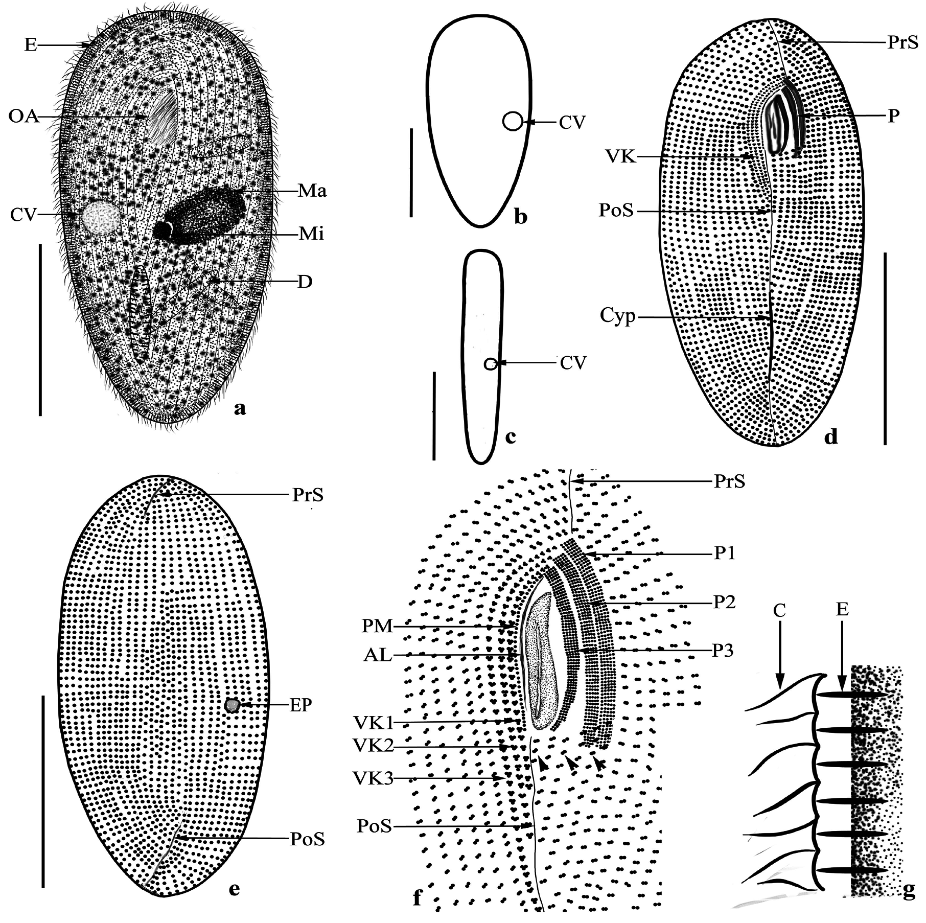
Description of the Turkish population: Size about 130–220 × 50–80 µm, but usually about 170 × 70 µm in size. Body outline is elongated oval with a slightly narrowed posterior half, dorso-ventrally compressed about 3:1 ( Table 1 View TABLE 1 ; Figs. 5 View FIGURE 5 a–e; 6a–e). Single oval macronucleus, 20–50 × 10–30 µm in size and located mid body. Micronucleus 5–8 µm in diameter, mostly globular rarely oval in shape, and located in an indentation of the macronucleus ( Table 1 View TABLE 1 ; Figs. 5a View FIGURE 5 ; 6a, f View FIGURE 6 ). Single contractile vacuole without a collecting channel located mid body, single excretory pore lo- cated on the right dorso-lateral of the cell ( Figs. 5–c View FIGURE 5 ; 6a, c, d, g View FIGURE 6 ). The shape, size, and arrangement of the extrusomes are similar to the previous species. Cytoplasm colorless, food vacuoles contain diatoms, algae, and bacteria ( Figs. 5a View FIGURE 5 ; 6a, b, f View FIGURE 6 ). Somatic cilia 8–9 µm long and arranged in 80–130 meridional rows of kinetosomes, form a suture in the ventral midline of the cell ( Fig. 5g View FIGURE 5 ). Postoral kineties counted as 4–6 ( Table 1 View TABLE 1 ). Oral apparatus typical of the genus, about 1/5–1/6 of the body length, located in the anterior 1/3 of the cell. Three vestibular kineties, 3 peniculus, each consisting of 4–5 rows of kinetosomes. Paroral membrane is in a single row and surrounds the right side of the buccal cavity ( Figs. 5d, f View FIGURE 5 ; 6e, h View FIGURE 6 ).
Frontonia anatolica Yıldız & Şenler, 2013
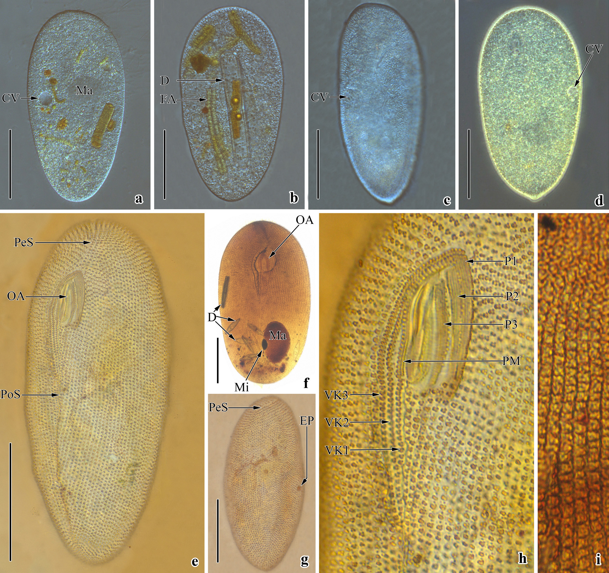
Description of the Turkish population: Body size 100–135 × 50–70 µm, usually about 120 × 60 µm in vivo, ratio of length to width about 1:2, elliptical outline when viewed from the ventral or dorsal side, dorso-ventrally compressed about 2:1 ( Table 1 View TABLE 1 ; Figs. 7a–e, 8a–e). Single macronucleus is usually oval, rarely globular, or elliptical, about 30 × 20 µm in size. Micronucleus is ellipsoidal to globular, located in an indentation of the macronucleus, about 6 × 5 µm in size ( Table 1 View TABLE 1 ; Figs. 7a; 8a, b, d, g, h). Two contractile vacuoles, each with long collecting canals,
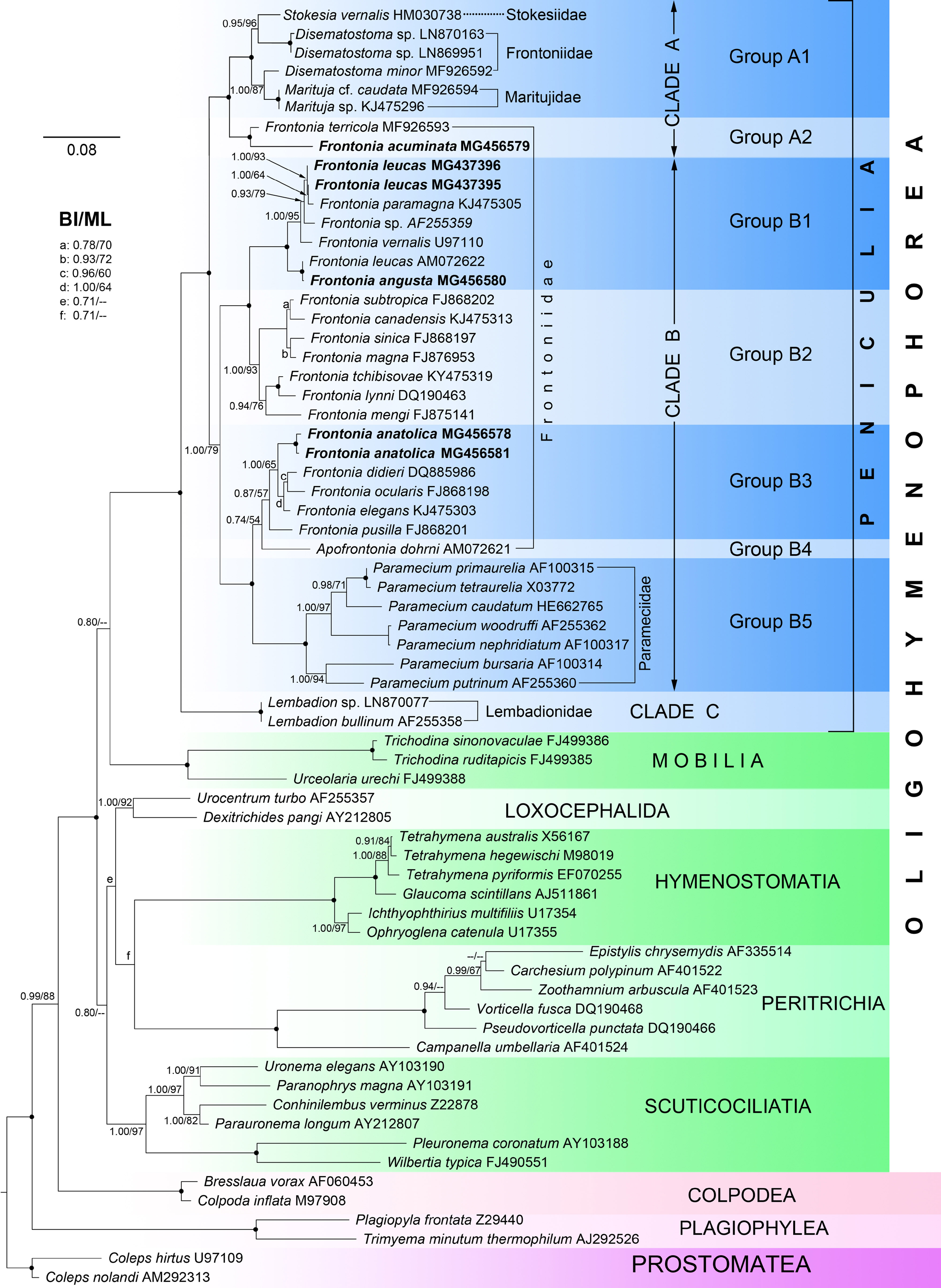
The topologies of the ML and BI trees were similar; therefore, the topology of the ML tree was shown with support values from both algorithms on the branches ( Fig. 9 View FIGURE 9 ). Peniculia formed a fully supported clade in the class Oligohymenophorea (BI/ML, 1.00, 100%). Within the Peniculia clade, the family Lembadionidae (clade C) was basal to the other peniculines included in the analyses (BI/ML, 1.00, 100%) (clades A and B). Clades A and B included 4 families: Stokesiidae , Frontoniidae , Maritujidae , and Parameciidae . Frontonia species were located in both clades A and B. Clade A was formed by group A1 ( Stokesia vernalis , Disematostoma minor , Disematostoma sp., Marituja cf. caudate, Marituja sp.) and group A2 ( Frontonia terricola and F. acuminata ) (BI/ML, 1.00, 100%). Clade B was separated into 2 subclades and consisted of 5 groups. The first subclade was formed by groups B1 and B2 and contained only Frontonia species. The second subclade consisted of group B3 ( F. anatolica , F. didieri , F. ocularis , F. elegans , F. pusilla ), group B4 ( Apofrontonia dohrni ), and group B5 ( Paramecium spp.). In the phylogenetic tree, F. acuminata and F. terricola were included in group A2, and were more closely related the members of the genera Stokesia , Disematostoma , and Marituja than the other Frontonia species. Similarly, F. anatolica ( MG456578 View Materials , MG456581 View Materials ), F. didieri , F. ocularis , F. elegans , and F. pusilla were more closely related the species of the genera Apofrontonia and Paramecium than the other Frontonia members.
No known copyright restrictions apply. See Agosti, D., Egloff, W., 2009. Taxonomic information exchange and copyright: the Plazi approach. BMC Research Notes 2009, 2:53 for further explanation.
|
Kingdom |
|
|
Phylum |
|
|
Class |
|
|
Order |
|
|
Family |
|
|
Genus |
Frontonia angusta angusta Foissner, Berger & Agatha, 2002
| Kizildag, Sibel & Yildiz, Ismail 2019 |
Frontonia angusta
| angusta Foissner, Berger & Agatha 2002 |
Frontonia acuminata var. angusta
| Kahl 1931 |
Frontonia angusta
| Kahl 1931 |