
Phaenicocleus, Štys, Pavel & Ř, Petr Ba Ň A, 2009
|
publication ID |
https://doi.org/ 10.5281/zenodo.186281 |
|
DOI |
https://doi.org/10.5281/zenodo.6224257 |
|
persistent identifier |
https://treatment.plazi.org/id/03C26853-FF98-194F-FF55-1EB7A41055E6 |
|
treatment provided by |
Plazi |
|
scientific name |
Phaenicocleus |
| status |
gen. nov. |
Phaenicocleus View in CoL n. gen.
Etymology. Anagram of Enicocephalus ; gender: masculine.
Type species: Phaenicocleus sabahensis Štys & Baňař , n. sp., by present designation.
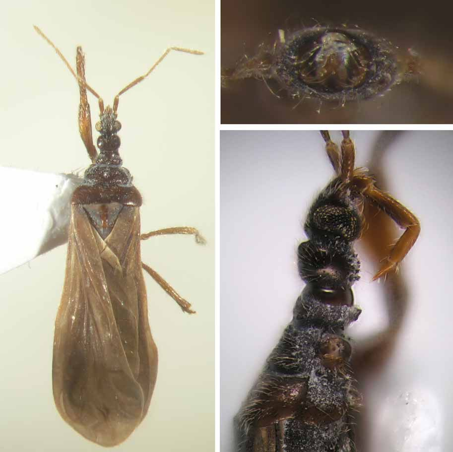
Description (valid for males only). Small-sized (3.9–5.2 mm), macropterous. Body slender, not depressed.
Texture of cuticle of head and pronotum mostly rugulose, but shiny in P. schwendingeri . Some areas of head and pronotum always with setigerous tubercles, at least on posterior lobe of head and collum ( P. schwendingeri ), often also on epicranium, and midlobe, prosupracoxale, and hindlobe of pronotum; cephalic and pronotal tubercles ranging from very minute, lens-shaped, bluntly columnar to large and sharply triangular. Conspicuous setigerous tubercles also on forelegs, on ventral faces of forecoxa, foretrochanter, and forefemur (two tubercles on basalmost part of forefemur in P. schwendingeri ). Vestiture formed by whitish straight or curly trichoid setae ( Figs. 1–3 View FIGURES 1 – 3 ), only sparsely distributed in P. schwendingeri . Longer, curly setae occure mostly on foretibiae. Texture of cuticle rugulose to smooth ( P. schwendingeri ). Coloration light brown to blackish, without particular colour patterns.
Head short, antenniferous tubercles diverging immediately in front of eyes, no part of genae visible in dorsal view. Epicranium strongly convex, anteclypeus by ca. half of its length exceeding apices of antenniferous tubercles. Eyes medium-sized, close to a deep postocular constriction, facettes small and separately rounded. Posterior lobe of head transverse, rounded, its posterior margin convex, separated by a sharp constriction from the neck; median shallowly concave, without impression. Ocelli large, mutually widely distant.
Scape with a distinctly separated prescapite. Pedicel slightly and regularly incrassate distalwards, antennal segments III and IV much thinner than pedicel, but not flagelliform, III terete, IV elongately subfusiform. Mutual length ratios of antennal segments II, III, IV variable, but differences small, pedicel shorter to about as long as the first flagellomere.
Labium thin, geniculate, directed posterad, apex reaching basis of posterior lobe of head, segment II strikingly long and thin, about once to 1.3 times as long as III. Labial formula (the longest segment first) II – III – IV – I.
Pronotum of distinct three lobes; collum long and robust, its dorsal architecture species-specific, in lateral outline separated from the midlobe by a deep gulf. Midlobe with a species-specifically constructed central fossette and a pair of indistinct lateral impressions; median species-specific; posterior margin entire, more or less triconvex. Lateral margins of midlobe either free or partly covered by anterolateral extensions of posterior lobe ( P. schwendingeri ). Hindlobe ample, much broader than midlobe, posterior margin broadly concave. Central fosette on midlobe and medians of mid-and hindlobes species-specific.
‘Proepimeral lobes’ reaching about half the length of fore coxa, nearly or fully closing fore acetabula from behind, but their ventralmost apices distant for about a length of fore coxa. Anterior terminations of prosupracoxale shifted mesad, right and left prosupracoxale converging and reaching or nearly reaching the posteromedial euprosternal species-specific structure.
Eumesosternum with an impressed, percurrent linear median provided with both anterior and posterior transverse linear bars. Eumetasternum with a median raised keel, its shape species-specific.
Mesoscutellum triangular, the apex extended in an obtusely rounded and separately convex mucro.
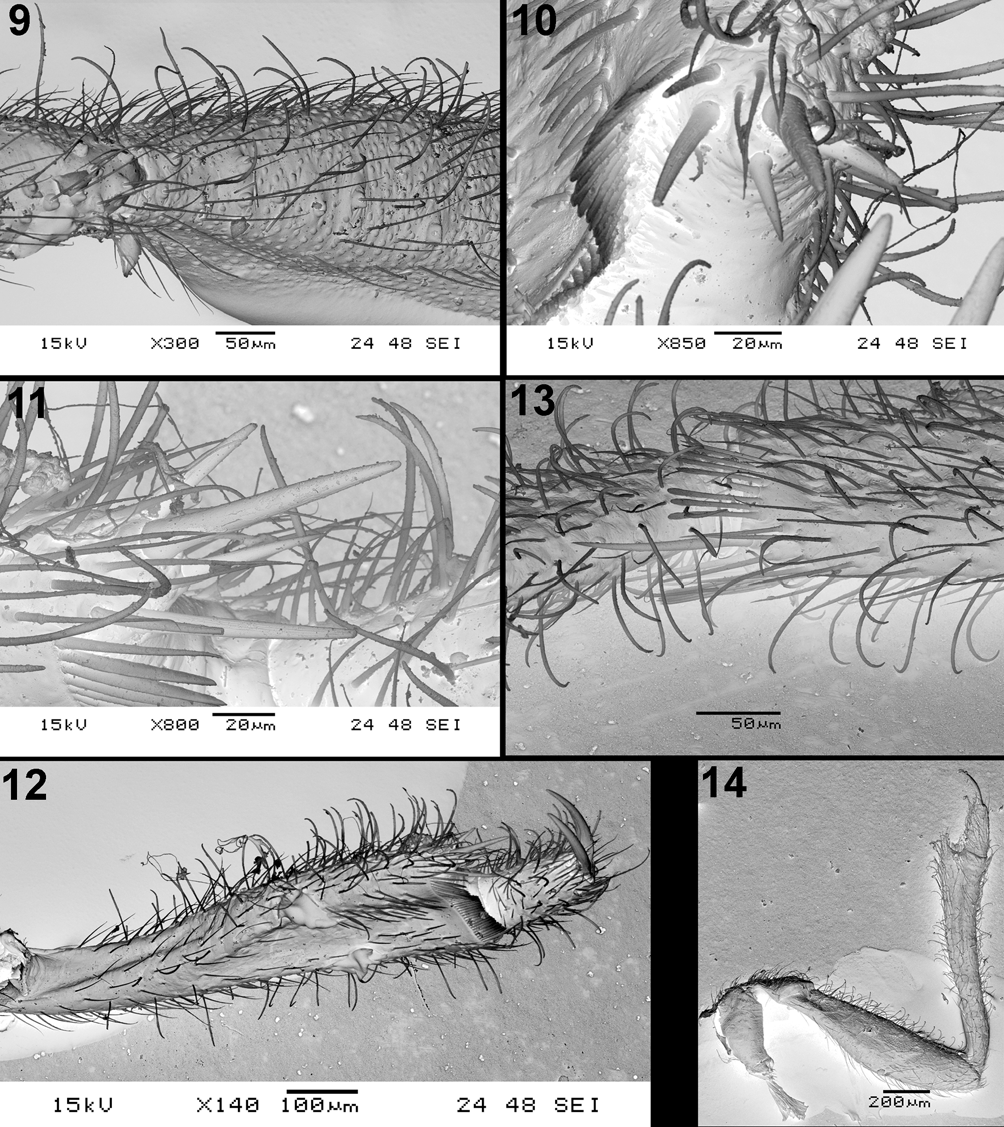
Forelegs. Coxa and trochanter without particulars. Femur stouter in P. sabahensis and P. minor , more slender in P. schwendingeri . Slender foretibia markedly dorsoventrally curved in P. schwendingeri ; stouter and normal shape in P. sabahensis and P. minor . Tibial armature consists from group of four spiniform setae, situated on apicitibial process (very conspicuous in P. schwendingeri ). Tarsal armature almost missing, with an exception of a slender short spiniform seta in the middle of ventral face of foretarsus. One isolated seta, adjoining the ventralmost seta of the bristle comb always present. Foretarsal claws subequal in length.
Middle and hind legs long and slender, without particulars. Apex of tibiae postero- and anteroventrally with a short comb of setae each, consisting of 9–10 setae each and provided with two long spiniform setae ( Fig. 13 View FIGURES 9 – 14. 9 – 13 ). Both middle and hind tarsi with extremely short first tarsal segment, resembling short ringlet only. Claws asymmetrical, the anterior ones much shorter (about one half of length) than posterior ones.
Forewing venation complete, modal, without basal cell1 and with a strikingly long and narrow, closed discal cell. Clavus with a percurrent AA1+2 separating at its basis from the marginal AA3+4; the latter distinct in the proximal third of clavus only, the distal fusion of both veins and formation of a claval cell absent or indistinct. AP short and distinct on ventral surface only. No macrotrichiae on wing membrane.
Pregenital abdomen. Architecture varying segmentally (as well as occurrence of transverse apodemes), in extreme case (see Discussion) all potential components distinct: mediotergite, pair of dorsal laterotergites, membranous connexival line, pair of ventral spiracle-bearing laterotergites, pair of laterosternites, sternum (sometimes subdivided in two hemisternites). Mediotergites 1 and 2 fused, ventrite 1 large and distinct. Segment 8 without any special modifications.
Terminalia (see Discussion). Pygophore strikingly strongly sclerotized, forming a complete, depressed
1. See the remark under P. schwendingeri View in CoL . and Discussion.
and transverse ring. Guide high, exceeding tergum 10 in posterior view, basally broadest, without a shaft, inversely U-shaped or inversely V-shaped (with rounded apex), always provided with an internal, basimedial sclerotized structure (shape species-specific, minutely triangular to long, tongue-shaped) associated with the ventral margin of posterior foramen. Parameres complex, immobile sclerites, situated beneath and outside the dorsolateral parts of guide arms, associated by apodemes with the guide, pygophore margins and tergum 10. The areas of posterior foramen ventrad to parameres and laterad to the guide sclerotized; no unpaired sclerotized element between the parameres present.
Segment 10 represented by smooth, sclerotized tergum only, the latter free, not fused with the pygophore, lid-shaped, convex, associated with two flaps representing segment 11. Ventral parts of both segments not sclerotized.
Differential diagnosis. Phaenicocleus n. gen. belongs to that group of genera of Enicocephalomorpha characterized by the absence of a basal cell and the presence of a closed discal cell; and it would fall in Štys´s (2002: 352) group 1.2.1.3 and would key under the couplets 33–37 in his key to the genera of Enicocephalomorpha of the World. The occurrence of this group is restricted to Enicocephalidae : Enicocephalinae: Enicocephalini, partim. The diagnosis below is preliminary and rather superficial since all the Eastern Hemisphere genera mentioned will be thoroughly revised in next future; also their relationships will be analyzed at another opportunity.
Phaenicocleus View in CoL differs from the American (Sonorian, broadly conceived) male-based genera Urnacephala Wygodzinsky & Schmidt, 1991 and Lysenicocephalus Wygodzinsky & Schmidt, 1991 by the normally developed, ample hindlobe of the pronotum and the absence of their diagnostic autapomorphies (Wygodzinsky & Schmidt, 1991; Štys, 2002). It differs from the Australian Usingeriella Wygodzinsky, 1950 View in CoL and a new, closely related Australian genus (Štys in MS) – both strikingly speciose although only one species was formally described – by a normally developed midlobe of the pronotum (not subquadrate), but shares with both of them the presence of many setigerous tubercles over the body and large ‘proepimeral lobes’. The minute species of Nesenicocephalus Usinger, 1939 View in CoL ( Australia, Philippines, Oceania, New Guinea – distribution based also on undescribed species) lacks in contrast to Phaenicocleus View in CoL any median and central structures on the pronotum, and their females are universally caducous (unknown in Phaenicocleus View in CoL ). The Madagascan Schenchiella Villiers, 1969 is based on one female lacking wings, and the general facies ( Villiers, 1969; Štys, unpublished) of its minute and depressed type species has nothing in common with Phaenicocleus View in CoL .
Phaenicocleus View in CoL would run in Štys´s (2002) generic key to Pseudohenschiella Villiers, 1958, an endemic Madagascan genus with 5 species ( Baňař & Štys 2006). The species differ from Phaenicocleus View in CoL species by their smaller size (total length under 3 mm), depressed body, dorsal part of the genae present, flagellum of the antennae filiform, labium directed anterad and its 2nd segment very short, posterior lobe of the pronotum with no indication of a median, discal cell in the forewings strikingly short and claval cell (= anal loop) well developed.
No known copyright restrictions apply. See Agosti, D., Egloff, W., 2009. Taxonomic information exchange and copyright: the Plazi approach. BMC Research Notes 2009, 2:53 for further explanation.
|
Kingdom |
|
|
Phylum |
|
|
Class |
|
|
Order |
|
|
Family |
Phaenicocleus
| Štys, Pavel & Ř, Petr Ba Ň A 2009 |
Usingeriella
| Wygodzinsky 1950 |
Nesenicocephalus
| Usinger 1939 |