
Macrobiotus huziori, Michalczyk & Kaczmarek, 2006
|
publication ID |
https://doi.org/ 10.5281/zenodo.2645581 |
|
publication LSID |
lsid:zoobank.org:pub:68009476-AFD2-4133-A7B5-F7303D38AE2F |
|
persistent identifier |
https://treatment.plazi.org/id/03C27D02-783F-FF90-5F4D-B5EEFCA413C9 |
|
treatment provided by |
Plazi |
|
scientific name |
Macrobiotus huziori |
| status |
sp. nov. |
Macrobiotus huziori View in CoL sp. nov. ( Figs. 1–5 View FIGURES 1–2 View FIGURES 3–5 , 7–43 View FIGURES 6–7. 6 View FIGURES 8–11 View FIGURES 12–15 View FIGURES 16–21 View FIGURES 22–23 View FIGURES 24–32 View FIGURES 33–36 View FIGURES 37–38 View FIGURES 39–42 View FIGURE 43–44 )
Material examined. 8 adults, 1 infant and 20 eggs (prepared for both, DIC and SEM) from the type locality and from Cartago ( Costa Rica; near the road from Pacayas to Turrialba, 27.5 km before Turrialba; moss from soil; 17.12.2002; leg. Ł. Kaczmarek) .
Description. Adult (measurements of the holotype): Body length 455.0 ( Fig. 1 View FIGURES 1–2 , see also Fig. 2 View FIGURES 1–2 ). Body transparent/white, eyes present. Cuticle smooth and without pores. Fine but evident, regular granulation present on all legs but developed better on IV pair ( Figs. 10–11 View FIGURES 8–11 ). Granules have a complex structure and with SEM they are visible as aggregations of small granules or cones (2 to 10 granules/cones on each aggregation) ( Fig. 11 View FIGURES 8–11 ).
Buccopharyngeal apparatus of Macrobiotus type ( Figs. 3–5 View FIGURES 3–5 ). Mouth anteroventral, surrounded by ring of 10 peribuccal lamellae. Oral cavity armature with three bands of teeth ( Figs. 3–4 View FIGURES 3–5 ). Teeth of first band are smaller than those of the other two bands and are in the shape of small cones/granules. They are present in anterior portion of oral cavity just behind peribuccal lamellae and on them. This band of teeth is continuous and looks the same on all oral cavity walls. The teeth of the second band are intermediate in size between those of the first band and those of the third band of teeth. They are in the shape of small ridges parallel to the main axis of the buccal tube (ventral teeth are little larger than dorsal). They are positioned in the posterior portion of the oral cavity just behind the ring fold and just before the third band of teeth. The second band of teeth is continuous and arranged in one row. Some of the teeth are uniform and regular, in the shape of ridges but the rest are joined onebyone. Joined teeth are H, V and Wshaped. The teeth of the third band are larger than those in the other two bands and there are usually six. They are in the shape of transverse ridges/baffles. Teeth in this band are positioned in the rear of the oral cavity just behind the second band of teeth and just before the buccal tube opening. Usually this band is not continuous and is divided into two series: ventral and dorsal. Both series consist of one median and two lateral teeth. Dorsal teeth are more barshaped and ventral are more rectangular shaped. Medioventral tooth is very often divided into 2–5 smaller teeth.
Buccal tube 54.0 long and 12.0 [22.2] wide with one bend in anterior part of tube (visible in lateral view). Stylet supports inserted on buccal tube at 42.0 [77.8]. Pharyngeal bulb slightly oval with apophyses and three macroplacoids ( Figs. 3, 5 View FIGURES 3–5 ). Pharyngeal apophyses distinct, triangular, wider and indented posteriorly. First macroplacoid thinner anteriorly, 9.0 [16.7] long, second oval or rectangular 7.0 [13.0] long, both without constriction (second microplacoid may sometimes have slight constriction in anterior part). Third macroplacoid 12.0 [22.2] long, with constriction in subterminal part. Microplacoid absent, but there are faint cuticular matchlike structures growing out of the posterior ends of the third macroplacoids ( Fig. 6 View FIGURES 6–7. 6 , visible also on Figs. 1–5 View FIGURES 1–2 View FIGURES 3–5 ). These matchlike structures are slightly wider in their terminal parts (see also ‘Differential diagnosis’). Macroplacoid row 31.5 [58.3] long.
Claws of hufelandi type, slender with very narrow bases ( Figs. 8–10 View FIGURES 8–11 ). Primary branches with distinct accessory points. Lunules on all legs smooth, better developed on IV pair of legs. Primary branch of external claw (pb) of II pair of legs 13.0 [24.1] long, secondary branch (sb) 10.0 [18.5] long; III pb 13.0 [24.1], sb 10.0 [18.5]; IV pb 16.0 [29.6], sb 12.0 [22.2]. Bars on legs absent but cuticular thickenings below claws present ( Fig. 8 View FIGURES 8–11 ).
Eggs. White, laid freely ( Figs. 12–21 View FIGURES 12–15 View FIGURES 16–21 , see also Fig. 2 View FIGURES 1–2 ). Spherical, areolated, with 9–11 processes on circumference. Processes generally in the shape of cones, elongated in terminal part, however the shape is highly variable ( Figs. 24–36 View FIGURES 24–32 View FIGURES 33–36 , see also Figs. 12–21 View FIGURES 12–15 View FIGURES 16–21 ). The elongated terminal part may be missing or be thin and long or split in the end. If the terminal part is present it is usually covered by irregular tubercles ( Figs. 30–32 View FIGURES 24–32 and 34– View FIGURES 33–36 36). Processes consist of double wall with transverse supporting walls forming ‘cells’ visible in DIC as a dense reticular design (‘reticulation’ slightly elongated vertically) ( Figs. 22–23 View FIGURES 22–23 ). External walls of processes smooth or slightly wrinkled (wrinkles in form of rings around a process), without pores. Internal walls strongly porous (visible in SEM only). Surface between processes areolated. Areolation complex ( Figs. 37–44 View FIGURES 37–38 View FIGURES 39–42 View FIGURE 43–44 ). Areolae in the shape of holes which branch into smaller holes closer to the egg surface (see Figs. 43– 44 View FIGURE 43–44 and 37–38 View FIGURES 37–38 ). This kind of areolation has not been described so far.
(DIC, scale on 23 same as on 22).
Remarks. Adults: Results of simple statistical analysis of measurements and pt values of selected morphological structures for six specimens are given in Table 1 View TABLE 1 . Eggs: Statistics for all measurable eggs are provided in Table 2 View TABLE 2 .
Type locality. Costa Rica, Cartago Province, slope of the Irazu Volcano; Irazu Volcano National Park ; moss from a tree; ca. 3400 m a.s.l.; 17.12.2002; leg. Ł. Kaczmarek.
Type depositories. Holotype and 16 paratypes (6 adults, 1 infant hatching out of egg and 9 eggs) are deposited in the Natural History Collections, Faculty of Biology , A. Mickiewicz University, Umultowska 89, 61–614 Poznań, Poland.
Etymology. The new species was named after the first author’s dear friend Paweł ‘ ihs ’ Huzior.
Differential diagnosis. The eggs of Macrobiotus huziori sp. nov. have a unique, never described before, type of multilevel areolation ( Figs. 37–44 View FIGURES 37–38 View FIGURES 39–42 View FIGURE 43–44 ) unlike other species’ eggs which have areolation in the form of ovals or polygons with just one bottom (‘one level’ areolation). This character excludes the new species from any of the known species groups within the genus Macrobiotus (like the areolatus group, for example) and makes M. huziori sp. nov. easily distinguishable from all the known species. However, the adults of the new species are similar to Macrobiotus areolatus Murray , Macrobiotus crenatus Maucci, 1991 , Macrobiotus csotiensis Iharos, 1966 and species of the Macrobiotus richtersi group. Also the eggs of these species (except for M. csotiensis ) are similar to the eggs of M. huziori sp. nov. with the respect to the conical shape of the processes and the presence of areolation between the processes.
Adults of M. huziori sp. nov. are similar to adults of M. areolatus , M. crenatus and M. csotiensis by the presence of three rodlike macroplacoids and lack of the microplacoid. However, the adults of the new species differ from M. crenatus and M. areolatus by the absence of teeth on the lunulae of IV pair of legs. Due to the poor description of M. csotiensis we were unable to find any differences in the adult morphology, but the eggs of M. csotiensis are very different from the eggs of the new species (hemispherical ornamentations positioned very near to each other covered with a hyaline layer, which is concaved between the processes) ( Iharos 1966). Additionally M. huziori sp. nov. has larger eggs than M. areolatus (80.0–100.0 in M. areolatus and 120.0–160.0 in the new species) ( Ramazzotti & Maucci 1983).
It is also worth noting that the matchlike structures growing out of the third macroplacoids in M. huziori sp. nov. and in M. areolatus terminate where the microplacoid is placed in the species of the Macrobiotus richtersi group (see Figs. 6–7 View FIGURES 6–7. 6 ). In the richtersi group the microplacoid is connected with the third macroplacoid by a thin cuticular yarnlike structure. This, and also similarity in the buccal apparatus structure and the egg shell appearance may suggest that both M. huziori sp. nov. and M. areolatus are closely related to the richtersi group but have probably lost the microplacoids and the matchlike structures are the microplacoids remnants.
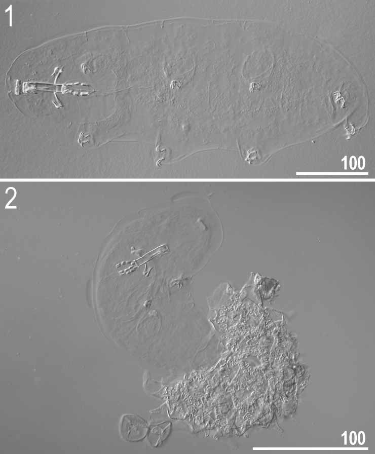


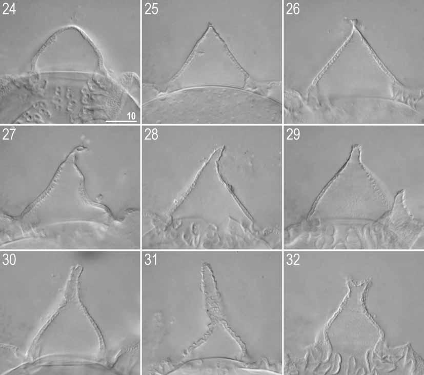


TABLE 1. Measurements [in µm] and pt values of selected morphological structures of specimens of Macrobiotus huziori sp. nov. mounted in Hoyer’s medium (Range refers to the smallest and the largest structure found among all measured specimens; N, number of specimens/structured measured, SD, standard deviation).
| CHARACTER | N | RANGE | MEAN | SD | |||||
|---|---|---|---|---|---|---|---|---|---|
| µm | pt | µm | pt | µm pt | |||||
| Body | 6 | 315.0– | 455.0 | 787.5– | 913.0 | 394.2 | 836.5 | 59.6 | 45.4 |
| Buccal tube | 6 | 40.0– 54.0 | – | 47.0 | – | 5.7 | – | ||
| Stylet support insertion point | 6 | 31.5– | 42.0 | 77.8– | 80.8 | 37.1 | 78.9 | 4.7 | 1.1 |
| Buccal tube external width | 6 | 8.0– | 12.0 | 17.3– | 22.2 | 9.3 | 19.8 | 1.5 | 1.6 |
| Macroplacoid 1 | 6 | 5.5– | 9.0 | 13.8– | 17.4 | 7.6 | 16.0 | 1.5 | 1.5 |
| Macroplacoid 2 | 6 | 4.0– | 7.0 | 10.0– | 13.0 | 5.8 | 12.2 | 1.1 | 1.1 |
| Macroplacoid 3 | 6 | 6.0– | 12.0 | 15.0– | 22.2 | 8.9 | 18.7 | 2.2 | 2.5 |
| Macroplacoid row | 6 | 19.0– | 31.5 | 47.5– | 58.3 | 24.8 | 52.6 | 4.4 | 3.6 |
| Claw 1 primary branch | 3 | 10.0– | 12.5 | 25.0– | 26.1 | 11.5 | 25.5 | 1.3 | 0.5 |
| Claw 1 secondary branch | 3 | 8.0– | 10.0 | 20.0– | 20.7 | 9.2 | 20.4 | 1.0 | 0.3 |
| Claw 2 primary branch | 5 | 10.0– | 13.0 | 24.1– | 26.1 | 11.6 | 25.3 | 1.3 | 0.8 |
| Claw 2 secondary branch | 5 | 8.0– | 10.0 | 18.5– | 21.3 | 9.2 | 20.1 | 0.9 | 1.1 |
| Claw 3 primary branch | 5 | 10.0– | 13.0 | 24.1– | 28.3 | 12.0 | 26.1 | 1.4 | 1.6 |
| Claw 3 secondary branch | 5 | 8.0– | 10.0 | 18.5– | 21.7 | 9.2 | 20.0 | 1.1 | 1.2 |
| Claw 4 primary branch | 5 | 13.0– | 16.0 | 28.8– | 32.7 | 15.0 | 31.1 | 1.2 | 1.8 |
| Claw 4 secondary branch | 5 | 9.5– | 12.0 | 21.2– | 24.5 | 10.9 | 22.6 | 1.1 | 1.3 |
No known copyright restrictions apply. See Agosti, D., Egloff, W., 2009. Taxonomic information exchange and copyright: the Plazi approach. BMC Research Notes 2009, 2:53 for further explanation.