
Asterocheres halichondriae Stock, 1966
|
publication ID |
https://doi.org/ 10.1080/00222933.2012.742588 |
|
publication LSID |
lsid:zoobank.org:pub:1507EC09-372A-4C75-9DD3-6AE64A90DF70 |
|
persistent identifier |
https://treatment.plazi.org/id/03C27E20-FFD7-6566-FE3E-021CFC4BFC23 |
|
treatment provided by |
Felipe |
|
scientific name |
Asterocheres halichondriae Stock, 1966 |
| status |
|
Asterocheres halichondriae Stock, 1966
( Figure 2 View Figure 2 )
Material examined
( a) Holotype female (ZMA-Co.100.951c), allotype male (ZMA-Co.100.951a) and 23 females and one male paratype (ZMA-Co.100.951b) associated with the sponge Halichondria symbiotica Levi from Flic en Flacq Lagoon ( Mauritius) at 1 m depth, collected 13 February 1964 by J.H. Stock; (b) 20 females (ZMA-Co.100.952) associated with Halichondria symbiotica Levi from Black River Bay ( Mauritius) at 1.5 m depth, collected 24 January 1964 by J.H. Stock; (c) two females (ZMA-Co.100.953) associated with Halichondria symbiotica Levi from Pointe aux Sables ( Mauritius) at 1.5 m depth, collected 26 January 1964 by J.H. Stock.
Description
Female. Body cyclopiform with oval cephalothorax and cylindrical urosome (fig. 2A; Stock 1966). Mean body length 480 µm (455–510 µm) and maximum width 265 µm (250–285 µm), based on five specimens. Prosome comprising cephalothorax (fully incorporating first pedigerous somite) and three free pedigerous somites. Urosome four-segmented, comprising leg-5 bearing somite, genital double-somite and two free abdominal somites (fig. 3A; Stock 1966). Dorsal surface of free abdominal somites and posterior part of genital double-somite with epicuticular spinules. Genital doublesomite about as long as wide; paired genital apertures bipartite, each comprising lateroventral copulatory pore and dorsolateral gonopore; lateral margins with long spinules in distal half, posterior to genital apertures (fig. 3A; Stock 1966). Each genital area with two small naked setae.
Caudal rami about as long as wide (fig. 3A; Stock 1966), trapezoidal with inner margin shorter than outer; with six setae; seta I absent, setae III–VI plumose (middle setae very stout) and setae II and VII slightly displaced onto dorsal surface and smooth.
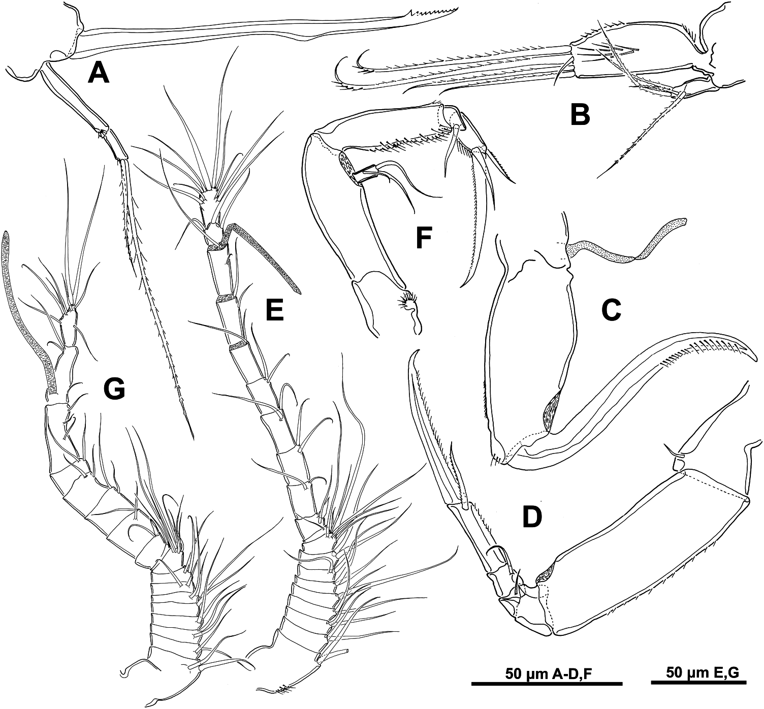
Antennule 20-segmented ( Figure 2E View Figure 2 ), about 265 µm long. Segmental fusion pattern as follows: 1(I)-2, 2(II)-2, 3(III)-2, 4(IV)-2, 5(V)-2, 6(VI)-2, 7(VII)-2, 8(VIII)-2, 9(IX-XII)-7, 10(XIII)-1, 11(XIV)-1+1 spine, 12(XV)-2, 13(XVI)-2, 14(XVII)-2, 15(XVIII)-2, 16(XIX)-2, 17(XX)-2, 18(XXI)-2+1 aesthetasc, 19(XXII-XXIII)-3 and 20(XXIV-XXVIII)-9. Segment 10(XIII) reduced and partly overlapped by distal expansion of compound segment 9(IX-XII). All setae smooth.
Antenna biramous ( Figure 2F View Figure 2 ), about 175 µm long. Coxa small with fan-like spinular tuft. Basis elongate, unarmed. Exopod one-segmented, about twice as long as wide, with two long terminal setae and one shorter and lateral seta. Endopod threesegmented; proximal segment elongated with spinules in inner margin; middle segment
produced distally on medial side but articulating with distal segment proximally on lateral side, bearing one distal smooth seta (longer than segment); distal segment with two subterminal setae, one of them plumose, and distal claw with minute spinules on inner margin.
Siphon short, about 125 µm long, and conical. Reaching to posterior margin of maxilliped insertion.
Mandible ( Figure 2A View Figure 2 ) comprising stylet-like gnathobase and slender twosegmented palp. Proximal segment of palp longer, with fan-like spinular tuft in distal part; distal segment shorter, with one distal spinule and two slightly plumose, unequal terminal setae. Stylet located in oral cone, with denticulate margin subapically.
Maxillule bilobed ( Figure 2B View Figure 2 ). Inner lobe almost three times longer and wider than outer one. Praecoxal endite with five distal setae, one of them minute and naked, with tuft of long spinules medially and row of shorter spinules laterally. Palp with two terminal and two subterminal setae; all slightly plumose.
Maxilla two-segmented ( Figure 2C View Figure 2 ) but with partial transverse surface suture on syncoxa (proximal segment) possibly marking plane of praecoxa–coxa fusion; praecoxal portion bearing flaccid aesthetasc-like element medially, representing tubular extension of external opening of maxillary gland. Coxal portion ornamented with spinules on outer margin being longer at apical part. Basis claw-like with spinule row in distal half as figured.
Maxilliped five-segmented ( Figure 2D View Figure 2 ). First segment with one short smooth seta on inner distal margin. Second segment elongate, with small spinules on outer margin. Third segment compound, partial suture marking original separation of two ancestral segments, with (2,1) armature formula. Fourth segment short, with one smooth seta on inner medial part. Fifth segment with small spinules on inner margin, one plumose subterminal seta and a terminal claw with spinules on inner margin.
Swimming legs 1–4 biramous (fig. 4A–D; Stock 1966), with three-segmented rami and each with intercoxal sclerite. Legs 1–4 as described and illustrated by Stock (1966). Spine and seta formula as follows ( Table 1):
Fifth leg (fig. 3A; Stock 1966) with protopod incorporated into somite. Exopod elongate, ornamented with small spinules on margins and armed with three terminal setae, the two longer plumose. Smooth protopodal seta on somite longer than entire exopod.
Sixth leg represented by two short smooth setae in genital area (fig. 3A; Stock 1966).
Male. Body cyclopiform, with cephalothorax oval and cylindrical urosome. Mean body length 406 µm (380–430 µm), based on three specimens. Urosome fivesegmented, comprising leg 5-bearing somite, genital somite, and three free abdominal
somites. Genital somite about as long as wide bearing genital apertures posterolaterally on ventral surface (fig. 5A; Stock 1966). Most appendages as for female except for antennules, maxillipeds, and legs 2 and 6.
Antennule 18-segmented ( Figure 2G View Figure 2 ), geniculate with geniculation positioned between segments 16(XIX-XX) and 17(XXI-XXIII). Segmental fusion pattern as follows: 1(I)-2, 2(II)-2, 3(III)-2, 4(IV)-2, 5(V)-2, 6(VI)-2, 7(VII)-2, 8(VIII)-2, 9(IX- XII)-7, 10(XIII)-1, 11(XIV)-1+1 spine, 12(XV)-2, 13(XVI)-2, 14(XVII)-2, 15(XVIII)- 2, 16(XIX-XX)-3, 17(XXI-XXIII)-3+1 aesthetasc and 18(XXIV-XXVIII)-9. Segment 10(XIII) reduced and partly covered by distal expansion of compound segment 9 (IX-XII). All setae smooth.
Maxilliped five-segmented (fig. 5C; Stock 1966), very similar to that of female but with tooth-like process in proximal half of second segment.
Second leg (fig. 5D; Stock 1966) showing sexual dimorphism in third endopodal segment.
Fifth leg (fig. 5A; Stock 1966) as for female but all exopodal setae smooth.
Sixth leg (fig. 5A; Stock 1966) represented by opercula closing off genital apertures; each with two smooth setae and rows of fine spinules.
Remarks
Asterocheres halichondriae was collected by Stock at Flic en Flacq ( Mauritius) in 1964 where it lives associated with the sponge Halichondria symbiotica Levi. The re-examination of the holotype and allotype has revealed some differences with the original description. (1) The antennule is 20-segmented in the female and not 19- segmented as Stock described it. (2) The antennary exopod carries one medial seta in addition to the two terminal setae described by Stock. The second endopodal segment has a subterminal seta much longer than the seta illustrated by Stock. (3) The second segment of the mandibular palp has two plumose terminal setae. (4) The inner lobe of maxillule has five terminal setae, all of them plumose except for the shorter seta, and the two longer setae have three or four stout spinules distally. (5) The maxilla bears a flaccid element medially, representing tubular extension of external opening of maxillary gland. (6) The third segment of maxilliped has three elements instead of the two elements described by Stock. (7) The antennule is 18-segmented in the male and not 17-segmented as Stock described it .
Asterocheres halichondriae belongs to the group of Asterocheres species with 20- segmented antennules in females, which comprises 21 species. These species are: A. aesthetes Ho, 1984 , A. boecki ( Brady, 1880) , A. bulbosus Malt, 1991 , A. complexus Stock, 1960 , A. corneliae Schirl, 1973 , A. crinoidicola Humes, 2000 , A. dentatus Giesbrecht, 1897 , A. galeatus Kim, 2010 , A. indivisus Kim, 2010 , A. maxillatus Stock, 1987 , A. neptunei Johnsson, 2001 , A. oricurvus Kim, 2010 , A. planus Kim, 2010 , A. sensilis Kim, 2010 , A. simplex Schirl, 1973 , A. stimulans Giesbrecht, 1897 , A. stocki Nair and Pillai, 1984 , A. tenuipes Kim, 2010 , A. tricuspis Kim, 2010 , A. trisetatus Kim, 2010 and A. ventricosus ( Brian, 1927) .
Asterocheres halichondriae differs from eight of these 21 species ( A. planus , A. sensilis , A. indivisus , A. bulbosus , A. boecki , A. corneliae , A. aesthetes and A. stocki ) in the possession of a one-segmented mandibular palp, in contrast to the two-segmented mandibular palp shown by the present species ( Brady 1880; Schirl 1973; Ho 1984; Nair and Pillai 1984; Bandera and Conradi 2009b; Kim 2010).
As for the body shape, A. halichondriae has an oval cephalothorax and cylindrical urosome, whereas A. tenuipes has a very broad, almost rounded prosome, and a very small urosome and A. galeatus has a large, helmet-shaped cephalothorax ( Kim 2010). Stock (1987) described the cephalosome of A. maxillatus as a rounded shield that covers metasomite 3 and urosomite 1, and A. neptunei has a dorsoventrally flattened prosome ( Johnsson et al. 2001).
Among the remaining 10 species, A. oricurvus and A. stimulans can be easily separated from A. halichondriae by the length of the oral cone. The siphon reaches leg 4 in A. oricurvus and A. stimulans , whereas in A. halichondriae it extends only to the insertion of maxillipeds ( Giesbrecht 1899; Kim 2010).
The shape of the caudal rami serves to separate A. halichondriae from A. complexus and A. simplex . The caudal rami in these two last species are longer than wide, in contrast A. halichondriae has caudal rami that are wider than long ( Stock 1960; Schirl 1973).
From the point of view of the genital double-somite, A. halichondriae shows a regularly rounded contour, whereas A. dentatus , A. ventricosus and A. tricuspis have one- or four-denticulated processes at posterolateral corners of broad anterior part ( Giesbrecht 1899; Kim 2010). Although the description made by Brian in 1927 for A. ventricosus (as Ascomyzon ventricosum ) is incomplete, the illustration of the urosome shows a tooth-like process on the genital double-somite ( Brian 1927).
Finally, the remaining two species, A. crinoidicola and A. trisetatus can be differentiated from A. halichondriae by the maxillule. In A. halichondriae , the inner lobe is almost three times longer and wider than the outer and the lobes are provided with five distal setae and two terminal and two subterminal setae, respectively. However, in A. crinoidicola , the inner lobe of maxillule is twice as long as wide; and A. trisetatus has only three distal setae on the outer lobe of the maxillule ( Kim 2010).
No known copyright restrictions apply. See Agosti, D., Egloff, W., 2009. Taxonomic information exchange and copyright: the Plazi approach. BMC Research Notes 2009, 2:53 for further explanation.
|
Kingdom |
|
|
Phylum |
|
|
Class |
|
|
Order |
|
|
Family |
|
|
Genus |