
Asterocheres maxillatus Stock, 1987
|
publication ID |
https://doi.org/ 10.1080/00222933.2012.742588 |
|
publication LSID |
lsid:zoobank.org:pub:1507EC09-372A-4C75-9DD3-6AE64A90DF70 |
|
persistent identifier |
https://treatment.plazi.org/id/03C27E20-FFDE-657D-FE43-00DEFDF3FB44 |
|
treatment provided by |
Felipe |
|
scientific name |
Asterocheres maxillatus Stock, 1987 |
| status |
|
Asterocheres maxillatus Stock, 1987
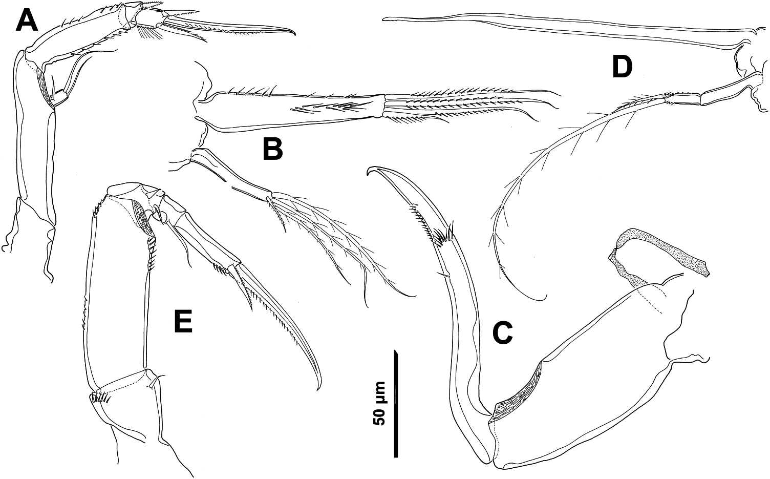
( Figure 5 View Figure 5 )
Material examined
Holotype female (ZMA-Co.102.745c) and one paratype female (ZMA- Co.102.745a+b) associated with Manicina areolata (L.) f. mayori. In Curaçao (500 m west off Piscadera Bay) at 4 m depth, collected 7 January 1974 by J.H. Stock.
Description
Female. Body cyclopiform, consisting of dorsoventrally flattened prosome with rounded cephalothorax, and cylindrical urosome (fig. 1A; Stock 1987). Total length 610 µm and maximum width 420 µm. Prosome comprising cephalothorax (fully incorporating first pedigerous somite) and three free pedigerous somites. Cephalothorax and pedigerous somite 2 and 3 forming a rounded shield covering pedigerous somites 4 and 5 and even half of genital double-somite dorsally (fig. 1A; Stock 1987). Epimeral areas of cephalothorax and somite bearing leg 2 with posterolateral angles pointed backwards. Urosome four-segmented, comprising leg-5 bearing somite, genital double-somite, and two free abdominal somites (fig. 1B; Stock 1987). Genital double-somite wider than long, with paired genital apertures bipartite, each comprising lateroventral copulatory pore and dorsolateral gonopore. Each genital area with one long seta and one stout spine (fig. 1B; Stock 1987). Prosome and urosome with numerous integumental pores and sensilla.
Caudal rami longer than wide, with six terminal setae. Seta I absent; setae II and VII slightly displaced onto dorsal surface; setae III–VI arranged around posterior margin and plumose (fig. 1B; Stock 1987).
Antennule 20-segmented, as described and illustrated by Stock (fig. 1D; Stock 1987).
Antenna biramous ( Figure 5A View Figure 5 ), about 220 µm long (including terminal claw). Coxa short and basis elongate, both unarmed. Exopod one-segmented, 1.5 times longer than wide, with one proximal short seta and two terminal setae, unequal in length. Endopod three-segmented; first segment elongate, with spinules rows as figured; second segment produced distally on medial side but articulating with distal segment proximally on lateral side, with short barbed seta; and third segment with row of long setules on inner margin, two plumose subterminal setae, and terminal claw with spinule row on inner margin.
Siphon conical about 190 µm long, reaching to maxilliped insertion.
Mandible ( Figure 5D View Figure 5 ) comprising stylet-like gnathobase and two-segmented palp. Stylet located in oral cone, with denticulate margin subapically. First segment of palp elongate, unarmed; second segment short, with spinules in lateral margins and apically, and two plumose setae, one short.
Maxillule bilobed ( Figure 5B View Figure 5 ). Both lobes very long and narrow, but inner lobe twice as long as outer. Inner lobe with spinules row in lateral margin and medially and four plumose terminal setae, unequal in length but all shorter than entire segment. Outer lobe with short barbed subterminal seta and three long plumose terminal setae.
Maxilla two-segmented ( Figure 5C View Figure 5 ) but with partial transverse surface suture on syncoxa (proximal segment) possibly marking plane of praecoxa–coxa fusion; praecoxal portion bearing flaccid aesthetasc-like element medially, representing tubular extension of external opening of maxillary gland. Coxa robust and unarmed and basis claw-like with fan-like tuft of spinules and spinule rows in distal half.
Maxilliped five-segmented ( Figure 5E View Figure 5 ), comprising short syncoxa, long basis and three-segmented endopod. Syncoxa with short distal seta on inner margin and spinule row on outer distal margin. Basis elongate, with spinules on margins. First endopodal segment compound, partial suture marking original separation of two ancestral segments, with (1,1) armature formula. Second endopodal segment short, with one smooth very long seta medially. Third endopodal segment with minute spinules on inner distal margin, one plumose subterminal seta, and terminal claw with spinule row on inner margin.
Swimming legs 1–4 biramous (fig. 2C–F; Stock 1987), with three-segmented rami and each with intercoxal sclerite. Spine and seta formula as follows ( Table 3):
Fifth leg with protopod incorporated into somite (fig. 1B; Stock 1987). Free segment (exopod) three times longer than wide, with two smooth terminal setae and one subterminal seta. Somite with outer smooth basal seta displaced to laterodorsal surface and longer than entire exopod.
Sixth leg (fig. 1B; Stock 1987) represented by one long smooth seta and one spine on each genital area.
Male. Unknown.
Remarks
This species lives associated with Manicina areolata (L.) f. mayori. Stock collected it in Curaçao in 1974. Comparison with Stock’s text and illustration revealed a number of discrepancies: (1) the antennary exopod has two distal setae and one proximal seta, which was overlooked by Stock; (2) the second segment of the mandibular palp carries rows of spinules; (3) the inner lobe of the maxillule has long spinules medially; (4) the maxilla bears a flaccid element medially, representing tubular extension of external opening of maxillary gland; (5) the setae present on the third and fifth maxilliped segments are plumose.
This species belongs to the group of species with 20-segmented antennules in the female. This group is composed of 21 species, however A. maxillatus can be separated from all of them by the shape of the body. In this species the cephalosome and metasomites 1 and 2 form a rounded shield, dorsally covering metasomite 3, urosomite 1 and the anterior half of urosomite 2 (genital double-somite) ( Stock 1987). No other species in this group shows this feature.
No known copyright restrictions apply. See Agosti, D., Egloff, W., 2009. Taxonomic information exchange and copyright: the Plazi approach. BMC Research Notes 2009, 2:53 for further explanation.
|
Kingdom |
|
|
Phylum |
|
|
Class |
|
|
Order |
|
|
Family |
|
|
Genus |