
Orientocreadiidae Yamaguti, 1958
|
publication ID |
https://doi.org/ 10.5281/zenodo.189374 |
|
DOI |
https://doi.org/10.5281/zenodo.5697818 |
|
persistent identifier |
https://treatment.plazi.org/id/03C3225A-FF86-873E-CC82-50BFFD2FF89D |
|
treatment provided by |
Plazi |
|
scientific name |
Orientocreadiidae Yamaguti, 1958 |
| status |
|
Orientocreadiidae Yamaguti, 1958 View in CoL
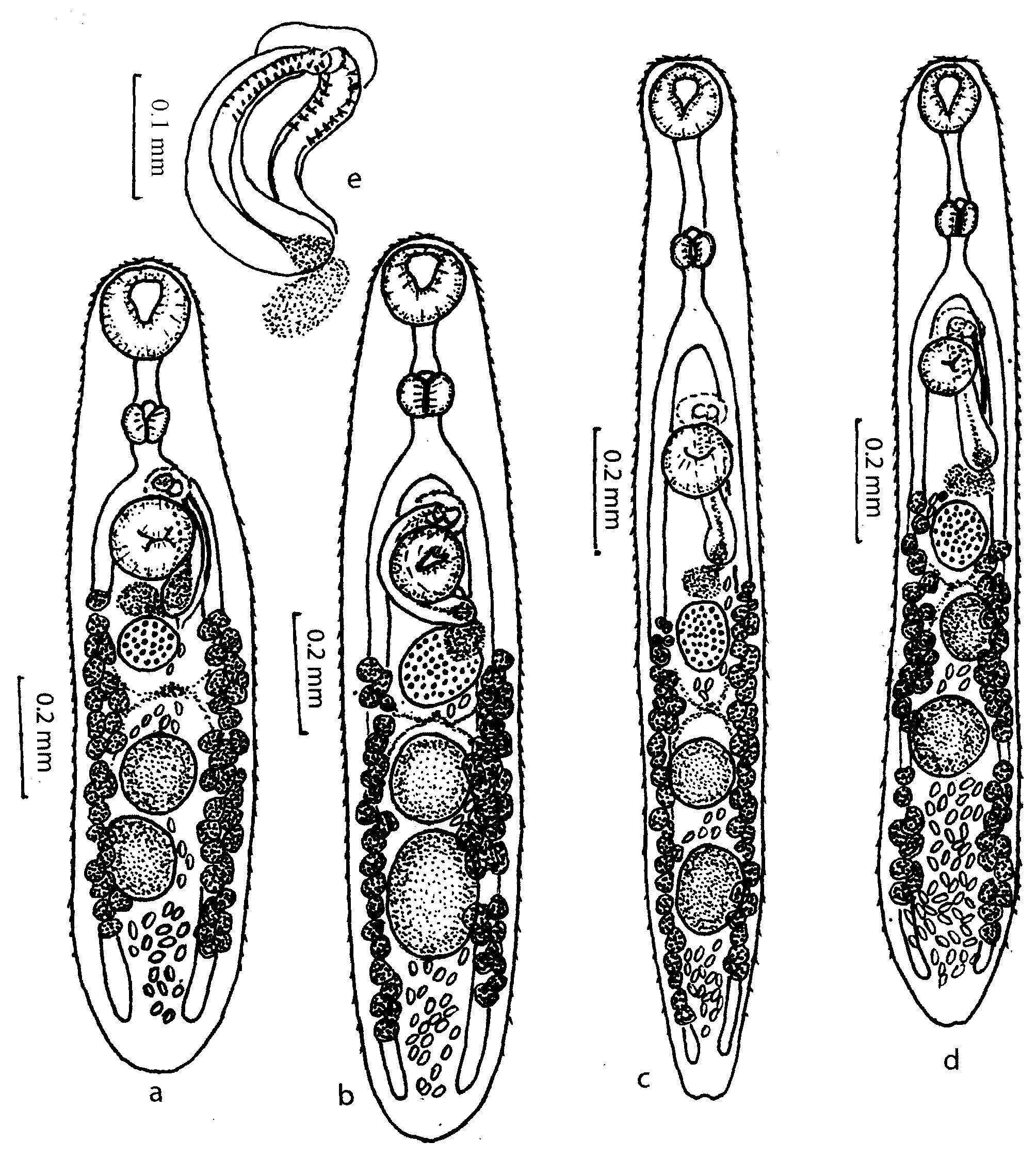
Orientocreadium pseudobagri Yamaguti ( Figs. 1A, 1 View FIGURE 1. A B, 1E, 2A, 3)
Definitive hosts: Osteichthyes— Perccottus glehni (Eleotridae) , Pelteobadrus fulvidraco (Bagridae) .
Site of infection: rectum.
Adult description ( Table 1 View TABLE 1 , Figs. 1A View FIGURE 1. A , B, E): Body spindle-shaped, ends attenuated. Tegument covered with spines. Oral sucker subterminal. Oral and ventral suckers equal size. Pharynx with cruciform indentation. Prepharynx and oesophagus well-developed. Intestine divides immediately anterior to ventral sucker; caeca 2, terminate blindly near posterior end of body.
Testes 2, spherical or oval, along body midline. Cirrus-sac at level of ventral sucker. Seminal vesicle bipartite; external seminal vesicle large; internal seminal vesicle small. Cirrus tubular, with curved spines anteriorly. Genital atrium shallow, immediately anterior to ventral sucker. Genital pore median, anterior to ventral sucker, one third or slightly less of body length posterior to anterior body margin.
Ovary single, entire, in midline or slightly right of midline, posterior to ventral sucker. Mehlis’ gland posterior to ovary, Laurer’s canal present. Uterus coiled, extends between ventral sucker and posterior margin of body; seminal receptacle uterine. Metraterm 12–20 long, lined with spines, opening into genital atrium. Eggs operculate, small 34–39 х 17–22. Vitellarium follicular, extends in lateral fields from level of ovary, or immediately anterior to ovary, to half way between posterior testis and posterior extremity of body, mostly continuous, fields do not extend to posterior margin of body.
Excretory vesicle Y-shaped, anterior margin immediately posterior to posterior testis; excretory pore terminal.
Remarks: Besprozvannykh (1984) described some aspects of the life cycle of O. pseudobagri . He observed that in the Khanka Lake basin the first intermediate host is a range of Lymnaea spp., the second intermediate hosts are species of pulmonate gastropods and 3 species of fish and that the definitive host is the odontobutid fish P. glehni . It was later discovered that tadpoles and prosobranch gastropods (particularly Cipangopaludina ussuriensis ) were also viable second intermediate hosts of this parasite. The miracidium remains in the egg and the first intermediate host is infected passively when the eggs are eaten. Cercariae of O. pseudobagri actively penetrate the second intermediate host and form a cyst in muscle or on the external surface the body. In the wild, intensity of O. pseudobagri in P. glehni (n=50) was (3–28), prevalence 70%.
In the current study, six of 12 specimens of O. pseudobagri from P. fulvidraco displayed a gap in the vitelline follicles at the level of the posterior testis. As summarized by Shimazu (1990), O. pseudobagri has vitelline follicles that do not reach the posterior end of the body, placing it in a group with O. chaenogobii Shimazu and O. siluri (Bychowsky & Dubinina) to the exclusion of other Orientocreadium spp. Orientocreadium chaenogobii , however, has an oral sucker that is smaller than the ventral sucker and an ovary that lies proximal to the ventral sucker. Orientocreadium pseudobagri can be differentiated from O. siluri by possessing spines on the cirrus and metraterm and the ovary being located in the posterior half of the body.
First intermediate hosts: Lymnaea schubinae Kruglov, Starobogatov & Zachvatkinin ; L. coreana (Martens) ; L. ussuriensis Kruglov & Starobogatov ; L. amurensis Kruglov, Moskvicheva & Starobogatov (Gastropoda, Lymnaeidae ).
Localities: Razdolnaja River basin, Khanka Lake basin, small nameless lakes and ponds near Vladivostok, Russia.
Sporocyst: sac-shaped, 0.36 х 0.16 mm, with terminal birth pore.
Site of infection: digestive gland.
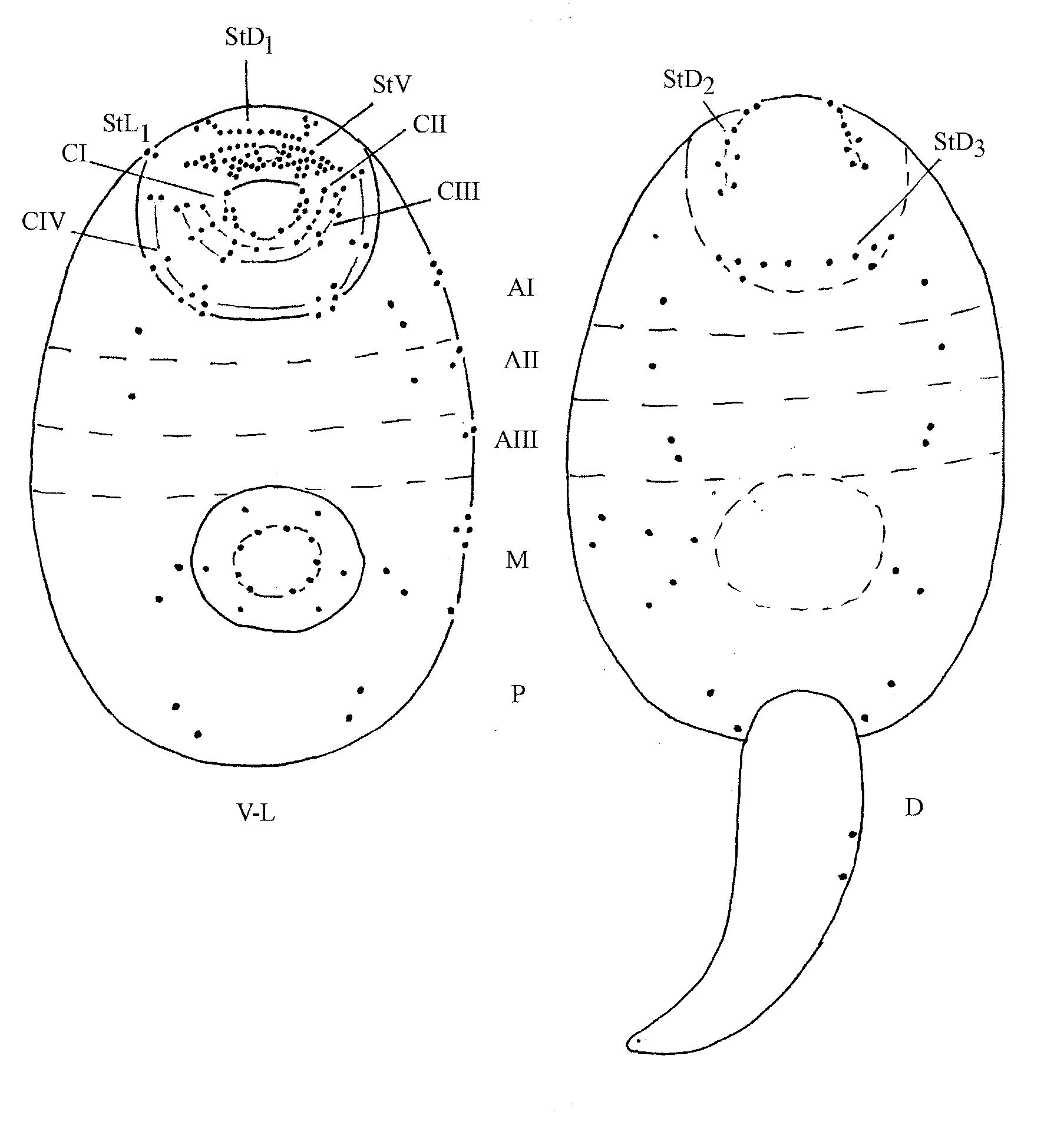
Cercaria ( Fig. 2A View FIGURE 2. A ): body oval, 210–240 × 90–110, with spines from anterior end to level of ventral sucker. Eyespots absent. Oral sucker 46–48 × 42–48, with stylet 19–21 × 2–3. Pharynx 16–21 × 13–19, preceded by prepharynx, anteceded by oesophagus; oesophagus shorter than prepharynx. Intestine bifurcates near anterior margin of ventral sucker; intestinal caeca short, terminating blindly near middle of ventral sucker. Three pairs of penetrating gland cells present on each side of ventral sucker; associated ducts opening at anterior of body, on each side of stylet. Ventral sucker 42–48 × 37–48, lies 100–120 from anterior end of body. Genital primordia located between ventral sucker and Y-shaped excretory bladder. Flame cell formula: 2[(3+3+3)+(3+3+3)]=36. Tail 160–165 х 21–27. Sensory apparatus ( Fig. 3 View FIGURE 3 ): CI= 1V 1, 5 V2, 1 V; CII= 1V 1, 1 V2, 1 V3, 1 V4; CIII= 2V 1, 3 V2 (or 2V 1, 2 V2, 2 V 3 in half of cercariae); CIV= 2V 1, 3 V2, 4 V3; St= 21–26V, 10D1, 4DL1 7D2, 5D3; AI=1– 2 V, 4L, 1–2D; AII= 1V, 0–1D, 2L; AIII=1– 2 V, 1L, 0–1D; M=0– 2 V, 2–6D; PIII= 3V, 1–2D; S=9S1, 5S2; U=2.
Pattern of emergence: Cercariae are shed from snails gradually throughout the day. Emergence does not occur at night. Released cercariae swim in various directions; then, after 3–4 hours, active swimming alternates with periods of immobility and settling on the bottom. The lifespan of cercariae in water is 16 hours at 20–22o C. Secondary intermediate hosts do not illicit changes in cercarial behaviour; cercariae appear to find hosts by random movement.
Second intermediate hosts (shown experimentally): Gastropoda— Helicorbis sujfunensis Starobogatov (Planorbidae) , Physa fontinalis (L.) ( Physidae ), Cipangopaludina ussuriensis (Gerstfeldt) (Bellamyidae) ; Bivalvia – Pisidiidae undescribed genus; Amphibia – Rana dybowskii Günther , juvenile ( Ranidae ); Osteichthyes – Perccottus glehni Dybowski (Odontobutidae) , Cyprinus carpio haematopterus Temminck & Schlegel and Phoxinus lagowskii Dybowski (Cyprinidae) , Misgurnus anguillicaudatus (Cantor) (Cobitidae) .
Metacercaria: cyst oval, rarely spherical, 14–19 × 11–16, covered with spines. Oral sucker 39–42 × 58–64. Pharynx oval, 320 × 350, with cruciform indentation on terminal surface. Prepharynx and oesophagus well-developed. Caeca 2, blind, short. Ventral sucker rounded, 61–65 in diameter. Body anterior end with 10 glandular ducts; glands not observed. Excretory bladder containing granules. After second intermediate host penetration, cercaria loses stylet, stylet remains visible inside cyst cavity.
Site of infection: muscle and mesenteries.

No known copyright restrictions apply. See Agosti, D., Egloff, W., 2009. Taxonomic information exchange and copyright: the Plazi approach. BMC Research Notes 2009, 2:53 for further explanation.