
Crocodylus depressifrons, BLAINVILLE, 1855
|
publication ID |
https://doi.org/ 10.1111/j.1096-3642.2008.00478.x |
|
persistent identifier |
https://treatment.plazi.org/id/03C37773-E07A-FF84-CEFF-F9F0D1B2F882 |
|
treatment provided by |
Felipe |
|
scientific name |
Crocodylus depressifrons |
| status |
|
‘CROCODYLUS’ DEPRESSIFRONS BLAINVILLE, 1855
Referred material and localities: IRSNB R251 (IG 1567), fragmentary skull, Erquelinnes ( Figs 5 View Figure 5 , 6A View Figure 6 , 7A View Figure 7 ); IRSNB R252 (IG 9912A), fragmentary skull and lower jaw, Orp-le-Grand ( Figs 6B View Figure 6 , 7B View Figure 7 , 8 View Figure 8 , 9 View Figure 9 ); IRSNB R253 (IG 9912B), fragmentary skull and lower jaw, Orp-le-Grand ( Figs 10 View Figure 10 , 11 View Figure 11 , 12 View Figure 12 , 13 View Figure 13 , 14A View Figure 14 ); IRSNB IG 9875, 21 vertebrae or vertebral fragments, ilium, ischium, pubis, several metapodials, nine ribs, four gastralia, 40 osteoderms or osteoderm fragments, Orp-le-Grand; IRSNB R254 (IG 8368), incomplete skeleton, Leval ( Figs 7C View Figure 7 , 14B View Figure 14 , 15 View Figure 15 , 16 View Figure 16 ); IRSNB IG 8558, dorsal vertebra, Leval; IRSNB IG 8699, cervical vertebra, Leval; IRSNB IG 8792, four caudal vertebrae and one large phalanx, Leval; IRSNB IG 8979, femur and seven fragmentary vertebrae (and 10 fragments), Leval; IRSNB IG ‘uncatalogued’, highly fragmentary humerus, Leval; IRSNB R255 right maxilla, Dormaal ( Fig. 17A View Figure 17 ); IRSNB R256 right maxilla, Dormaal ( Fig. 17B View Figure 17 ); IRSNB R257 parietal, Dormaal ( Fig. 17C View Figure 17 ); DIIA R50RS parietal and supraoccipital, Dormaal; IRSNB R258 ectopterygoid, Dormaal; IRSNB R259 caudal vertebra, Dormaal ( Fig. 17D View Figure 17 ); IRSNB R260 osteoderm, Dormaal ( Fig. 17E View Figure 17 ). (See online supplementary information for further figures.)
BELGIUM FRANCE East South de group Ipper Kortrijk Fm. Montagne Laon group Mont Cuise Notre- Fm. Mutigny Avenay NP11 MP 8+9 EOCENE Ypresian group facies Dame Soissonnais Fm Fm.. Soissonnais lignites Argiles du à NP NP 10 10b a Bernon Sparnacian " Tienen Fm. Orp-le-Grand Trieu de Leval Mont Vaugirard Mortemer Fm. Meudon Mb. NP9b " group Dormaal Mb. Erquelinnes Mb. Fm. Marnes de Try MP 7 PALEOCENE Thanetian Landen Bois-Gilles Hannut Fm Fm.. Vesles group Bracheux Châlons-sur- Vesle Moulin Fm Fm.. Cernay - Berru NP NP 9 8 a MP 6 NP7 compensé Fm. NP6
Age: Tienen Formation, Early Ypresian, earliest Eocene.
Emended diagnosis: A basal generalized crocodyloid characterized, as in the other Asiatosuchus -like taxa, by an elongated dentary symphysis (reaching the sixth alveolus), but differing from Asiatosuchus grangeri Mook, 1940 (the type species of Asiatosuchus ), as well as from ‘ Asiatosuchus ’ germanicus Berg, 1966 and ‘ Crocodylus ’ affinis Marsh, 1871 because of the large medial jugal foramen; from ‘ A. ’ germanicus by the fronto-parietal suture not entering the supratemporal fenestrae, the intermediate (not overbiting) occlusal pattern, and the splenial not participating to the symphysis; and from ‘ C. ’ affinis by the exposure of the postorbital on the lateral surface of the skull table.
Moreover, this basal crocodyloid is also characterized by the presence of an evident depression on the lateral surface of the jugal, a shallow depression located anteriorly to the choana on the ventral surface of each pterygoid, absence of caecal recesses in the narial canal of the maxillae, anterior ectopterygoid process not deeply forked, and a particular trapezoidal shape of the external naris. There are five teeth on the premaxillae, 13 teeth on the maxillae and 16–17 teeth on the dentaries (but maybe up to 18 on the left dentary of the Leval specimen). Such additional characters, at least in part shared by other Asiatosuchus -like taxa, could be useful for the identification of Palaeogene fragmentary remains mixed with those of other taxa.
Description
The following descriptions are focused on the incomplete skull from Erquelinnes ( IRSNB R 251), which is by far the most informative specimen. The specimens from Orp-le-Grand, Leval, and Dormaal will be briefly commented on in order to prove their conspecificity with the one from Erquelinnes; their descriptions will also focus on the morphological characters that are not available on the Erquelinnes skull (or the minor incongruences).
SPECIMEN IRSNB R 251 FROM ERQUELINNES
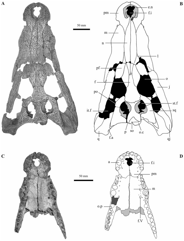
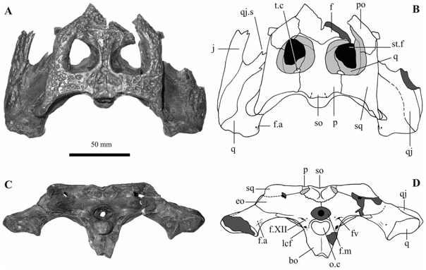
Skull ( Figs 5 View Figure 5 , 6A View Figure 6 )
Preservation, form, and general features: The skull is largely preserved with only the posterior sector of infratemporal fenestrae, the occipital and posteriorpalatal areas severely damaged or incomplete. The only major breakage crosses the snout transversally at the level of anterior tips of the prefrontals: a few millimetres of bone are lacking along this fracture. The preserved elements of the skull do not show any sign of deformation but several bones are separated along the sutures (that are therefore generally well visible), allowing the appreciation of their internal morphology (such as the narial canal of the maxillae). Small restoration integrations with a gypsum-like substance were carried out in the past in order to join some fragments. The estimated length of the skull (from the tip of the premaxillae to the quadrates) is about 400 mm.
The skull outline is markedly festooned: in dorsal view, concavities correspond to the premaxilla– maxilla suture and to the eighth alveolus, whereas convexities correspond to the last premaxillary teeth and the fifth dentary tooth; in lateral view, the convexities correspond to the fourth premaxillary and fifth maxillary teeth (but also the last maxillary teeth) and concavities correspond to the premaxilla– maxilla suture and the seventh interalveolar space on the maxilla.
With the exception of the moderate convexity on the maxillae corresponding to the fifth tooth, the dorsal skull surface is particularly smooth, not showing any median boss, canthi rostralii, or preorbital ridges. The skull table and the sloping interorbital area are rather flat (although not coplanar).
In dorsal view, the skull table has rather long squamosal prongs and nearly straight lateral edges, not parallel but diverging posteriorly about 30°, whereas in posterior view, its dorsal edge is approximately planar.
All the dorsal and lateral surfaces, with the exceptions of the quadrates, are markedly ornate with irregular pits that are particularly well defined, wide and deep, on the frontal and the skull table in general.
Cranial fenestrae and openings: The naris is rather large, about as wide as long, with nearly straight and slightly divergent lateral sides and an anteriorly convex rim. It can be defined as dorsally orientated although the anterior rim is located slightly ventrally to the posterior one. The posterior rim of the naris hosts the tip of the nasals, whose precise development inside the naris cannot be evaluated but seems to be modest. The lateral walls of the naris are not vertical but markedly sloping. The foramen incisivum is anteriorly incomplete but the preserved portion is nearly circular in shape, rather small and placed relatively near to the tooth row. The orbits have a vaguely semicircular shape (with a deeply concave medial margin and a nearly straight lateral one) and are characterized by having margins flush with the skull surface. Although the anterior portion of both orbits is partly damaged, the anterior edge seems to be not vertical but slanting. Palpebral bones are not preserved and, although there are no traces of ligamentary attachment on the prefrontals (such as those visible in extant Osteolaemus spp. ), their presence has not been excluded a priori. The infratemporal fenestrae were much smaller than the orbits; their posterior and medial rims are now broken off on both sides. The supratemporal fenestrae are not completely preserved because the anterior edges and the posterior (postero-medial) walls are partly missing. Their preserved rims do not overhang the fossae and their medial and anteromedial walls are markedly sloping and not pierced by any foramen. The anteromedial corner of each supratemporal fenestra is smooth. The otic aperture is preserved only on the right side of the skull. Although the dorsal sector of its posterior margin is broken, it is clear from the remaining (ventral) part, made by the quadrate, that the posterior margin was not straight. The ventral surface of the otic aperture shows a median convexity. Information on the shape of suborbital fenestrae and the relationships among the surrounding elements is offered by the fragments representing the margins of the fenestrae. The lateral margins are nearly straight or slightly concave. The preserved posterior corner of the right fenestra is not large enough to assess the presence or absence of a notch. However, a modest convexity of the anterior edge of the preserved ectopterygoid could suggest the former presence of such a notch (as testified by the material from Orp-le- Grand). The anterior corners of suborbital fenestrae reach the seventh interalveolar space. An inner section of the trigeminal foramen (= foramen ovale) is visible on the right side of the skull. Because the lateral surface of the braincase is not preserved, it is not possible to assess the configuration of the bones around the foramen. Post-temporal fenestrae and fossae are not entirely preserved: their medial sector is inclined at about 45° and they are likely to have been relatively small. The median Eustachian foramen is only partly preserved but, because of its position, it is clear that it opened ventrally to the small, slit like, right lateral Eustachian foramen (the only one preserved). The foramen magnum is represented by its lower rim, made by the occipital condyle of the basioccipital. With the exception of a small exoccipital fragment of its left lateral edge, all of the bones around the foramen are missing. The cranioquadrate passage opens under a blade-like development (slightly eroded) of the exoccipital and is visible in occipital view; the other end of the passage reaches the postero-ventral corner of the otic recess and is not visible in lateral view.
Skeletal elements: The premaxillae are nearly complete because only a small area anterior to the foramen incisivum (and not reaching the tooth row) is damaged. The premaxillary surface at the posterolateral corner of the external naris is not raised in any particular ridge. A modest, very low, convexity is located on both sides of the skull along the premaxilla–maxilla suture. A deep lateral notch is developed where the premaxilla–maxilla suture reaches the lateral edge of the bone. The premaxillae preserve all the five alveoli and some of them host the remnant of a tooth (there are no crowns). The fourth premaxillary alveolus is by far the biggest. The dorsal premaxillary processes are moderately long and reach the level of the third maxillary alveolus; the ventral ones are not elongated being simple convexities reaching the level of the first maxillary alveolus.
The maxillae are rather well preserved. The right one shows a partial restoration on the dorsal surface. The left maxilla is complete and hosts 14 alveoli, the left one is posteriorly broken and hosts 13 alveoli. Seven teeth are preserved on each maxilla and nearly all have crowns. The largest alveolus is the fifth and it corresponds to a fairly marked convexity (although not as developed as in some extant crocodylid taxa) on the dorsal surface of the maxillae. A shallow concavity is present on each maxilla between this boss and the mild convexity developed along the premaxillary– maxillary suture. Thanks to the fact that maxillae become disarticulated from each other ventrally and from the nasals dorsally, it is possible to see clearly the smooth lateral wall of the narial canal in both maxillae; no caecal recesses are present, but a large foramen (actually two foramina confluent in one single opening) is visible approximately at the level of the fourth tooth. On the palatal surface, the foramen for the palatine ramus of the fifth cranial nerve opens posteriorly to the fifth maxillary alveolus and is significantly larger than the other foramina aligned lingually to the maxillary tooth row. The left foramen (the right one is partly occluded because of the restoration) is nearly as big as the smallest alveolus (the first one; therefore character 111 has been tentatively scored as 1).
Nasals extend from the frontal process (they laterally extend a little posteriorly to the anterior median tip of the frontal process) to the external naris, which they enter slightly. Although their anterior tips are broken off, it is highly probable they did not bisect the naris (as confirmed by the remains from Orp-le- Grand, IRSNB R 252, and Leval, IRSNB R 254). For most of their length they are rather wide compared to the width of the snout, and roughly symmetrically reduce in size in the proximity of both the anterior and posterior tips.
The lacrimals constitute the anterior edge of the orbits and, although they are not perfectly preserved, an elliptic, dorsoventrally depressed lacrimal duct is present medially to the anterior tip of the orbit. The lateral suture with the jugal could correspond to the edge of the preserved lacrimal, whereas the suture with the maxilla is not perceivable with confidence except close to the lacrimals’ anterior tip; however, it is clear that they extend anteriorly more than the prefrontals.
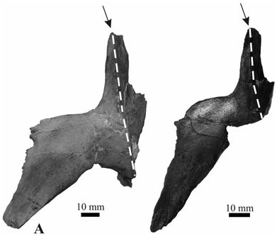
The prefrontals are widely separated by nasals anteriorly and by the frontal process posteriorly. The prefrontal pillars are broken off at mid height but their preserved portions show that they were dorsally expanded in an anteroposterior direction; their medial processes are not preserved.
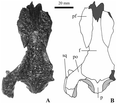
The dorsal surface of the frontal is heavily pitted and nearly flat (almost imperceptibly concave). Its lateral edges, forming the medial margin of the orbits, are not raised. The frontal process (the region anterior to the suture with prefrontals) is approximately as long as half of the total length of the frontal. Its anterior tip is tripartite on the dorsal surface. Although the anterior edges of both the supratemporal fenestrae are not complete, it is possible to state with confidence that the fronto-parietal suture develops entirely on the skull table because the frontal, which is not firmly joined with the parietal, clearly shows a posterior edge entirely represented by sutures (therefore the frontal does not constitute the anterior rim of the supratemporal fossae). The frontoparietal suture is slightly concave anteriorly.
The postorbital bar is slender and presents a small and low spine in an anterolateral position in its dorsal sector. The postorbital is exposed above the anterior process of the squamosal on the lateral surface of the skull table.
The squamosals are particularly flat without dorsolateral convexities (the so-called ‘horns’); they markedly overhang posteriorly on the occipital surface. In lateral view, the dorsal and ventral rims of the squamosal are approximately parallel. The anterior process of the squamosal, along the lateral margin of the skull table, does not reach the middle of the postorbital bar and is not dorsally developed.
The parietal is nearly complete (its posterior left lateral area is broken off). The area exposed on the skull table is X-shaped, with small anterior branches and wider and longer posterior branches. The lateral margins of the parietal, constituting the medial vertical wall of the supratemporal fossae, widen in a ventrolateral direction forming a gently sloping surface. The anterolateral edges merge rather gradually with the sloping anteromedial surface of the supratemporal fossae. On both lateral sides of the large supraoccipital, the parietal constitutes a small section (about 5 mm long) of the posterior edge of the skull table. The posterior region of the dorsal surface, along with the dorsal surface of the supraoccipital, is slightly concave. Very small ridges are developed along the medial rim of the supratemporal fenestrae (not significantly altering the nearly flat aspect of the skull table).
The right quadrate is better preserved than the left one, although its condyle is medially eroded. Its dorsal surface is nearly entirely devoid of any ornamentation but shows some rugosities in its medial sector. Anteriorly to the otic aperture, it hosts a rather large foramen (foramen aerum; after Iordansky, 1973). The left condyle has an expanded medial hemicondyle. The foramen aerum on the dorsal surface of the element is rather small and placed close to the medial edge.
Only the posterior part of both the quadratojugals is preserved. Although the right element is better preserved than the left one, we are not able to assess its relationships with the jugal at the posterior corner of the infratemporal fenestra, the position and development of the quadratojugal spine, and the development of the quadratojugal anterior process along the lower temporal.
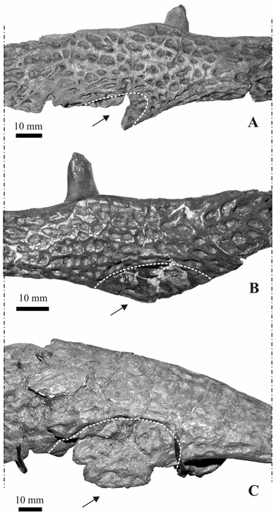
The right jugal is better preserved than the left, whose caudal region is missing. The medial jugal foramen is extremely large (it is about 14 mm long) on the left jugal and is represented by two large openings on the right one. The jugal extends on the lateral surface of the postorbital bar up to the spine placed on its anterolateral surface. The postorbital bar is inset from the lateral surface of the jugal; it originates from the medial surface of the jugal and is separated from the dorsal edge of the latter by a gutter. In lateral view, the dorsal edge of the jugal is approximately straight anteriorly to the postorbital bar and is modestly concave at the level of and posterior to the bar itself, without developing a distinct notch. A very well-marked depression ( Fig. 7A View Figure 7 ) is located on the lateral surface of the jugal at the base of the descending process that reaches the ectopterygoid. This depression is at least dorsally and posteriorly limited by a continuous evident ridge (like the ridges separating the pits on jugal outside the depression but larger). The ornamentation of the jugal surface inside the depression is represented by large and shallow pits contrasting with the smaller and deeper ones on the rest of the jugal surface.
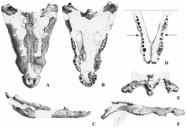
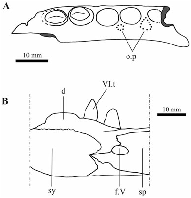
The supraoccipital is perfectly preserved. Its dorsal exposure is considerable because although it does not exclude the parietal from the posterior edge of the skull table, it is a triangle with a base of 28 mm and a height of 15 mm. Its posterior edge markedly overhangs on the posterior (occipital) surface (note that, despite not being delimited by sutures, the area corresponding to the supraoccipital exits from the posterior profile of the skull in the original drawing by Blainville, 1855, here reproduced in Fig. 1A View Figure 1 ). The occipital exposure of the supraoccipital is rather wide and narrow (about 45 mm wide and approximately 15 mm tall); it hosts two small lateral concavities symmetrically placed, but not a marked longitudinal ridge.
The vomer is not exposed on the palate.
Only the right palatine is preserved. It is rather narrow and its lateral edge is approximately straight and slightly flared anteriorly (not producing any lateral process) but not posteriorly. The palatine–pterygoid suture is developed anteriorly to the posterior end of the suborbital fenestra (although laterally not far from the posterior corner of the fenestra, character 85 has been tentatively scored as 1). The anterior tip is missing and therefore the shape of the maxillary–palatine suture cannot be evaluated, but because of the presence of part of such a suture on the left maxilla it seems that it was broad and rounded. The palatine appears not to have extended anteriorly to the suborbital fenestrae because the maxillae extend (medially) beyond the anterior corner of the fenestrae.
The left ectopterygoid is nearly completely preserved ( Fig. 6A View Figure 6 ), but as its posteroventral tip is broken off and the corresponding pterygoid is not preserved, it is not possible to evaluate the relationships between these two elements. The anterior process of the ectopterygoid nearly touches the last three alveoli of the maxillary: its tip, pointed and not deeply forked, reaches the anterior edge of the third last alveolus and it is quite close to the last two alveoli (without forming their medial rim). The dorsal ectopterygoid process is considerably developed along the postorbital bar and it forms its medial sector.
The pterygoids are represented by two fragments only. One is a fragment of the left pterygoid coming from the anteromedial sector, close to the sutures with the right pterygoid and the left palatine. It preserves the posteromedial corner of the left suborbital fenestra and shows a marked concavity on its ventral surface. The second fragment belongs to a right element and represents a small area lateral to the Eustachian foramen and the internal choana; the posterior pterygoid process is preserved and is rather small, as high as it is long, and points in a posteroventral direction. The pterygoid–ectopterygoid suture is visible on the left ectopterygoid where it represents the medial edge of the preserved fragment: it is approximately straight with a modest lateral bend in the anterior sector (because the bend is only weakly developed, character 116 has been scored as 0).
The basisphenoid is not preserved ventrally to the medial Eustachian foramen but as the braincase is anteriorly truncated on the transversal section, this element shows the two symmetric openings of the foramen caroticum anterius. A lamina of the basisphenoid is preserved on the right lateral side of the basioccipital.
Only a small fragment of the right laterosphenoid is preserved above the section of the trigeminal foramen. As a result of the presence on the frontal ventral surface of the scars for the suture of the laterosphenoid capitate processes, it is possible to state that these processes were anteroposteriorly directed.
There is no trace of a boss in the area lateral to the opening of the cranio-quadrate passage. As a result of the incompleteness of the paroccipital processes it is not possible to assess their relationships with the squamosal caudal projections. A fragment of the exoccipital is attached to the left side of the occipital condyle. The exoccipitals are not involved in the occipital tubera.
The basioccipital is represented by two nonadjacent fragments joined together during the restoration of the specimen. A fragment forms the occipital condyle (which is wider than tall) and its base, the other constitutes a part ventral to the occipital condyle. The latter is not laterally expanded and hosts a pronounced vertical crest.
Teeth and dentition pattern: Nearly all of the preserved teeth crowns are markedly worn. The only tooth showing the original morphology is the one in the third right maxillary alveolus. This tooth is sunk in the alveolus and does not show any sings of wear: it could be a substitution tooth in the process of replacing the lost functional one. It is conical, rather slender and pointed, with two marked mesiodistal ridges delimiting a small lingual and a large labial surface. The rest of the teeth are characterized by extensive apical wear so that the apexes are flattened and the dentine is exposed (the enamel is completely eroded). All of these teeth show evident mesiodistal ridges. The tooth surface (not the basal region of the crown) is ornate with wrinkles also partly developed on the mesiodistal ridges. The exposed roots are not smooth but have a markedly crumpled surface.
Very large and deep occlusal pits are developed on both premaxillae caudally and medially to the first interalveolar space (such pits nearly reach the dorsal skull surface within the naris, whose anterior bottom is broken off). Other pits are preserved before and after the fourth premaxillary alveolus: they are developed in the medial sector of the interalveolar space and partly lingually to it (their position is slightly less lingual in the left element than in the right one). On the maxillae, a small quasi-interalveolar pit is present after the first alveolus. Two other small pits develop partly in the interalveolar area but mostly medially to it after the second and third alveoli on the left maxilla and after the second alveolus on the right one. The wide seventh maxillary interalveolar spaces host a very large pit (about double in size if compared to the surrounding alveoli). Much smaller pits are located somewhat lingually (but not completely lingually) to the tooth row after the sixth tooth on the left maxilla and eighth on the right one (the condition was probably symmetrical but because of the poor state of preservation it is not possible to affirm this with confidence). The maxillary tooth row is approximately straight after the large interalveolar pit that fills the seventh interalveolar foramen. The last alveoli have an approximately round section.
SPECIMEN IRSNB R 252 FROM ORP- LE- GRAND
This specimen is represented by a partially preserved skull ( Figs 6B View Figure 6 , 7B View Figure 7 , 8 View Figure 8 , 9 View Figure 9 ) and by the posterior region of the lower jaw.
Skull
Preservation, form and general features: The skull is nearly complete but split into several fragments. In the case of the snout, the general shape is slightly distorted and the bones are slightly separated from each other. The sutures among the skeletal elements of the skull are not visible with confidence on the lateral sides of the braincase and on the occipital surface (with the exception of the limits of the supraoccipital): the interpretative drawings of these areas are not presented because the absence of clearly visible sutures strongly decreases their informativeness.
Cranial fenestrae and openings: Both of the posterior walls of the supratemporal fossae are perfectly preserved and host the strongly elongated and inclined opening (about 14 mm long) of the temporal canal. The quadrate reaches the temporal canal and therefore separates the frontal from the squamosal on the posterior wall of the fossae. Although the infratemporal fenestrae are not completely preserved because of the incompleteness of the postorbital bars and jugals, they seem to be rather small and their posterior medial corner is constituted by the quadratojugals. In addition, the suborbital fenestrae are incomplete but by the shape of the isolated ectopterygoids showing an anterior edge (forming the posterolateral rim of the fenestra) that is distinctly convex, it is possible to state that the fenestra had a posterior notch. On the right maxilla, the suborbital fenestra reaches anteriorly the posterior end of the seventh interalveolar space (where a large interalveolar occlusal pit is developed). The foramen magnum is slightly wider (19.3 mm) than the occipital condyle (18.0 mm). Ventrolateral to the foramen magnum there are the three foramina: the medial one is the small foramen for the cranial nerve XII, the large lateral one is the foramen vagi and the ventral one (only slightly smaller than the latter) represents the lateral carotid foramen that is placed dorsally to the basisphenoid exposure on the lateral side of the braincase.
Skeletal elements: There is some matrix covering the anterior edge of the right orbit but on the left lacrimal it is possible to see an elongated lacrimal duct (developed entirely on the lacrimal) to the right of the anteriormost angle of the orbit. The lateral edge of the lacrimals is not visible. On both lacrimals it seems that there is a depressed elongated nearly parasagittal area placed anteriorly to the anterior end of the orbit.
The frontal dorsal surface is not perfectly flat because of the presence of a modest concavity, but does not develop any ridge along the orbits’ rim. The frontoparietal suture is not entirely visible with confidence but it is not developed inside the perimeter of supratemporal fenestrae and seems to be markedly concave.
The parietal clearly constitutes the anteromedial corner of supratemporal fenestrae. The underlying wall of this corner is not vertical but is markedly sloping. Parietal and postorbital are clearly in contact for about 2 mm on the skull table (this is visible on the left side only because a fracture develops on the right side).
At the dorsal angle of the infratemporal fenestra, the postorbital probably contacts not only the quadratojugal but also the quadrate. The postorbital is visible on the lateral surface of the skull table, posteriorly to the postorbital bar and dorsally to the anterior process of the squamosal.
On the lateral surface, the jugals have a depression ( Fig. 7B View Figure 7 ), dorsally rather well delimited, that occupies the ventral expansion for the suture with maxilla and ectopterygoid. This depression is caudally not as well delimited as in the specimen from Erquelinnes.
The quadratojugals constitute the entire medial rim of the infratemporal fenestrae and their posterior corner. The quadratojugal spine is perfectly preserved in the left infratemporal fenestra where it is placed approximately halfway between the posterior and the dorsal corner of the fenestra; it is prominent but rather small (its medial side is 3 mm long). The left quadratojugal sends a rather long process (about 10 mm) along the lower temporal bar.
The boundaries of the supraoccipital are not visible with confidence on the dorsal skull surface and therefore it is not completely represented in the interpretative drawing in Figure 8 View Figure 8 . Conversely, its sutures are the only ones clearly visible on the occipital surface of the skull, where it is wider than tall and shows two symmetric depressions separated by a median ridge.
The maxillo–palatine suture is not visible with confidence but it is clear that it is not significantly placed anteriorly to the anterior rim of the suborbital fenestra. This suture is probably placed in a highly fractured area immediately posterior to the anterior end of the suborbital fenestrae, and can be tentatively defined as rounded and broad. The palatine– pterygoid suture is not preserved. The lateral edges of the palatines show a modest lateral expansion that is not symmetric on both sides: more posteriorly placed and smooth in the left element, rather irregular on the right element (because of the modest development of this structures and their position, the status of character 94 has been scored as 0). The palatines are posteriorly parallel. It is clear that the palatines are not developed posteriorly to the posterior corner of the suborbital fenestrae and they did not even reach this corner. However, it seems that the zig-zag process of the palatine–pterygoid suture is caudally directed and therefore the caudal extension of the suture is not so far from the corner of the fenestra.
The ectopterygoids do not reach the ventral tip of the pterygoid flanges. Their anterior process grazes the maxillary tooth row and has unbifurcated tips ( Fig. 6B View Figure 6 ).
The pterygoids are not completely preserved but the right one retains the anteromedial corner of the internal choana which therefore opens completely within the pterygoids ( Fig. 9 View Figure 9 ). The pterygoid surface is markedly concave in the area anterior (and maybe also lateral) to the choana. The small (few millimetres long) preserved margin of the choana is not flush with the (concave) ventral surface of the pterygoid indicating the probable presence of a neck surrounding the choana.
The squamosal–quadrate suture cannot be entirely followed in the otic recesses because of the incompleteness of the region. However, the right squamosal is nearly completely preserved in the right otic recess: it ends with a suture showing that the quadrate makes the dorsal edge of the otic recess. The squamosal does not extend ventrolaterally as the lateral extent of the paraoccipital process. On both the sides of the skull, the anterior process of the squamosal reaches the middle of the postorbital bar without excluding the postorbital from the lateral wall of the skull table.
Lower jaw
Preservation, form and general features: The lower jaw is represented by the posterior regions of a right and left dentary, by a left splenial, and a left angular, and by the still-joined left surangular and articular.
Openings: As a result of the presence of a rather well-preserved left angular, it is possible to assume that the (partially preserved) external mandibular fenestra is not anteriorly developed sufficiently to reveal the foramen intermandibularis caudalis in lateral view.
Skeletal elements: The posterior region of the right dentary corresponds to the dentigerous area of the last ten alveoli followed by nearly all of the posterior edentulous area. Interpreting a wide interalveolar space as the diastema corresponding to the eight interalveolar space, the largest alveolus is the 11th, and the total number of teeth of the lower jaw should have been 16. No teeth are preserved.
The left dentary preserves the last seven tooth positions and the posterior edentulous area, with teeth preserved in the first four alveoli. Interpreting the largest alveolus (in this case the second preserved tooth that is larger and taller than the others) as the 11th, the total number of teeth should have been 16.
The right angular delimits the ventral, caudal, and part of the dorsal rim of the large foramen intermandibularis caudalis. The angular process dorsal to the foramen is broken off (in the other angular the foramen is even less preserved).
The surangular reaches the tip of the lateral wall of the glenoid fossa and nearly that of the retroarticular process. The development of the surangular in the ventral direction allows us to suppose that the surangular–angular suture reaches the articular toward its ventral tip. The lingual foramen for the articular artery is not visible with confidence (and therefore character 45 has not been scored).
The articular gradually slopes on the surangular without a sulcus between them. The articular forms the retroarticular process, which is posterodorsally directed. The left articular is rather complete and shows no evidence of an anterior process that hides the medial surface of the surangular. The medial expansion of the retroarticular process is poorly developed because it does not surpass the width of the condyle. The shape of the articular–surangular suture inside the glenoid fossa cannot be evaluated because of the presence of matrix; however, the shape may be ‘bowed’ as in the left lower jaw of the other specimen from Orp-le-Grand because, despite the presence of a brownish painting-like cover, a ‘bowed’ linear depression could possibly represent the sutural line.
Dentition: The only preserved maxillary tooth (on an isolated fragment) is particularly labiolingually compressed, short and with a nearly flat ‘apex’ (it is not significantly worn) so that its shape is approximately rectangular in lateral view (basic measurements are: mesiodistal diameter: 7.1 mm; labiolingual diameter: 3.5 mm; crown height: about 3 mm). Its crown surface is not entirely wrinkled or worn.
The teeth on the left dentary (no teeth are preserved on the right one) are quite different, being pointed (therefore not apically flattened) and approximately triangular in lateral view. The crowns are distinctly labiolingually compressed and bear sharp mesiodistal ridges. Small longitudinal crests and wrinkles are variably expressed on the crown surfaces. Only the last tooth has a crown that is markedly wrinkled (their basic measurements are as follows: first: not measurable because it is embedded in matrix; second: mesiodistal diameter: 8.8 mm, labiolingual diameter, 6.1 mm, crown height: about 9.3 mm; third: mesiodistal diameter: 8.5 mm, labiolingual diameter: 5.0 mm, crown height: 7.4 mm; fourth: mesiodistal diameter: 6.8 mm, labiolingual diameter: 3.9 mm, crown height: estimated as about 5 mm).
Moreover, there are 21 isolated teeth. Their crown shape varies from an elongated and slender shape to a markedly depressed one. Wrinkles are variably expressed.
SPECIMEN IRSNB R 253 AND ASSOCIATED REMAINS FROM ORP- LE- GRAND
This specimen is represented by a partial skull ( Figs 10 View Figure 10 , 11 View Figure 11 ) and lower jaw ( Fig. 12 View Figure 12 ). Several postcranial elements (bearing the same collection number as number IRSNB IG 9875) are associated to these cranial remains and in most of the cases could possibly belong to the same specimen.
Skull
Cranial fenestrae and openings: The small fragment of the posteroventral sector of the braincase clearly shows (on its left side) that the lateral Eustachian foramen opens dorsally to the median one.
Skeletal elements: The premaxillae are anteriorly incomplete. The right one is preserved in the sector corresponding to the last four alveoli, the left one to the last three. Both the premaxillae retain a single tooth: the penultimate. It is likely that both the premaxillae had five alveoli before breakage.
The maxillae are morphologically congruent with those already described for Erquelinnes. The right element corresponds to the first seven alveoli, the left one to the first five. Altogether, six teeth are preserved. A more posterior fragment of the right maxilla (shown in Fig. 13A View Figure 13 ) corresponds to about seven alveoli (five of which preserved) in the area of the suborbital fenestra. The last preserved alveolus and half of the previous one correspond to the sutural area with the ectopterygoid (that therefore is quite close to the maxillary tooth row). Two teeth are preserved in the third and second alveoli from last positions.
The frontal dorsal surface is somewhat more concave than in the larger specimen from the same locality, with lateral edges slightly raised (but with a flat dorsal surface) along the orbits’ rim. The anterior process (anterior to the suture with prefrontal) is about as long as the rest of the bone; its anterior tip is roughly tripartite as in the Erquelinnes specimen. The frontoparietal suture is deeply concave (with a posterior median tip) and entirely developed on the skull table.
The jugal is represented only by the region ventral and posterior to the postorbital bar and shows the
In occipital view the basisphenoid is not visible between the basioccipital and the pterygoids of the isolated fragments of this specimen because of its reduction. A transverse suture is visible inside the median Eustachian foramen. The basisphenoid is visible only on the left side of the basioccipital.
Lower jaw
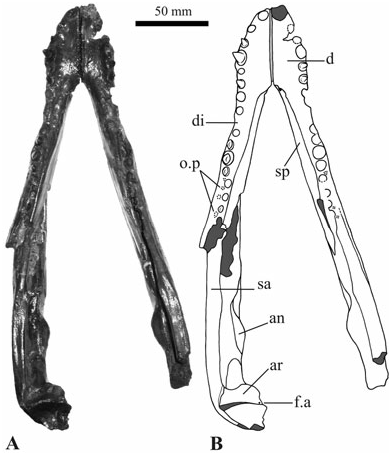
The lower jaw is nearly complete (only the coronoids are missing) but partly damaged (the articular of the right jaw and the retroarticular process of the left are missing). In dorsal view, the general shape of the lower jaw is rather narrow (although some degree of deformation is the result of several breakages with partial displacement of the surrounding elements).
posterior area of the depression described above for the other specimens from Erquelinnes ( IRSNB R 251) and Orp-le-Grand ( IRSNB R 252). The depression is dorsally limited by a well-marked ridge, like that of the Erquelinnes specimen, but not so well developed posteriorly.
The anterior process of the left squamosal reaches the middle of the postorbital bar without excluding the postorbital from the lateral wall of the skull table. Openings: The external mandibular fenestra is present and rather small. It is nearly entirely preserved in the right dentary but its relationship with the foramen intermandibularis caudalis cannot be evaluated because the latter is not preserved. The dentary constitutes the dorsal edge of the fenestra and ends slightly anteriorly to the posterodorsal corner of the fenestra itself.
Skeletal elements: Both the dentaries are not completely preserved but they are somehow complementary in terms of available information. They are markedly festooned in lateral view (convexities corresponding to alveoli four and 11). Their tooth rows are damaged or not complete posteriorly but it seems likely that the maximum number of alveoli is 16 on both sides (a depression of the medial surface of the dentary external wall located after the 16th alveolus does not correspond to a flexure on the dentary dorsal surface and is here not considered as evidence for s 17th alveolus). The first alveoli do not preserve teeth but it is clear that they were directed anterodorsally. The largest alveoli are the first, the fourth, and the 11th. Alveoli three and five are much smaller than the fourth. A diastema is clearly present after the eighth tooth on both the dentaries. The symphysis nearly reaches the posterior rim of the sixth alveolus. Teeth are preserved in positions five and six on the right element and three, four, six, ten, 11, and 12 on the left one. Although the last alveoli are slightly labiolingually compressed (compression possibly partly a result of post-mortem deformation), their shape can be defined as rounded. The splenial constitutes the medial wall of the last alveoli. The dentary upper edge has an evident concavity between the fourth and the tenth alveolus. The dentary–angular and the dentary–surangular sutures reach the external mandibular fenestra nearly at its ventral and dorsal corners, respectively.
Neither of the splenials preserve their posterior region. They reach the symphysis without participating in it. The right splenial, better preserved than the left one, allows us to see that its anterior tip passes ventrally to the Meckelian groove ( Fig. 13B View Figure 13 ) and that the mandibular ramus of cranial nerve V exits through a perforation of the dentary located between the dorsal and ventral anterior splenial processes. As a result of the fact that the anterior tips of the splenial do not join anteriorly to the foramen, it is possible to state that this element does not possess any anterior perforation for mandibular ramus of cranial nerve V.
The angular–surangular suture reaches the external mandibular fenestra at its caudal corner.
The anterior processes of the surangular are not the same length; the dorsal one is longer than the ventral but does not reach the tooth row (assuming that 16 alveoli are present). There is no sulcus between the surangular and articular.
The foramen aerum is placed close to the medial edge of the glenoid caudal boundary.
Teeth and dentition pattern: Premaxillary and maxillary teeth are slender and elongated, with sharp ridges. Their crown surfaces are rather smooth, devoid of any particular ornamentation such as crests or wrinkles. The two maxillary teeth preserved in the third and second from last positions are different because their surfaces are wrinkled.
On the premaxillae ( Fig. 10 View Figure 10 ), occlusal pits are evident on the right element in interalveolar spaces three and four. The first pit is more medially placed than the second, which is entirely developed in the interalveolar position. On the maxillae, all the visible occlusal pits have a position that can be generally defined as interalveolar: they are present in interalveolar spaces number one, two, six, and seven on the right maxilla and one, two, and three on the left one. Only the anterior pit on the right maxilla is significantly (but partly) developed in the medial direction. In contrast, the fragment of a right maxilla corresponding to the suborbital fenestra shows more medially placed occlusal pits ( Fig. 13A View Figure 13 ).
Evident occlusal pits are present in the dentaries lateral to the tooth row. In particular, pits are visible laterally to the 13th alveolus (both in the anterior and the posterior sector), to the 14th alveolus (exactly in lateral position) as well as laterally to the 15th interalveolar space of the left dentary, and laterally to the 12th, 15th, and 16th interalveolar spaces on the right. More pits could be present but because of the dentary preservation they are not visible with confidence.
Postcranial skeleton: A couple of humeri of the same size (130 mm) probably belong to this specimen. The right one is much more damaged than the left one because the proximal epiphysis is nearly completely broken off. A single longitudinal scar for the common insertion of the M. teres major and M. dorsalis scapulae is present on the posterodorsal surface of both the humeri in the proximal epiphysis region, just dorsally to the deltopectoral crest. The latter structure is completely preserved only in the right element, where it is quite sharp and slightly concave.
The ulna is only slightly damaged in its distal epiphysis. Its length (101 mm) matches with the size of the humeri. The large olecranon process is rounded and rather flattened and is markedly constricted at the base.
An isolated left femur (145 mm long) may belong to the same specimen. Although fractured and with the epiphyses partly covered by a matrix rich in pyrites, its morphology is rather well preserved, with the exception of the scars of the M. caudofemoralis, which are not visible with confidence.
Twenty-two vertebrae or vertebral fragments are preserved: one axis odontoid process, two cervical vertebrae, three cervical centra, three postcervical centra, seven dorsal or lumbar vertebrae, one pair of sacral vertebrae, two neural arches, one neural spine, and one isolated chevron bone.
All the presacral vertebrae are procoelous. The neurocentral suture of the cervical vertebrae is invariably open, whereas the postcervical elements have fused neurocentral sutures. The cervical with the fused neural arch has probably been glued. The second vertebra is partly damaged because the right neural arch is missing. The largest dorsal or lumbar vertebra is heavily pyritisized. The pair of sacral vertebrae are characterized by the articular surfaces of the centra: the first one has a centrum anteriorly concave and posteriorly flat, the second one has the opposite condition. The tuberculum and capitulum of the first sacral vertebra are equally developed so that in dorsal view the capitulum is nearly not visible.
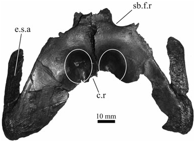
A right ilium is nearly perfectly preserved ( Fig. 14A View Figure 14 ). The acetabulum is wide and deep. An evident narrow scar for muscular insertion is located dorsally to the acetabulum. The iliac blade is approximately rounded and has a very shallow indentation on its dorsal edge. The process located on the anterior edge of the ilium, anteriorly to the dorsal margin of the acetabulum, is weakly developed.
The right ischium lacks the distal anterior portion. The major one of the proximal articular surfaces has a V-shaped articular surface for the ilium, delimiting laterally a smooth surface that represents the ventrolateral surface of the acetabulum.
The pubis does not have the proximal end preserved; only the majority of the flaring distal part and its basis.
A few small elongate bones represent several fragmentary elements of the acropodium, nine ribs or rib fragments, and at least four fragmentary gastralia.
Thirty-nine osteoderms (26 complete or nearly complete and 13 fragments) are preserved. At least the more symmetric ones can be tentatively considered as midline elements. All of them are approximately rectangular (the best preserved has a length of 31 mm, and width of 42 mm), possess a medial keel and have a smooth anterior edge not showing any anterior process. The osteoderms are rather thin and lightly built. The dorsal ornamentation consists of a few small pits and rare furrows (no ridges are present) not completely filling the surface that therefore has large, nearly smooth areas. All of the preserved osteoderms have keels that are not high or sharp. Nearly all have a small anterior smooth gliding surface and sutures are developed on both the lateral sides. The few elongated and roughly oval-shaped osteoderms (the lateral ones) do not show the anterior edge and the lateral sutures, as they were isolated from other osteoderms.
A further osteoderm having the same collection number (so it is the 40th) is quite different in shape and colour (the most of its surface is not as dark as the rest of the material from Orp-le-Grand) and probably belonged to a different specimen. It is smaller (22 mm length, 20 mm width), proportionally more thick, with a large anterior gliding stripe, with many more pits than the osteoderms described above, and with pits larger and deeper and with only a hint of keel. It is perfectly preserved and shows sutures on the sides.
Three other remains bearing the same collection number do not match in size with the remains previous described. A poorly preserved right femur is much bigger (total length of about 180 mm) than the previous one (it could possibly belong to the specimen IRSNB R 252). A small portion of a left femur diaphysis and a rather large and massive tibia (130 mm long) match in size with it.
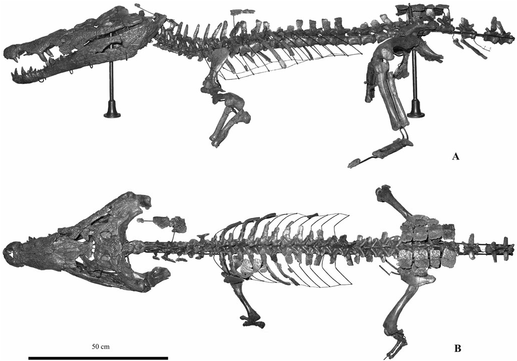
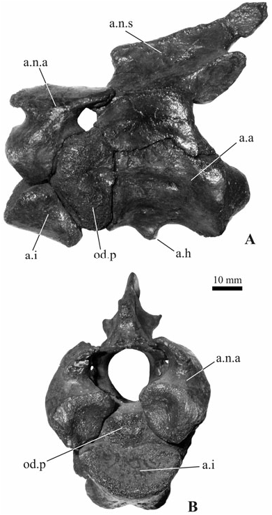
SPECIMEN IRSNB R 254 FROM LEVAL
The specimen from Leval ( Fig. 15 View Figure 15 ) is represented by a nearly complete skeleton (with skeletal elements belonging to one single individual) preserving the skull (damaged and incomplete in the palatal area) and lower jaw, as well as several vertebrae ( Fig. 16 View Figure 16 ), part of the limb bones, and some osteoderms.
The specimen has been mounted on a metal support and thus offers a good chance to observe the general anatomy of this taxon. However, the fact that many cranial sutures are not clearly visible hinders a detailed description. At present, the mounted skeleton is 1760 mm long but only five caudal vertebrae are present (with gaps between them).
Further material coming from the same locality but at least partly not belonging to the mounted specimen is available (see the Referred material and localities section for a list).
Skull
The general shape of the skull in dorsal view is comparable to that figured by Berg (1966: fig. 4) for ‘ Asiatosuchus ’ germanicus , with a wide posterior region and a significantly narrower snout (skull length from the level of quadrates to the tip of the snout: 500 mm). All the available characters match with those already discussed with the other specimens from Erquelinnes and Orp-le-Grand.
A major discrepancy concerns the tooth numbers because the lower jaw has 17 teeth on the right side and possibly 18 on the left one (five premaxillary teeth are visible on both the premaxillae and 13 teeth are probably present on the right maxilla; the left maxillary tooth row is incomplete). It is worth noting that the dentary symphysis reaches the sixth alveolus, but the diastema is not located after the eight alveolus as in the smaller specimen from Orp-le- Grand (and the most of the living crocodylids) but after the ninth: the eighth and ninth teeth are preserved on both the rami and they are fairly small and extremely near (so that that their alveoli could be confluent; however, such a condition cannot be proven because some bone fragments seem to be present between these teeth at least on the left ramus). The largest dentary teeth are the first, fifth, and 12th.
The splenials clearly show an abrupt dorsoventral reduction about 15 mm before the symphysis and it seems that the anterior tip of the splenial nearly reaches, but does not enter, the symphysis.
Although the supratemporal fenestrae do not seem to be intersected by the fronto-parietal suture, the latter is not visible with confidence on the skull table because of several breakages and the restoring plaster.
On the lateral side of the skull table, the postorbital is visible above the anterior process of the squamosal that reaches the postorbital bar.
On both the maxillae, there are very deep occlusal pits before and after the seventh alveolus. They are both interalveolar but the anterior one is placed closer to the lingual edge of the interalveolar space, whereas the posterior one, which is more large and deep, is more centred in the interalveolar space.
As in the smaller skulls previously described from Orp-le-Grand and Erquelinnes, there are neither significant depressions on the skull table or anteriorly to it, nor significant upturned edges of orbits and supratemporal fenestrae. The squamosals have a and the parapophysial processes are weakly developed. The axis neural spine is dorsally damaged but it seems likely that the anterior half of the neural spine was sloping and that its posterior half was not upturned (the corresponding characters, 11 and 12, have been tentatively scored). The neural arch does not seem to have any lateral process. The axis hypapophysis is a prominent tubercle located slightly behind one third of the centrum length; it can be defined as forked. The third cervical vertebra (following Brochu, 1997, here considered as the first after the axis) is well preserved and shows a distinct hypapophysis, and a rather long neural spine whose dorsal tip is about as long as half of the centrum without the cotyle. The neurocentral sutures of all cervical vertebrae are closed. The hypapophysis is present up to the 12th vertebra posterior to the atlas.
The limbs lack almost all the autopodia. The left fore limb (the right one is missing) is shorter than the hind limbs (not considering the missing autopodia) but equally robust. Although the dorsal edge of the preserved scapula is broken off, it is clear that it flares dorsally, whereas it seems likely that the damaged deltoid crest was rather thin. The scapula and the coracoid are not joined together suggesting that the synchondrosis closed later during ontogeny.
The ilia are slightly different from the element from Orp-le-Grand because, although their dorsal notch is modest like in the latter (and unlike in extant Crocodylus ), their outline is dorsally depressed and not rounded at all ( Fig. 14B View Figure 14 ). Nevertheless, there is some asymmetry in the same specimen because the right element is particularly narrow posteriorly.
About a dozen osteoderms are preserved. Those placed along the midline are rectangular and keeled as are some of those described for the Orp-le-Grand specimen, but larger, more heavily built than them, and with a higher number of pits.
nearly flat dorsal surface. The depressions on the lateral surface of the jugals ( Fig. 7D View Figure 7 ) have a morphology only slightly less continuously delimited than in the Erquelinnes specimen.
Postcranial elements: The atlas and axis are rather well preserved ( Fig. 16 View Figure 16 ). The atlas is represented by both the two separated neural arches and the intercentrum (no proatlas preserved), the axis by the odontoid process, the neural arch, and the centrum. The atlas intercentrum is wedge-shaped in lateral view
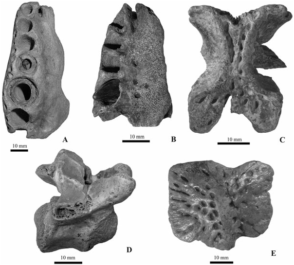
SPECIMEN IRSNB R 255-260 AND DIIA R 50 RS FROM DORMAAL
Two incomplete right maxillae from Dormaal, IRSNB R 255 and R 256 ( Fig. 17A, B View Figure 17 ), are characterized by the following characters: fourth and fifth alveoli completely separated (their interalveolar space is approximately equal to the interalveolar space between the other preserved alveoli) and different in size, with the fourth alveolus being much smaller than the fifth (the fifth alveolus is only partly preserved in specimen IRSNB R 256 but it is clearly larger than the fourth). In both cases, occlusal pits are located towards the lingual side of the interalveolar spaces from one to three. In both specimens, the preserved portions of the medial wall of the caviconchal recess are smooth.
The isolated fragmentary parietal IRSNB R 257 ( Fig. 17C View Figure 17 ) possesses thick anterolateral processes indicating that the frontoparietal suture did not enter the supratemporal fenestrae. Its dorsal surface is moderately thickened and raised in correspondence to the rim of the supratemporal fenestrae and weakly concave in the area posterior to the fenestrae.
The skull table fragment DIIA R 50 RS preserves the area corresponding to the supraoccipital and posterior region of the parietal. The morphology of the preserved part of the parietal is similar to that of the remains previously described and the supraoccipital is significantly exposed on the skull table.
The incomplete first caudal vertebra IRSNB R 259 ( Fig. 17D View Figure 17 ) has a centrum 30 mm long and a closed neurocentral suture.
The isolated dorsal osteoderm IRSNB R 260 ( Fig. 17E View Figure 17 ) is comparable in size (width 45 mm, length 35 mm) and general morphology to the osteoderms of the skeleton from Leval.


| MP |
Mohonk Preserve, Inc. |
| IRSNB |
Institut Royal des Sciences Naturelles de Belgique |
| R |
Departamento de Geologia, Universidad de Chile |
| IG |
Institute of Geology |
| V |
Royal British Columbia Museum - Herbarium |
No known copyright restrictions apply. See Agosti, D., Egloff, W., 2009. Taxonomic information exchange and copyright: the Plazi approach. BMC Research Notes 2009, 2:53 for further explanation.
|
Kingdom |
|
|
Phylum |
|
|
Class |
|
|
Order |
|
|
Family |
|
|
Genus |