
Amurinocellia sinica Liu, H. Aspöck & U. Aspöck, 2009
|
publication ID |
https://doi.org/ 10.5281/zenodo.190814 |
|
DOI |
https://doi.org/10.5281/zenodo.6221954 |
|
persistent identifier |
https://treatment.plazi.org/id/03C3878A-3572-8C1A-FF71-241FFDD1FCC0 |
|
treatment provided by |
Plazi |
|
scientific name |
Amurinocellia sinica Liu, H. Aspöck & U. Aspöck |
| status |
sp. nov. |
Amurinocellia sinica Liu, H. Aspöck & U. Aspöck View in CoL , sp. nov.
( Figs. 1–3 View FIGURES 1 – 3 , 6–15 View FIGURES 6 – 15 )
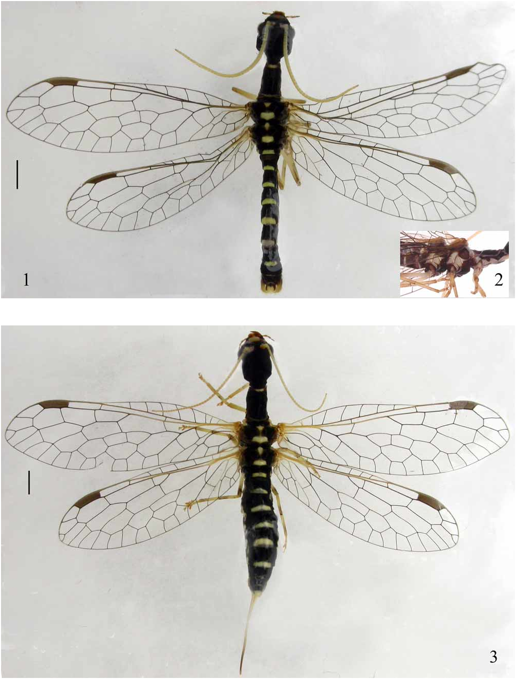
Diagnosis. Eidonomically this species is characterized by the yellowish antennae and by a series of yellowish stripes on the meso- and metanota as well as the terga of the pregenital segments. The male ninth gonocoxites roundly produced on posterodorsal corners, with the pseudostyli (gonapophyses 9) obliquely directed dorsad.
Description. Male. Body length 10.5 mm; forewing length 8.0–8.5 mm, hindwing length 7.0–7.5 mm.
Head ( Fig. 1 View FIGURES 1 – 3 ) subquadrate, black, with anterior clypeus pale yellow. Compound eyes blackish brown. Antennal sclerite (torulus) and antennae pale yellow throughout. Mouthparts yellowish brown except for mandibles blackish brown with distal half reddish brown; maxillary palpi slightly darkened toward apex, labial palpi mostly blackish brown.
Thorax blackish brown; anterior margin of pronotum pale yellow; meso- and metanota anteriorly with a yellowish median spot, posteriorly with a yellowish transverse marking on scutellum, and lateral portions of meso- and meta thorax each with a broad yellowish marking ( Fig. 2 View FIGURES 1 – 3 ). Legs yellow with yellowish setae except for coxae blackish brown; apices of femora and middle portions of tibiae slightly darker. Wings hyaline, pterostigma brown, veins blackish brown, longitudinal veins proximally pale yellow except for anal veins. Anterior branch of Rs with one forked vein and one simple vein running to wing margin.
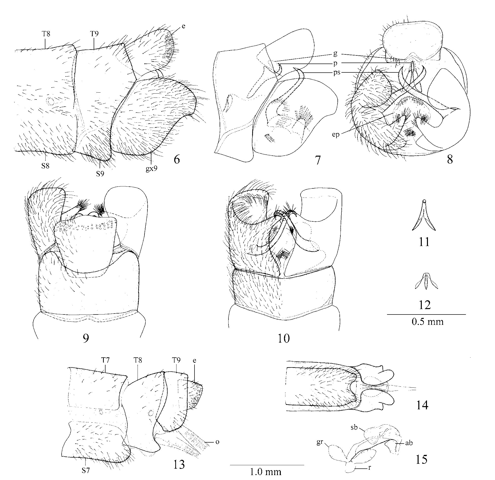
Abdomen blackish brown; each pregenital segment posteriorly with a yellowish band-like stripe; genital segments blackish brown with ninth gonocoxites pale yellow ( Fig. 1 View FIGURES 1 – 3 ). Ninth tergite ( Figs. 6, 9 View FIGURES 6 – 15 ) with anterior margin slightly incised medially, posterior margins with wide arcuate incision. Ninth sternite ( Figs. 6, 10 View FIGURES 6 – 15 ) arcuate, with anterior margin slightly prominent. Ninth gonocoxite ( Figs. 6–10 View FIGURES 6 – 15 ) nearly as long as ninth tergite, posteriorly with a broad semicircular dorsal lobe and a digitiform ventral process. Pseudostyli (gonapophyses 9) forming a pair of long hook-like sclerites between gonocoxites and obliquely directed dorsad with subproximal portion strongly curved; in lateral view posterodorsal corner of gonocoxites 9 distinctly and roundly prominent, ventral process invisible; in ventral view posterior margin nearly truncate, ventral process with a tuft of bristles at tip, anteroventral portion with a tuft of bristles. Fused parameres (gonocoxitegonapophyses-gonostyli complex 10) ( Figs. 7–8 View FIGURES 6 – 15 ) with proximal portion flattened and strongly expanded ventrad, forming a subtriangular plate, ventrally incised into a pair of narrow lobes; distal process slender and hook-like. Gonarcus (gonocoxites 11) ( Figs. 7–8 View FIGURES 6 – 15 ) membranous, almost obliterated. Endophallus ( Figs. 7–8 View FIGURES 6 – 15 ) short, with a pair of bristle tufts laterally and numerous bristles at tip. Ectoproct ( Figs. 6, 9 View FIGURES 6 – 15 ) subquadrate, slightly widened toward apex, in lateral view posterodorsal corner roundly prominent. Hypandrium internum ( Figs. 7, 12 View FIGURES 6 – 15 ) well developed, lateral lobes posteriorly widened and twisted.
Female. Body length with ovipositor 15.0 mm, body length without ovipositor 11.0 mm; forewing length 10.0 mm, hindwing length 8.5 mm.
Coloration similar to male ( Fig. 3 View FIGURES 1 – 3 ); genital segments with ectoproct and ovipositor pale yellow.
Seventh sternite ( Figs. 13–14 View FIGURES 6 – 15 ) in lateral view subtrapezoidal with posteroventral corner rounded, in ventral view posterior margin with distinct incision, forming a pair of obtuse processes. Eighth tergite ( Figs. 13–14 View FIGURES 6 – 15 ) strongly expanded ventrad, nearly enveloping ventral portion of eighth abdominal segment, with posteroventral corners slightly produced into a pair of obtuse processes. Subgenital plate absent. Ectoproct ( Fig. 13 View FIGURES 6 – 15 ) in lateral view subquadrate, slightly narrowed toward apex. Atrium bursae ( Fig. 15 View FIGURES 6 – 15 ) rounded, slightly pointed anteriorly; sacculus bursae with apex bulbiformed; receptaculum seminis short, with a pair of Material examined. Holotype ♂ [preserved in alcohol], CHINA: Henan Province, Neixiang County, Baotianman, 33°25'N, 111°53'E, 16.V.2006, Weihai Li (CAU). Paratypes 1♂ [preserved in alcohol], CHINA: Henan Province, Neixiang County, Baotianman, 1300 m, 28.V.2006 (CAU); 1Ƥ [preserved in alcohol], CHINA: Henan Province, Neixiang County, Baotianman, 1300 m, 25.V.2006, Yusi Cui (CAU).
Distribution. China (Henan).
Etymology. The specific epithet ‘ sinica ’ (adjective, feminine) refers to the geographic distribution; possibly the new species is endemic to China.
Remarks. At first glance the new species differs from A. calida by the male ninth gonocoxites with the posterodorsal corners roundly prominent, the gonapophyses 9 obliquely directed dorsad, the presence of a pair of bristle tufts on the anteroventral portions of the male ninth gonocoxites, and essentially by the gonocoxitesgonapophyses-gonostyli complex 10 with the proximal portion strongly expanded ventrad, and the female seventh sternite with a pair of obtuse posterior processes. In the holotype of A. calida , the male ninth gonocoxite has the posterodorsal corner produced into a triangular lobe, however after having studied additional material of this species (H. Aspöck et al. 1991), considerable variation of the apex of the gonocoxites 9 was found: in all these specimens the apex is more or less rounded and not triangular as in the holotype. Another discrepancy concerns the pseudostyli (gonapophyses 9) appearing horizontally directed, and without tuft on its anteroventral portion. In the additional materials mentioned above the gonapophyses 9 rather resemble those of A. sinica , this might reflect true variability or just an effect of position or swelling by maceration. The most stable character seems to be the proximal portion of the parameres (gonocoxitegonapophyses-gonostyli complex 10) which is not expanded ventrad, in the holotype of A. calida , and in the additional specimens studied. The posterior incision of the female seventh sternite is V-shaped, forming a pair of triangular posterior processes.
No known copyright restrictions apply. See Agosti, D., Egloff, W., 2009. Taxonomic information exchange and copyright: the Plazi approach. BMC Research Notes 2009, 2:53 for further explanation.